




IntelliPaper
Abstract
Intracranial complications of rhinosinusitis in children are rare. Symptoms are nonspecific, and diagnosis relies on a high index of suspicion and early imaging evaluation. Treatment should be multidisciplinary and may involve antibiotics alone or in combination with surgery to drain the sinus focus and intracranial abscess. We describe four cases of intracranial complications in pediatric patients who were successfully managed with a multidisciplinary approach.
Explore Digital Article Text
I. INTRODUCTION
Intracranial complications of sinusitis are uncommon and may include epidural and subdural empyema, meningitis, cerebral abscess, and venous sinus thrombosis. Approximately 4% of sinusitis cases in children result in intracranial complications . Management of these complications requires a multidisciplinary approach.
Due to the rapid onset of intracranial complications, nonspecific symptoms, and the absence of neurological signs at the onset of the disease, a high index of suspicion is necessary to diagnose an intracranial complication resulting from a sinus infection.
We describe four clinical cases of pediatric patients with intracranial complications secondary to sinusitis who were successfully treated with a multidisciplinary approach.
II. CLINICAL CASES
Case 1: Epidural Abscess and Orbital Cellulitis An 11-year-old girl presented with pain during eye movement and mild upper eyelid swelling. She had a history of hospitalization at another centre 15 days prior for sinusitis complicated by orbital cellulitis. Blood cultures isolated Group A Streptococcus. She was initially treated for nine days with Ceftriaxone 1g/day and Clindamycin 2.7g/day, then switched to Amoxicillin-Clavulanic Acid 600mg/5ml for seven days.
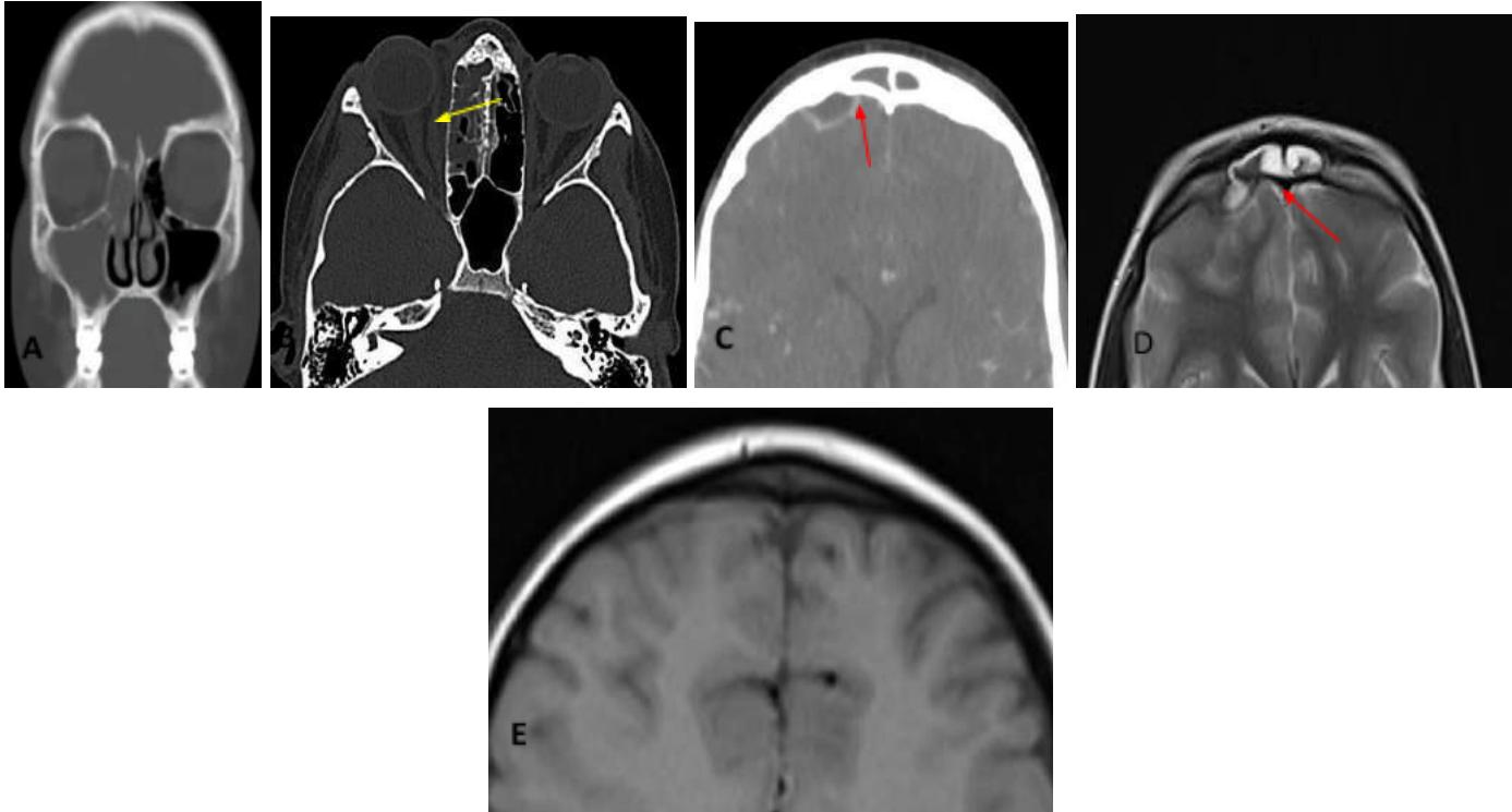
Hospitalization was recommended; her complete blood count and ophthalmological examination were average. Computed tomography (CT) with intravenous contrast revealed right-sided sinusitis and thickening of the soft tissues in the medial aspect of the right orbit. At the same time magnetic resonance imaging (MRI) showed a hypodense lesion consistent with an epidural abscess.
The multidisciplinary decision was to treat with Ceftriaxone 1500mg every 12 hours and Ornidazole 720mg/day intravenously for six weeks via a peripherally inserted central catheter (PICC) to complete the treatment on an outpatient basis.
Follow-up MRI four months later showed complete resolution of the sinusitis and the epidural abscess without neurological sequelae. Fig. 1
 Figure 1: Epidural Abscess and Orbital Cellulitis
A: Computed tomography with intravenous contrast, coronal section: right maxillary-ethmoidal Sinusitis, B: CT, axial section: inflammatory thickening of the soft tissues in the medial aspect of the orbit (yellow arrow), C: Magnetic resonance imaging showing an epidural abscess with an anteroposterior diameter of 7 mm and a transverse diameter of 15 mm (red arrow), D: Post-treatment MRI showing resolution of the complication.
Case 2: Epidural Abscess and Longitudinal Sinus Thrombosis A 12-year-old patient presented with frontoparietal headache, nasal obstruction, and fever of 72 hours duration. Two days prior, the patient had been seen at another hospital and was prescribed symptomatic medication. Examination revealed a tender mass in the left temporal region.
Hospitalization was indicated. Blood cultures isolated Streptococcus anginosus (Streptococcus anginosus group) and Haemophilus influenzae. CT and MRI diagnosed sinusitis and a left subtemporal collection. Intravenous treatment was initiated with Ampicillin 100 mg-Sulbactam 500 mg every 6 hours.
Three days later, the patient experienced a decline in consciousness and required respiratory support. A follow-up CT revealed a left frontal subdural collection. MRI and magnetic resonance angiography of the brain showed a left frontal subdural empyema and partial thrombosis of the longitudinal sinus.
Antibiotics were switched to Vancomycin 300 mg every 6 hours for seven days and Ceftriaxone 1500 mg/day for six weeks, along with Dexamethasone 4 mg every 6 hours. Four days later, the patient was successfully extubated.
Imaging for control showed resolution of the sinusitis and recanalization of the longitudinal sinus but no reduction in the subdural abscess. Subsequent images revealed diffuse edema of the left cerebral hemisphere with characteristics of encephalitis, and ophthalmological examination detected bilateral papilledema.
A frontotemporal craniectomy and durotomy were performed to drain the empyema. The patient had a good recuperation and underwent cranioplasty with a prosthesis eight months later. There were no sequelae. Fig. 2
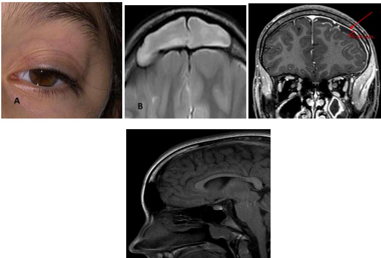 Figure 2: Subdural Abscess with Longitudinal Sinus Thrombophlebitis and Encephalitis
A: Edema in the left temporal region, B: MRI showing frontal sinusitis, C: MRI showing a subdural abscess with a maximum thickness of 5.66 mm, D: Post-treatment MRI showing resolution of frontal sinusitis and subdural empyema.
Case 3: Epidural Abscess, Potts' Tumor, and Frontal Osteomyelitis A 10-year-old girl presented with headache, fever, and cough of 24 hours' duration. Chest and craniocafial X-ray were normal. Nasal washes with hypertonic saline were recommended.
Four days later, she returned with persistent headache, fever, and a mass in the frontal region. There were signs of meningeal involvement, motor deficits, and the pupils were isocoric. CT diagnosed left-sided sinusitis with edema of the frontal soft tissue and an extradural empyema. MRI revealed a frontal extradural empyema and an anterior frontal subcutaneous collection. Blood cultures were negative.
The patient was admitted and treated with intravenous Ceftriaxone 2000 mg every 12 hours and Metronidazole 500 mg every 8 hours for four weeks. Due to persistent symptoms, Vancomycin 750 mg every six hours were added to complete a 6-week course of antibiotics.
Follow-up MRI showed persistence of the empyema and the anterior frontal subcutaneous collection. Bone scintigraphy with Tc99 diagnosed frontal osteomyelitis. An endonasal drainage of the affected paranasal sinuses and a frontal craniectomy were performed simultaneously to drain the empyema. The histopathological report of the resected bone confirmed osteomyelitis.
She continued with antibiotic treatment (Ceftriaxone 2000 mg/day and Teicoplanin 10 mg/kg/day) for 45 days. The patient had a good recovery and no residual infection or sequelae. Follow-up was done every two months, and cranioplasty was performed at eight months. Fig. 3.
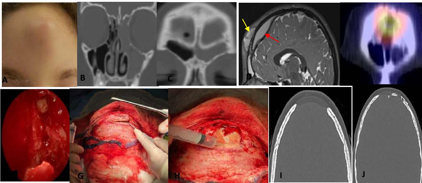 Figure 3: Epidural Abscess, Potts' Tumor, and Frontal Osteomyelitis
A: Frontal Potts' tumour, B and C: Facial CT, coronal sections showing left maxillary-ethmoidal and frontal Sinusitis, D: MRI with contrast showing an epidural abscess measuring 37 x 14 x 59 mm (red arrow) and a Potts' prefrontal abscess measuring 17 x 7 mm (yellow arrow), E: Scintigraphy with technetium showing frontal osteomyelitis, F: Endonasal view with the endoscope of the drainage of the left paranasal sinuses. G: Frontal craniectomy, H: Drainage of the abscess and sample collection for culture, I-J: CT showing resolution of the abscess and evidence of craniectomy and cranioplasty.
Case 4: Subdural and Cerebral Abscess A 15-year-old male patient presented with severe headache and sudden deterioration of consciousness, leading to referral and admission to the intensive care unit. He had been treated seven days earlier with Amoxicillin and Sulbactam for sinusitis.
CT and MRI diagnosed left-sided sinusitis and subdural abscesses. Treatment was initiated with Ceftriaxone 2000 mg/day, Metronidazole 500 mg every 8 hours, and Rifampicin 15 mg/kg/day intravenously.
Due to unfavourable progression after seven days, endonasal drainage of the affected paranasal sinuses and subdural empyemas was performed through a decompressive craniectomy. Direct examination identified positive cocci and gram-negative bacilli, but cultures were negative.
The patient remained on mechanical ventilation for 13 days. He completed seven weeks of antibiotic treatment and recovered without neurological sequelae. Fig. 4
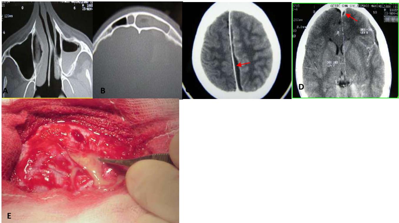 Figure 4: A-B: Facial CT with contrast, axial sections showing left maxillary-ethmoidal-frontal sinusitis, C-D: MRI showing frontotemporal and occipital subdural abscesses (red arrows), E: Craniectomy and drainage of intracranial abscesses
III. DISCUSSION
The mortality rate from intracranial complications of sinusitis decreased from 25.9% between 1950 and 1979 to 3.8% between 1980 and 2004 .
Another study conducted between 2006 and 2016 reported an increased rate of intracranial complications from sinusitis, rising from 2.2% to 4.3% . This rise may be attributed to increased antibiotic resistance, a higher prevalence of children with immune-suppressive conditions or treatments, poor medical management, or delays in surgical intervention.
Intracranial complications can arise from direct extension through dehiscences in the bony walls of the paranasal sinuses or through retrograde thrombophlebitis of the valveless veins of the diploë of the bone.
The frontal sinus is the most common source of intracranial complications.
In a study of 16 patients with intracranial complications from sinusitis, 56% (9/16) had a subdural abscess, 44% (7/16) had an epidural abscess, and 19% (3/16) had a cerebral abscess. Meningitis was diagnosed alone or in association with other abscesses in 19% of the patients. Additionally, 35% (5/16) had more than one complication, with multiple abscesses. Two patients had intracranial abscesses associated with frontal bone osteomyelitis (Potts' tumour) .
An epidural abscess is a purulent collection located between the skull and the dura mater. One study reported that 60% of patients with epidural abscesses had frontal bone osteomyelitis .
The management of epidural abscesses in children is controversial and generally involves intravenous antibiotic administration alone or in combination with surgical drainage. Some studies have shown that patients with epidural abscesses without neurological deficits or elevated intracranial pressure can be treated with antibiotics alone for six weeks without requiring neurosurgical drainage . Other studies have suggested medical treatment for abscesses \<1 cm and neurosurgical drainage for abscesses >1 cm .
In selected cases with epidural empyemas located behind the posterior table of the frontal sinus, endonasal drainage via endoscopes through a Draf II or III frontal approach may be feasible. In Case 3, the epidural collection was located in the superior region above the frontal sinus, making this approach unsuitable.
Additionally, bone scintigraphy confirmed frontal bone osteomyelitis, necessitating craniectomy to resect the infected bone and drain the abscess.
Subdural empyema (purulent collection between the dura mater and arachnoid) requires early neurosurgical drainage, along with drainage of the sinus focus and intravenous antibiotic administration, due to its rapidly fatal progression . In Case 2, craniectomy was deferred until the patient was stable after requiring respiratory support.
A study reported that 58% of children with intracranial abscesses were treated with otolaryngological and neurosurgical procedures .
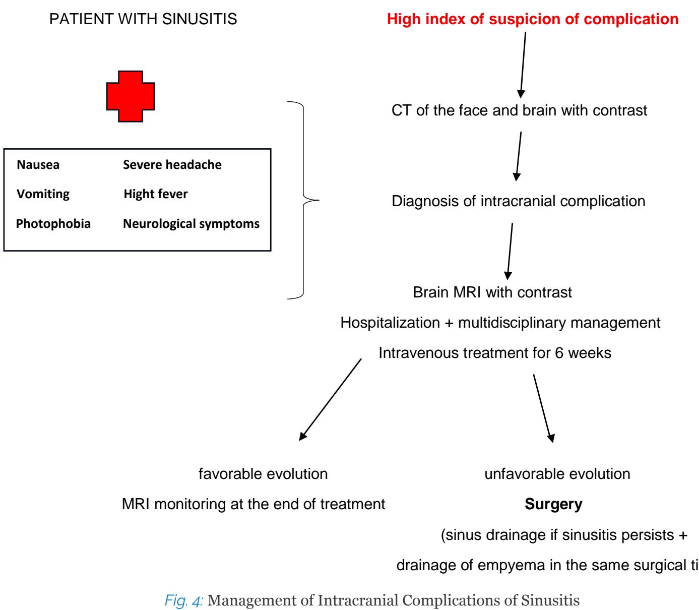
Another study compared patients under eighteen years old with complicated acute frontal sinusitis with non-contiguous intracranial abscesses. The study assessed whether frontal sinusitis treated via endonasal approach, intracranial access, or leaving the sinus undrained impacted the number of surgeries or complications. The authors concluded that frontal drainage did not reduce the number of surgeries or increase complications, and the benefit of frontal drainage was not clearly established . Potts' tumour is a subperiosteal abscess caused by osteomyelitis of the anterior table of the frontal sinus. It represents an extracranial complication of sinusitis but is associated with intracranial complications in 60-85% of cases . The treatment of Potts' tumour should include intravenous antibiotics and, if unresolved, abscess drainage and resection of the osteomyelitis bone . Fig.4
Conflicts of Interest
The authors declare no conflicts of interest.
Conflict of Interest
The authors declare no conflict of interest.
Ethical Approval
Not applicable
Data Availability
The datasets used in this study are openly available at [repository link] and the source code is available on GitHub at [GitHub link].
Funding
This work did not receive any external funding.
References
Cite this article
Related Research



Special Issue
Launch a focused special issue to highlight research, emerging trends, and expert insights in your academic field.
