


IntelliPaper
Abstract
The eyes are a set of sensory organs that play a crucial role in the visual system [1]. Visual stimuli from our environment are processed by a complex system of interconnected neurons that begins with the optic nerve in the eye and extends to the visual processing center in the forebrain - the visual cortex [2].
The eye is an extension of the brain and contains nerve tissue in the light-sensitive layer, the retina, which is related to brain tissue, and the outer membrane are an extension of the medulla [3]. The optic nerve has capillaries that have barrier properties due to the presence of dense interendothelial cell connections. In addition, the optic nerve has a well-developed, hemato-tissue barrier, which is formed by the pial vessels, the intermediate tissue of Kunt and the border tissue of Jacobi, located between the chorioidea and the optic disc (OD). All of these tissues are composed of astrocytes. However, the blood-tissue barrier has defects, which allows some substances, including antigens, to penetrate through it choriocapillary endothelial cells’ fenestrations also contribute to the penetration of various substances into the bloodstream [4, 5, 6].
Most of the biological material is freely filtered through the tissue of the Jacobi tissue border and enters the optic nerve prelaminar region [3, 7, 8]. Thus, there is a circulation of certain substances from eye’s certain compartments into the bloodstream and from the bloodstream into the eye’s compartments, including the visual analyzer tissues. Consequently, the nervous tissues of the eye are not adequately protected from the body's innate immune system.
The eye is one of several organs and tissues with immune privileges [9, 10]. The term “immune privilege” was introduced by Peter Medawar to show that the eye is exempt from the laws of transplantation immunology. Nevertheless, the impetus for the term came from the research of Dutch ophthalmologist van Doormal more than 150 years ago. In experiments on mice, he showed that the eye’s anterior chamber protects allografts from rejection by the body's immune system [18]. Anterior chamber’s this feature was the impetus for the study eye’s immune system and its interaction with the innate immune system [11, 12].
Immune privilege is an active process in which certain tissues and the innate immune system cooperate to protect the eye from autoaggressive damage. The mechanisms that contribute to immune privilege include primarily tolerance of peripheral T-cells. Antigens entering the eye are taken up by local antigen-presenting cells, which migrate through the blood and initiate an immune response. This initiates the specific antibodies’ synthesis in the spleen [11, 13, 14] and specific T-cells are formed in the thymus. Most of them are eliminated under the target antigen influence expressed by the eye tissues itself, but some of them - gets back into the bloodstream and, accordingly, into the visual analyzer system [6, 9, 10].
Thus, because of the visual analyzer tissues’ structure peculiarities, the eye antigens collide with the innate immune system. This encounter leads to the inevitable immune response development culminating in the specific immunoglobulins formation and sensitized lymphocytes that enter the bloodstream [11, 15, 16]. Consequently, there is a circulation not only of eye tissues’ antigens, but also of collision products with the innate immune system.
In this regard, it is legitimate to assume that because of visual analyzer tissues’ structure some features, eye antigens’ certain amount encounters the innate immune system. As a result, immunoglobulins and leukocytes specifically sensitized to the eye tissues and, among them, to the visual analyzer tissues’, inevitably appear in the circulation. Because immune responses are essential defense elements against foreignness and inflammation, the eye has developed distinct mechanisms that provide an immune response to avascular tissues' injury to the eye. It is now known that injury and/or pathology in the eye’s avascular regions triggers an immune system response that culminates in fibrosis that impairs vision [17, 18, 19].
Our early studies have shown that in the blood of practically healthy person (PHP), patients with glaucoma and keratitis circulates sensitized to the nerve, trabecular and lens tissues' antigens leukocytes. Their number clearly correlates with the pathology presence and its severity expression [20, 21, 22, 23, 24]. We explained this finding with the constantly occurring in almost all organs and tissues natural regeneration processes.
Since primary open-angle glaucoma (POAG) belongs to neurodegenerative eye diseases [10, 25, 26], we investigated the PHP and POAG patients’ circulating leucocytes sensitization degree to optic nerve (ON) and optic disc (OD) tissue antigens. Moreover, the study results showed the presence in the PHP and POUG patients’ peripheral blood leukocytes specifically responding in vitro to ON and OD tissue antigens [23]. As a result, the question arose about the PHP’ peripheral blood leukocytes sensitization to other tissue antigens of the visual analyzer.
Explore Digital Article Text
I. INTRODUCTION
The eyes are a set of sensory organs that play a crucial role in the visual system . Visual stimuli from our environment are processed by a complex system of interconnected neurons that begins with the optic nerve in the eye and extends to the visual processing center in the forebrain - the visual cortex .
The eye is an extension of the brain and contains nerve tissue in the light-sensitive layer, the retina, which is related to brain tissue, and the outer membrane are an extension of the medulla [3]. The optic nerve has capillaries that have barrier properties due to the presence of dense interendothelial cell connections. In addition, the optic nerve has a well-developed, hemato-tissue barrier, which is formed by the pial vessels, the intermediate tissue of Kunt and the border tissue of Jacobi, located between the chorioidea and the optic disc (OD). All of these tissues are composed of astrocytes. However, the blood-tissue barrier has defects, which allows some substances, including antigens, to penetrate through it choriocapillary endothelial cells' fenestrations also contribute to the penetration of various substances into the bloodstream .
Most of the biological material is freely filtered through the tissue of the Jacobi tissue border and enters the optic nerve prelaminar region . Thus, there is a circulation of certain substances from eye's certain compartments into the bloodstream and from the bloodstream into the eye's compartments, including the visual analyzer tissues. Consequently, the nervous tissues of the eye are not adequately protected from the body's innate immune system.
The eye is one of several organs and tissues with immune privileges [9, 10]. The term "immune privilege" was introduced by Peter Medawar to show that the eye is exempt from the laws of transplantation immunology. Nevertheless, the impetus for the term came from the research of Dutch ophthalmologist van Doormal more than 150 years ago. In experiments on mice, he showed that the eye's anterior chamber protects allografts from rejection by the body's immune system [18]. Anterior chamber's this feature was the impetus for the study eye's immune system and its interaction with the innate immune system [11, 12].
Immune privilege is an active process in which certain tissues and the innate immune system cooperate to protect the eye from autoaggressive damage. The mechanisms that contribute to immune privilege include primarily tolerance of peripheral T-cells. Antigens entering the eye are taken up by local antigen-presenting cells, which migrate through the blood and initiate an immune response. This initiates the specific antibodies' synthesis in the spleen and specific T-cells are formed in the thymus. Most of them are eliminated under the target antigen influence expressed by the eye tissues itself, but some of them - gets back into the bloodstream and, accordingly, into the visual analyzer system [6, 9, 10].
Thus, because of the visual analyzer tissues' structure peculiarities, the eye antigens collide with the innate immune system. This encounter leads to the inevitable immune response development culminating in the specific immunoglobulins formation and sensitized lymphocytes that enter the bloodstream [11, 15, 16]. Consequently, there is a circulation not only of eye tissues' antigens, but also of collision products with the innate immune system.
In this regard, it is legitimate to assume that because of visual analyzer tissues' structure some features, eye antigens' certain amount encounters the innate immune system. As a result, immunoglobulins and leukocytes specifically sensitized to the eye tissues and, among them, to the visual analyzer tissues', inevitably appear in the circulation. Because immune responses are essential defense elements against foreignness and inflammation, the eye has developed distinct mechanisms that provide an immune response to avascular tissues' injury to the eye. It is now known that injury and/or pathology in the eye's avascular regions triggers an immune system response that culminates in fibrosis that impairs vision [17, 18, 19].
Our early studies have shown that in the blood of practically healthy person (PHP), patients with glaucoma and keratitis circulates sensitized to the nerve, trabecular and lens tissues' antigens leukocytes. Their number clearly correlates with the pathology presence and its severity expression . We explained this finding with the constantly occurring in almost all organs and tissues natural regeneration processes.
Since primary open-angle glaucoma (POAG) belongs to neurodegenerative eye diseases , we investigated the PHP and POAG patients' circulating leucocytes sensitization degree to optic nerve (ON) and optic disc (OD) tissue antigens. Moreover, the study results showed the presence in the PHP and POUG patients' peripheral blood leukocytes specifically responding in vitro to ON and OD tissue antigens [23]. As a result, the question arose about the PHP' peripheral blood leukocytes sensitization to other tissue antigens of the visual analyzer.
Purpose of the Study: The purpose of this investigation was to determine in vitro the practically healthy young persons' circulating leukocytes' sensitization degree to visual analyzer tissue antigens.
II. MATERIALS AND METHODS
120 practically healthy volunteer students (PHS) of the M. Garryev State Medical University of Turkmenistan aged 18 to 25 years without vision problems were examined. All of them underwent a standard ophthalmologic examination at the International Eye Disease Treatment Center of Turkmenistan. Besides the peripheral blood leukocytes' sensitization degree to optic nerve's tissue antigen (ONTA), optic disc's (ODTA) and retina's (RTA) tissue antigens were determined.
The material for the antigens was obtained at males aged 18 to 22 years autopsies who died from accidental injury. The leukocytes' sensitization degree to tissue antigen (TA) was determined in a modified leukocyte's migration inhibition reaction (LMIR) [27]. Antigens were used as leucocytes' migration inducers and they were prepared by water-salt extraction method following the recommendations [28]. Antigens were dosed according to protein concentration, which was determined by the Lowry method [29]. The protein concentration in the antigens was at least . The antigens (1.0 mL) were stored in disposable microtubes in a freezer at -20 °C. All of TA were melted once before the study.
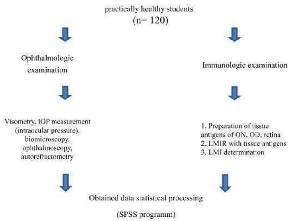
During the study, the 0.05 mL of antigens was added into the incubation medium of chambers with capillaries. An equal amount of 0.9% sodium chloride solution was added to the control chamber. The leukocytes' number that migrated into the control chamber (without antigens) was taken as 100% and on relation to this, the leukocyte's migration index (LMI) was calculated. The study design is presented in Fig. 1.
Approval was obtained from the local ethics committee (M. Garryyev State Medical University of Turkmenistan). The study was conducted in compliance with the ethical standards of the responsible institution on human subjects as well as with the Helsinki Declaration.
Study Results and their Discussion
Ophthalmologic examination showed that despite the absence of visual impairment complaints, it was not so of the all subjects (Fig. 2).
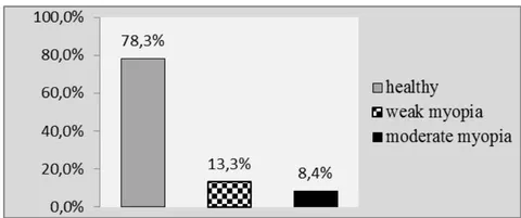
As can be seen on the diagram, visual acuity corresponding to the norm was detected only in 94 out of 120 PHSs (78.3%). Weak myopia was detected in 13.3% of PHSs and moderate myopia in 8.4%. In other words, in a quarter of the PHS cases there was hidden, undiagnosed myopia.
Immunologic examination showed that on the whole group all antigens - ONTA, ODTA, RTA in vitro modulates the PHS' peripheral blood leukocytes' migration activity, i.e., both stimulate and inhibit the leukocytes' migration from the glass capillary. This is evidenced by the fluctuations in the LMI values. The LMI value in the ONTA's presence ranges from 34 to 156, in the ODTA - from 29.3 to 121 and RTA - from 46 to
- The LMI average values depending on antigens are presented in the table (Table 1).
| Group | Tissues antigens | ||
| ONTA | ODTA | RTA | |
| PHS | |||
| Group | Tissues antigens | ||
| ONTA | ODTA | RTA | |
| Common (n=120) | 76,4 ±4,9 | 79,4 ± 5,0 t2, | 50,8 ± 3,8 |
| Healthy (n=94) | 47,3 ±4,9* | 64,3 ± 5,0* | 58,6 ± 3,8 |
| Weak myopia (n=16) | 81,3 ± 7,2** | 81,3 ± 5,1** | 46,6±3,1 |
| Moderate myopia (n=10) | 53,2 ± 6,7 | 98,3 ± 8,9** | 40,6 ± 5,1* |
From the table's data, we can see that in the group of students with normal vision the LMI value in the ONTA and ODTA presence are the minimal. Since the LMI value is below 100, the obtained results show, first of all, about peripheral blood leukocytes' migration inhibition in vitro in the visual analyzer' tissue antigens presence.
Secondly, obtained results show about the leukocytes presence specifically responding to these tissue antigens in the blood circulation of healthy young people, i.e. sensitized to them.
In the group of students with myopia the LMI value in the ONTA and ODTA presence is significantly increased (p<0.01), but in the RTA presence is slightly lower compared to the healthy students' group (p>0.05).
Differences in the LMI value depending on the tissue antigen type are illustrated by the diagram (Fig. 3), which clearly shows that with increasing myopia's severity the blood leukocytes' sensitization degree to the ONTA increases.
{"image_source":{"path":"images/e14b354d81500c6f71aa03c73b07c30c9d55c200f05b551cc0d8676e5dd07c2e.jpg"},"content":"","chart_caption":[{"type":"text","content":"Fig. 3: LMI Values Depending on the Antigen and Vision Type"}],"chart_footnote":[]}
At the same time, the leukocyte response to retinal antigen in vitro progressively decreases and at the myopia's average degree the difference becomes reliable in relation to healthy individuals (p<0.05). Immune response to ON antigen is significantly increased in myopia (p<0.05), but decreases in relation to the students' group with myopia (p<0.05), but practically does not differ from the control level (p>0.05). The increase in the LMI value in the ONTA and ODTA presence can be explained, the decrease in the LMI value in the presence of RTA is difficult to explain. Previously, we found that the increase LMI value in the presence of tissue antigens in vitro indicates the autoimmune attack development on the tissue in vivo, while the LMI value decrease, on the contrary, indicates the degenerative-sclerotic process development in the tissue [45]. In our opinion, the LMI value increasing in the RTA presence in myopia indicates the development of an inflammatory process in the tissue, whereas a LMI decreasing on increasing myopia's severity indicates the degenerative-sclerotic process development in the retina. It is even more difficult to explain the absence of positive correlation between the LMI value in the RTA, ONTA and ODTA presence (Table 3).
| Pirson's Correlation (r) | RTA | ODTA | ONTA |
| RTA | 1 | -,625 | -,815 |
| ODTA | -,625 | 1 | ,962 |
| ONTA | -,815 | ,962 | 1 |
As it can be seen from the table, the LMI value in the RTA presence is inversely correlated with ONTA (r=,815) and ODTA LMI (r=-,625), while the ONTA and ODTA LMI values are connected by a high direct correlation (r=,962).
III. CONCLUSION
The life of an organism, the work of its various systems, organs, cells, the diversity of its reactions to external influences are accompanied by the old structures replacement with new ones. The renewal process takes place at all tissues organization levels - organ, cellular, molecular. That is, we are talking about the constantly running regeneration process in the body. Regeneration is the material basis for the adaptation and compensation processes to disturbed functions. Control over all processes of organism renewal is carried out by the immunobiologic surveillance system (IBSS) [30, 31, 32]. As a result, eye's many areas have evolved mechanisms to deliver immune cells to dysgenesis sites, injury, or in response to many age-related pathologies [14]. Although the immune reactions' goal is reparative or protective, cytokines secreted by immune cells impair vision acuity by causing inflammation and fibrosis [33, 34, 35]. It is possible that RTA plays a role as a target antigen, that inhibiting the immune response and therefore preventing the autoaggression' development toward the ON and OND tissue antigens.
Thus, the results of our studies have shown that leucocytes, specifically sensitized to visual analyzer tissues' antigens - optic nerve, optic nerve disc and retina – circulates in the healthy persons' peripheral blood. It is possible, the LMI value in the RTA presence can be used as an early immunologic marker of myopia development. In our opinion, the negative correlation between the RTA, ONTA and ODTA LMI suggests that the RTA is a target antigen protecting the ON and OD from autoaggression. Studies in this direction seem to us promising, shedding light on the immune system nature involvement in the visual analyzer pathology development.
ACKNOWLEDGMENTS
None to declare
Financial Disclosure
The authors received no financial support from any funding agencies for this study.
Conflict of Interest
None to declare.
Informed Consent
Informed consent was obtained.
Author Contributions
This work was carried out in collaboration among all authors. All authors read and approved the final manuscript.
Data Availability
The authors declare that data supporting findings of this study are available within the article.
Abbreviations OD: optic disc; PHP: practically healthy person; POAG: primary open-angle glaucoma; ON: optic nerve; PHS: practically healthy students; ONTA: optic nerve's tissue antigen; ODTA: optic disc's tissue antigen; RTA: retina's tissue antigen; TA: tissue antigen; LMIR: leukocyte's migration inhibition reaction; LMI: leukocyte's migration index; IOP: intraocular pressure;
Conflict of Interest
The authors declare no conflict of interest.
Ethical Approval
Not applicable
Data Availability
The datasets used in this study are openly available at [repository link] and the source code is available on GitHub at [GitHub link].
Funding
This work did not receive any external funding.
References
Cite this article
Related Research

Special Issue
Launch a focused special issue to highlight research, emerging trends, and expert insights in your academic field.