

Abstract
BACKGROUND: Penetrating macular holes of the retina remain today one of the main reasons for a significant loss of central vision, especially in working age patients. At the same time, the mainstream problem are the validity and effectiveness of treatment of large and giant macular holes with a diameter of more than 500–1000 µm.
AIM: to optimize the treatment method for patients with large and long-standing macular holes. To analyze the results of treatment of patients with this condition.
MATERIALS AND METHODS: In 2023, 56 patients were operated on for idiopathic large macular hole. During vitrectomy, a new surgical technique was used for the macular hole bed tamponade with flaps of the internal limiting membrane in combination with the introduction of platelet-rich plasma.
RESULTS: After the resorption of the gas-air mixture (1–1.5 months after surgery), ophthalmoscopically and according to optical coherence tomography data, closure of the macular hole was observed in 51 of 56 operated patients, which amounted to 91.1%.
CONCLUSIONS: The surgical treatment technique used allows for closure of large macular holes in 91.1%.
Explore Digital Article Text
I. BACKGROUND
Penetrating macular holes (MH) of the retina remain one of the main reasons for a significant loss of central vision, especially in working-age patients. Penetrating MH - acquired disease, at which a full-thick-ness defect is observed in the foveolar part of the retina — from the internal limiting membrane to the exterior segment of the photoreceptor layer [1]. The yearly prevalence of this disease is 8.69 cases per 100,000 people [2]. The peak incidence falls on the sixth-seventh decade of patients' life; women. are susceptible to this disease 3 times more often than men. In 15–20% of cases, the macular hole develops in both eyes. The occurrence of full-thickness macular holes leads to progressing decrease of visual acuity, appearance of metamorphopsias, and this significantly reduces the patients' quality of life.
The primary method of treatment for macular hole patients is a surgical procedure aimed at the closure of the anatomical defect of the retina in any way, what predetermines future increase of visual functions. To this date, the micro-invasive 25G or 27G vitrectomy with staining and removal of the internal limiting membrane (ILM) to increase the mobility of macular hole's edges with subsequent air-gas tamponade is commonly believed to be a gold standard of treatment for MH patients. As a rule, this method alone allows good anatomical results in treatment of small macular holes (of a diameter up to 400 m) with disease duration up to 6 months.
Among additional mechanisms of intraoperative closure the retinal defect in the foveolar area, the following directions are highlighted [3]:
Use an inverted Internal Limiting Membrane (ILM) flap (flaps) or fragment, which is not entirely detached from the macular edge.
Mechanical opposition or approximation macular defect edges.
Use of bioadhesive substances - of platelet-rich plasma (PRP) or of autologous conditioned plasma (ACP), of autologous blood.
Without an additional use of above-mentioned methods in treatment of large (minimal diameter more than 400 m) and old (existing more than 6 months) macular holes, acceptable anatomical results are not always achieved. The detection rate of macular holes of III–IV stages (according to the classification by J.D. Gass), according to data of various authors, amounts to 86–93% [4]. In the case of recurrent failure of MH closure, after surgery, it often increases in dimensions, its edges become more rigid, and the patient's visual acuity worsens [5]. If surgical treatment of patients with MH diameter up to 400 m, according to the majority of the authors' data, is predictable and highly effective (the closure rate reaches 96–97%), the maximal problem today is the reasonableness and effectiveness of treatment of large and giant MH with diameter more than 500–1000 m. The anatomical success in such cases does not exceed 57–80% [4–9].
The use of an inverted ILM flap method and its variants described by various authors demonstrates a significant increase of the anatomical success in cases with large MH (more than 400 m) in comparison with the method of the ILM peeling and removal [10–13]. A formed free ILM flap is sufficiently mobile, could be easily divided from the foveolar edge when performing manipulations and by intraocular flows, and its fixation in the lumen of the hole demands from the surgeon performing additional measures. Some authors for the positioning of the ILM flap practice intraoperational introduction of perfluorinated compounds (PFCs), viscoelastics, autologous blood, use of silicone tamponade [6–9, 11, 14].
Thus, further studies and search for effective treatment modes for this surgical problem are actual and sought-after.
The aim of the study is to analyze the results of treatment of patients with large (minimal diameter more than 400 m) and long-term (more than 6 months) existing MH. To optimize the treatment method for patients with this disease.
II. MATERIALS AND METHODS
The distribution of patients according to the diameter of the hole base (maximal diameter) is presented in the diagram (Fig. 1).
{"image_source":{"path":"images/defb827a21dba220fc420ca67769ed4b0a1a969022071b8716250f8a13c5da27.jpg"},"content":"","chart_caption":[{"type":"text","content":"Fig. 1: Distribution of Patients According to the Diameter of the Macular Hole's Base"}],"chart_footnote":[]} In 22 patients (39%) and 23 patients (41%), MH dimensions were from 700 to 1,000 m and from 1,000 to 1,300 m at the base respectively; in 11 patients (20%), giant MH were noted - from 1,300 to 1,600 m.
Surgical procedures were performed by the same surgeon on the Constellation device (Alcon, USA) using the Lumera 700 microscope (Carl Zeiss, Germany). Into the study were not included patients with diabetic retinopathy, glaucoma of III–IV stages, high myopia, retinal vascular occlusions and their sequelae, advanced manifestations of macular degeneration. In all patients were carried on standardophthalmological examination, as well as optical coherence tomography (OCT) of the macular area in dynamics by the optical coherence tomograph Zeiss Cirrus HD-OCT 5000 (Germany). During the surgical procedure, a combination of the
After fluid-air exchange (at control of the position of ILM flaps, 2–3 drops of platelet-rich plasma were introduced on the macular areausing a 25 G tamponade of the hole bed by a flap (flaps) of the ILM and of the introduction of the platelet-rich plasma into the hole area after the fluid-air exchange was used.
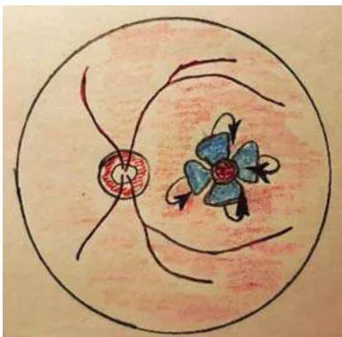
In all patients was performed posterior vitrectomy (25 G), with the removal of the posterior hyaloid membrane of the vitreous. After the ILM staining (Membrane-Blue-Dual dye, DORC, the Netherlands), its peeling was performed, concentrically to the hole edges as several flaps in such a way as to preserve the adhesion of them with macular hole edges. Hereafter, peripheral ends of the flaps were evened by vitrector and placed on the hole's bed using closed ends of endovitreal forceps, delicately, without efforts, producing the hole's tamponade. In our study was used the inverted ILM flap technique, according to N. Andrew, et al. [15](Fig. 2).
cannula; the substance was obtained from the patient's autologous blood using a centrifuge (Rotofix 32A, Hettich, Germany). At the end of the procedure, into the vitreous cavity was added, the CF gas (Alcon, USA), the volume being about 1 ml to obtain 20–25% gas-air mixture. There were no intraoperational complications noted.
In the post-op period, patients received standard anti-inflammatory therapy, they were recommended to stay in a face-down position or on the contra-lateral side during 3–4 days after the procedure.
III. RESULTS AND DISCUSSION
To solve the problem, we were guided the following principles:
The use of an ILM flap (flaps) is, to our mind, a requisite condition and a sufficient measure for the large diameter hole tamponade. As ILM is connected to the neurosensory retinal tissue, it does not pose a risk of pathological changes in its structure, at the same time, the ILM flap is transparent and does not reduce the transparency of optical media.
Not to use a mechanical approximation of the hole edges because of their rigidity and high risk of retinal tissue damage.
To stimulate the healing of the hole's edges and to increase the ILM flap stabilization during the post-op period, it was decided to use the platelet-rich plasma, obtained from the patient's autologous blood drawn immediately before surgery.
To exclude a toxic effect on the retina and the optic nerve, as well as to avoid re-operations, it was decided to refrain from the use of PFO and from the silicone tamponade.
During the early post-op period, there were no complications. Short-time rise of intraocular pressure, caused by the gas-air tamponade, was reversed by a local use of hypotensive medications. After the gas-air mixture resorption, ophthalmoscopically and according to OCT data, there was a MH closure in 51 out of 56 operated patients, making 91.1%. In 5 patients (8.9%), a residual hole in the macular area persisted. The visual acuity of patients before surgery and to the moment of complete gas-air mixture resorption in the vitreous cavity is shown in the table.
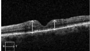
According to the data presented in the table, before surgery, in the overwhelming number of patients (85.8%), visual acuity was from 0.01 to 0.1, during the post-op period, in a significant number of patients (76.8%), visual acuity raised up to 0.2–0.3 and higher. As examples, pre- and postoperative OCT results of patients L. and G. and their visual functions are shown on Fig. 3 and 4, respectively.
| Visual acuity (with correction) | 0.01–0.05 | 0.06–0.1 | 0.2–0.3 | 0.4–0.8 | In total |
| Before surgery | 15 (26.8%) | 33 (59%) | 8 (14.2%) | 0 | 56 |
| After surgery | 0 | 13 (23.2%) | 29 (51.8%) | 14 (25%) | 56 |
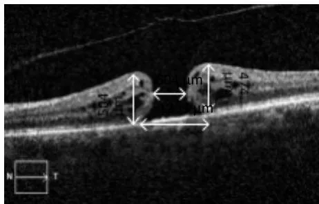
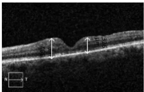
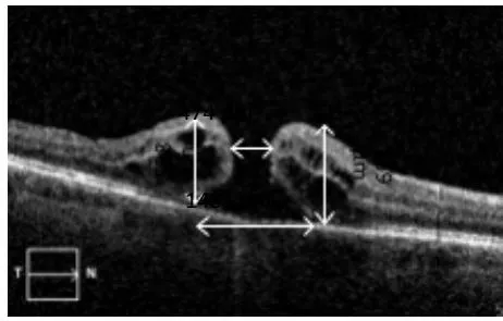
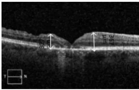
Most of patients after surgery noted an increase in quality of vision. However, despite the anatomical MH closure, many patients, along with a visual acuity increase, noted the preservation of metamorphopsias in one form or another, the necessity to "look closely" by visual acuity testing. We could explain this phenomenon by displacing the fixation point on the background of decreasing retinal oedema after the healing of MH edges. At that, a scotoma could be found in the area of the fovea itself, taking into account large dimensions of observed macular hole along with scar tissue formation.
Evaluating the anatomical outcome of surgery based on the OCT data, it has to be mentioned that in all studied cases. the ellipsoid zone of the foveola was deformed without significant dynamics after successful surgery. A complete MH closure was observed in 51 out of 56 patients (91%). When investigating the cases of macular hole closure, conspicuous is the fact that retinal oedemawas absent in 22 cases (42%), and in 30 eyes (58%), positive dynamics were observed in the form of oedema decrease, restoration of the structure of most of the macular area layers was visualized in 27 (53%), traces of the interior limiting membrane flap was absent in 33 patients (63%), foveolar pseudocysts were visualized in 11 cases (21%), fibrotic changes of external retinal layers at long-term (more than 8 months) were observed in 15 eyes (29%). Quoted data are of preliminary character, as the dynamic follow-up of patients continues.
The central retinal thickness in the foveal area is a new, highly informative index for the prognosis of the anatomical effect of macular hole surgical treatment, superior in prognostic value to most other criteria . In our sample, this index before surgery was , after surgery, the average thickness in the foveolar area decreased by a mean of , and amounted to (Fig. 5).
{"image_source":{"path":"images/0f4499a3e8826856772dd7a2b824408d1a9d789e6d02ea8184a4dae4e4095343.jpg"},"content":"","chart_caption":[{"type":"text","content":"Fig. 5: Distribution of Mean Retinal Thickness before (1) and after (2) Surgical Treatment of the Macular Hole"}],"chart_footnote":[]} In one female patient with moderate degree myopia, 2.5 months after surgery, the development of rhegmatogenous retinal detachment was observed, the cause for which was the appearance of a peripheral retinal tear. In this patient, cerclage scleral buckling was performed, resulting in a blockage of the tear and reattachment of the retina.
IV. CONCLUSIONS
The used surgical method of the MH bed tamponade with ILM flaps in combination with the introduction of platelet-rich plasma allows to achieve a closure of large MH in 91.1% of cases.
After surgical treatment, many patients mentioned the preservation of scotomata and metamorphopsias, this is due to the formation of scar tissue in the hole area.
Further investigations of this problem will serve as a reason for future publications.
Conflict of Interest
The authors declare no conflict of interest.
Ethical Approval
Not applicable
Data Availability
The datasets used in this study are openly available at [repository link] and the source code is available on GitHub at [GitHub link].
Funding
This work did not receive any external funding.
References
Cite this article
Related Research

Special Issue
Launch a focused special issue to highlight research, emerging trends, and expert insights in your academic field.