





IntelliPaper
Abstract
INTRODUCTION: Pseudo-papillary and solid tumors of the pancreas are rare tumours, of uncertain etiopathogenesis. We report the difficulty of managing a pseudopapillary tumor in our country, where resources were limited. Our objective was to discuss the therapeutic management in relation to the literature.
OBSERVATION: This is a 19 year old young women. View in consultation for chronic abdominal pain, without family neoplastic history. Abdominal palpation revealed an intra-abdominal mass syndrom. Abdominal computed tomography had shown a heterogeneous solidocystic tumor mass in the corporeo-caudal region, well encapsulated, well limited, without infiltration, without peripheral lymph nodes with homogeneous contrast uptake, without vascular invasion or remote secondary location. This allowed us to decide on pancreatic enucleation by laparotomy. The diagnosis was confirmed by histological study of the surgical specimen. The evolution was favorable without remote metastasis or recurrence after two years of follow-up.
Explore Digital Article Text
I. INTRODUCTION
Solid pseudopapillary tumor of the pancreas is a relatively rare tumor, most often found in young women[1,2]. In general, it is a tumor with a low potential for malignancy [3.4]. It is a tumor with clinical polymorphism. These tumors are now recognized with increased frequency thanks to the progress and improvement of imaging techniques. We report a case of pseudo-papillary tumor of the pancreas, which was treated by pancreatic enucleation, in a 19-year-old young woman seen at the Joseph Ravoahangy Andrianavalona University Hospital of Antananarivo (CHU-JRA). Our objective was to discuss therapeutic management compared to the literature.
II. OBSERVATION
Young 19-year-old woman, with no family history of neoplastic presenting with heaviness-type epigastric pain, transfixing, intense, without calming factors or triggering factors, evolving for a year before her admission, worsening, becoming more accentuated and sometimes associated with vomiting for two weeks. She was in good general condition, afebrile. Palpation of the abdomen showed a bulky, firm, rounded, well-defined epigastric mass, mobile in relation to the deep plane, and painful on palpation. The lymph node areas were accessible. Physical examinations revealed lipaemia 1.4 times normal, neutrophil-predominant hyperleukocytosis, AST 1.3 times normal, GGT 1.1 times typical, total bilirubin elevated three times everyday, and bilirubin conjugated at 1.8 times average, the other biological tests were standard (CA 19-9, fasting blood glucose, CRP, alkaline phosphatase, anti-nuclear auto-antibodies, anti-neutrophil cytoplasm auto-antibodies). Abdominal ultrasound revealed a solid intra-abdominal mass hypoechoic, heterogeneous, rounded, well limited, vascularized on Doppler, localized at the level of the left hypochondrium, of pancreatic origin.
Abdominal computed tomography showed a heterogeneous solid cystic tumoral mass in the corporeal-caudal region, well-encapsulated, well limited, without infiltration, without peripheral lymph nodes with homogeneous contrast uptake, and without vascular invasion or remote secondary localization. (Figure 1).
Surgical treatment was performed by midline supraumbilical laparotomy; on exploration: there was no peritoneal carcinomatosis. After the opening of the posterior cavity of the omentums, we found a rounded and well-defined tumor sitting at the corporate-caudal level of the pancreas with no visible lymphadenopathy. A frozen section histological examination was performed, showing a well encapsulated tumor. A lumpectomy by pancreatic enucleation was performed (Figure 2).
The anatomopathological study of the surgical specimens showed a brownish piece, firm, well encapsulated, measuring 12x8x7 cm, of heterogeneous brownish-white content with necrotic-hemorrhagic changes and a well-encapsulated lymph node of 1cm long axis without tumor invasion or vascular embolism. On histological examination, it was a solid pseudopapillary tumor of the pancreas with reactive lymph node hyperplasia.
The postoperative follow-up was simple after a postoperative hospital stay of seven days without adjuvant therapy without complications of pancreatic fistula. With a follow-up of 2 years, the patient is in good general condition with no clinical or CT scan recurrence.
III. DISCUSSION
The pseudopapillary tumor of the pancreas is a rare exocrine pancreatic tumor in the order of 2% and accounts for less than 5% of cystic tumors of the pancreas [4]. It was described by Frantz in 1959[5]. They are low-grade malignant tumors composed of poorly cohesive uniform epithelial cells forming solid, pseudopapillary structures. [4]. There are less than 1000 cases reported in the literature, mainly in the form of isolated instances. Similar to our patient, it is a predominantly female tumor with a sex ratio of 0.18. It occurs at a young age, with an average age of 29.3 years [6]. Its pathogenesis remains poorly understood; its female predominance may be linked to hormonal factors . Most of the series in the literature find a black or Asian ethnic origin, such as our patient .
The preferential localization of this tumor is corporeal-caudal in 64% of cases. However localizations at the level of the head of the pancreas or even extra-pancreatic can be found. [1,2]. In our case, the tumor was found in the body of the pancreas. It is a solid mass delimited by a capsule and associating areas of hemorrhage, necrosis, and calcifications. Symptoms are generally not specific, often incidental discovery of an abdominal mass, or abdominal pain with signs of digestive compression, depending on the size of the tumor [9] which explains the symptoms of our patient by compression. There are no specific biological signs of the pseudopapillary tumor of the pancreas. The immunohistochemistry examination, coupled with the histological examination, poses the diagnosis of certainty of the disease[10]. Immunohistochemistry is a very efficient examination but not routinely available in our country. Pancreatic pseudopapillary tumors are usually located in the tail of the pancreas. It is a large tumor containing solid and cystic components due to necrosis, hemorrhage, and cystic degeneration. The radiological aspect of the cancer shows a well-encapsulated, heterogeneous complex mass with solid and cystic components[1]. Abdominal ultrasound, most often finds a well-limited cystic mass with regular contours, poorly vascularized, with heterogeneous contents and no interior partitions, such as our case. Computed tomography is the most requested examination and better than ultrasound in terms of precision, which finds the same characteristics on ultrasound with partial enhancement in the periphery after injection of the contrast product without invasion of neighboring organs and possible intra-tumoral hemorrhage and calcification consistent with our case [1].
Despite the superiority of MRI over computed tomography and ultrasound, our patient could not benefit from magnetic resonance imaging because of the very high cost. In addition, the clinical symptoms associated with ultrasound and computed tomography were sufficient to establish the indication for primary surgery.
We proceeded to surgery alone without preoperative biopsy, thanks to anamnestic, clinical, and radiological evidence. According to the literature, the reference treatment is complete resection by cephalic duodenal-pancreatectomy, if the tumor is located in the head or body and partial pancreatectomy with partial splenectomy in the event of a tumor located in the tail of the pancreas, especially in case of capsular rupture or invasion of neighboring organs , In our case, a spontaneous, an impromptu histological examination was done intraoperatively which showed a well-encapsulated tumor which motivated us to perform pancreatic enucleation.
The advantage of our choice allows decreasing the morbidity and mortality compared to the cephalic duodenal pancreatectomy in front of a well-encapsulated tumor without capsular rupture or lymph node invasion. The risk of tumor recurrence varies from 0 to [11]; with a follow-up of 2 years, no reproduction was reported in our case. According to the literature, the place of chemotherapy and radiotherapy is in the managing of unresectable tumors with lymph nodes or distant metastasis [12]. Even in metastasis, the prognosis for long-term survival is good[6]. In our patient, we did not perform chemotherapy.
IV. CONCLUSION
The pseudopapillary tumor of the pancreas is a relatively rare, the symptomatology of which is not specific. The diagnosis of certainty is based on histology coupled with immunohistochemistry.
Imaging examinations make it possible to locate and help with tumor resection. A pancreatic enucleation can be practiced before a solid pseudo-papillary tumor of the pancreas, well encapsulated without lymph node invasion or capsular rupture on histological examination. With a low recurrence rate and a good survival prognosis.
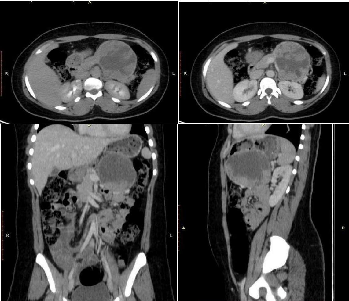 Figure 1: Abdominal CT Images of Our Patient in Transverse, Coronal, and Sagittal Sections Showing the Typical Characteristics of the Pseudopapillary Tumor of the Pancreas
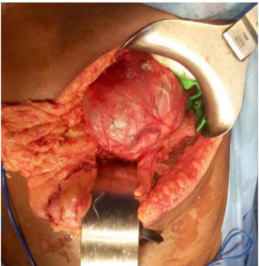
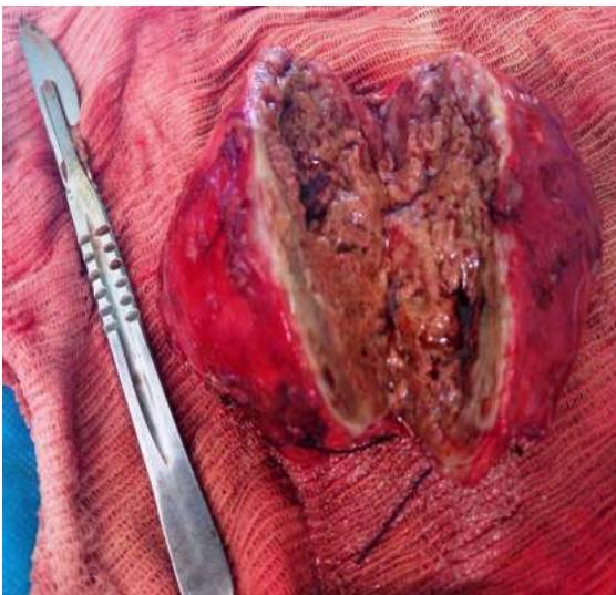 Figure 2: Intraoperative Image of Our Patient Showing Tumor's Location at the Level of the Tail of the Pancreas and the Intratumor Content
Conflict of Interest
The authors declare no conflict of interest.
Ethical Approval
Not applicable
Data Availability
The datasets used in this study are openly available at [repository link] and the source code is available on GitHub at [GitHub link].
Funding
This work did not receive any external funding.
References
Cite this article
Related Research


Special Issue
Launch a focused special issue to highlight research, emerging trends, and expert insights in your academic field.
