


IntelliPaper
Abstract
Introduction: Bilateral mastoiditis complicated by unilateral zygomaticitis is an exceptional clinical entity, rarely described in the literature. Although the incidence of complications of acute otitis media has decreased significantly with the introduction of modern antibiotic therapy, atypical forms of bone extension remain a diagnostic challenge. The association of bilateral mastoid involvement with zygomatic bone involvement can cause a non-specific clinical picture, delaying diagnosis and the establishment of appropriate treatment.
Materials and Methods: In this case study we present a 41-year-old male patient who suffered bilateral mastoiditis complicated by unilateral zygomaticitis following a chronic rhinosinusal pathology in exacerbation. He presented with accusations of: pronounced headache, bilateral otodynia, suppurative otorhea, swelling in the projection of the zygomatic bone on the left, bilateral hearing loss. The patient underwent bilateral petromastoid clearance surgery with incision and drainage of the zygomatic collection on the left.
Results: Epidemiological data from the literature indicate that acute mastoiditis in adults accounts for approximately 10–15% of all mastoiditis cases. The overall incidence is estimated at less than 0.5 cases per 100,000 adults annually in countries with access to modern health services. Although antibiotic therapy has dramatically reduced mortality associated with mastoiditis (from 20–30% in the pre-antibiotic era to less than 1% today), it remains a pathology with a significant risk of extracranial complications (subperiosteal abscess, apical-cervical mastoiditis, petrositis) and endocranial complications (meningitis, lateral sinus thrombosis, brain abscess). Although less common, acute mastoiditis remains a pathology of current interest, with a significant impact on public health, requiring a multidisciplinary and updated approach to the new infectious challenges. We present a rare case of such an otic pathology in a 41-year-old male patient who suffered bilateral mastoiditis complicated by unilateral zygomaticitis as a result of enduring a chronic acute rhinosinusal pathology.
Conclusions: Acute mastoiditis, although rare in the age of antibiotics, continues to be an otological emergency, with significant potential for endocranial and extracranial complications, especially in cases not properly treated.
Explore Digital Article Text
I. INTRODUCTION
Acute mastoiditis is one of the most common complications of acute otitis media, resulting from the spread of the infection to the mastoid cell system. Although predominantly affecting the pediatric population, adult cases account for approximately 10–15% of all reported cases of mastoiditis and present a distinct clinical profile [1, 2]. In countries with modern health systems, the overall incidence of acute mastoiditis has decreased significantly and is currently estimated at less than 0.5 cases per 100,000 adults per year, largely due to early diagnosis, widespread use of antibiotics, and vaccination programs [2, 3].
The introduction of antibiotic therapy has dramatically reduced mortality associated with mastoiditis, from 20–30% in the pre-antibiotic era to less than 1% in contemporary clinical practice [1, 4]. However, acute mastoiditis remains a potentially serious condition with a significant risk of endocranial and extracranial complications. These include the formation of subperiosteal abscesses, cortical bone erosion, petrositis, thrombosis of the lateral venous sinus, meningitis, and intracranial abscesses [4, 5]. Given its rarity in adults and the possibility of an atypical presentation, acute mastoiditis continues to represent a diagnostic and therapeutic challenge, requiring prompt recognition and a multidisciplinary approach in management [1, 5].
From a contemporary otological perspective, atypical forms of mastoiditis are classified based on the topographic extension of the infectious process and the affected bone structures, each subtype presenting characteristic clinical manifestations and a specific profile of complications, which requires a differentiated diagnostic and therapeutic approach.
Zygomaticitis (zygomatic mastoiditis)- is an unusual spread of the suppurative process from the mastoid to the zygomatic process of the temporal bone. This type occurs when mastoid pneumatization extends anteriorly to the zygomatic root, allowing the infection to infiltrate the spaces near the zygomatic bone and the retroauricular subcutaneous tissue.
Affected Region- Zygomatic process and adjacent tissues of the temporal bone, preauricular and zygomatic region.
Clinical Manifestations- Edema and pain in the temporal-zygomatic region, preauricular or facial swelling, displacement of the pinna of the ear in the antero-inferior, hyperemia and possible palpable fluctuation. The diagnosis is confirmed clinically and by imaging (CT).
Complications- Abscess formation, tissue necrosis, bone erosion and extension of the suppurative process to the facial region, possibly mimicking other entities (e.g., parotitis). Surgically, the approach can be complex due to extracranial extension [6].
Squamitis (Squamous Mastoiditis)- The purulent-necrotic form in which the infection spreads from the mastoid cell system to the squamous portion of the temporal bone. This can lead to destructive osteomyelitis located in the lateral region of the temporal bone.
Affected Region– The temporal bone scale, representing the external and lateral portion of the temporal bone above the external auditory canal.
Clinical Manifestations- Intense localized pain in the temporal region, edema and tenderness to palpation, possible local hyperthermia. The diagnosis is based on imaging (CT/MRI) which indicates extensive bone destruction.
Complications - Progressive bone erosion, deep infection and increased risk of intracranial or extracranial extension, requiring aggressive surgical approach [7, 8].
Petrositis Ppetrosal Mastoiditis/Gradenigo)-petrositis is the purulentnecrotic infection of the apex of the petrous portion of the temporal bone, traditionally associated with the Gradenigo triad: acute otitis media, trigeminal retroorbital pain and paresis of the abducens nerve (VI). This form represents the medial extension of the infectious process from the mastoid bone to the bony apex.
Affected Region– The petrous apex of the temporal bone, deep region of the base of the skull. Clinical manifestations – periorbital or retro-orbital pain, diplopia or paresis of the abducens nerve, persistent otorhea, fever and otalgia. Neurological signs may precede the obvious signs of mastoiditis.
Complications– Increased risk of thrombosis of the lateral venous sinus, meningitis, intracranial abscesses (epidural, subdural, cerebral), and other cranial neuropathies .
Cervical Apical Mastoiditis- (Bezold Abscess)-cervical apical mastoiditis-is an externalized form of mastoiditis in which purulent exudate extends from the tip of the mastoid process into the cervical plane, between the deep muscle planes, below the sternocleidomastoid muscle (Bezold abscess and similar variations).
Affected Region– The area of the mastoid tip and the deep cervical muscle spaces (under the sternocleidomastoid muscle, parotid or in the parapharyngeal space).
Clinical Manifestations- Edema and pain in the upper cervical region, limitation of neck mobility, fluctuating subcutaneous swelling, stiffness of the cervical muscles, possible fever. Imaging can reveal purulent accumulations under the muscles.
Complications– Deep cervicofacial abscesses, skin fistulas, local sepsis, risk of extension to the parapharyngeal space or mediastinum, as well as systemic complications if treatment is delayed [9].
Although, in the age of antibiotics, the incidence of mastoiditis has decreased significantly, severe and complicated forms continue to be encountered, especially in the context of delayed or inadequate treatment. Bilateral mastoiditis is a rare and severe pathology, indicating an extensive infectious process, with an increased potential for endocranial and extracranial complications.
Zygomaticitis is a rare bone complication of mastoiditis, characterized by the propagation of the infectious process at the level of the zygomatic bone, through pneumatic cells or pathways of anatomical continuity. The occurrence of unilateral zygomaticitis in the context of bilateral mastoiditis underlines the unpredictability of the dissemination of infection and requires thorough clinical and imaging evaluation. This pathological association is particularly important from a diagnostic and therapeutic point of view, as it can mimic other inflammatory or tumoral conditions of the facial region.
Early recognition of complicated mastoiditis, as well as understanding the mechanisms of infection spreading to neighboring bone structures, are essential for instituting appropriate treatment and preventing intracranial and extracranial complications. In this context, this article aims to address the issues related to classification, pathogenic mechanisms and the importance of the correct diagnosis of bilateral mastoiditis complicated by unilateral zygomaticitis.
II. MATERIALS AND METHODS
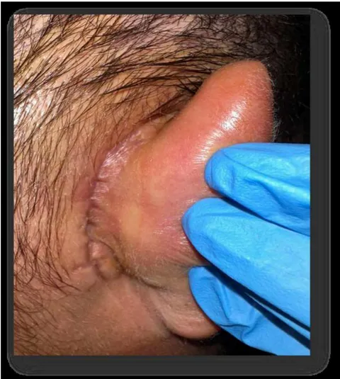
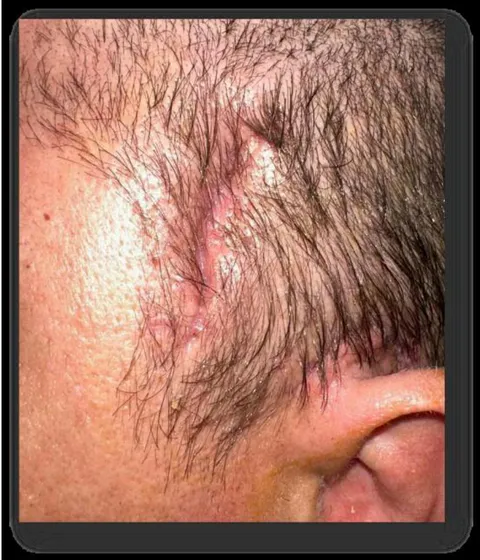
In this case study we present a 41-year-old male patient who suffered bilateral mastoiditis complicated by unilateral zygomaticitis following a chronic rhinosinusal pathology in exacerbation. The patient presented with complaints of: pronounced headache, bilateral otodynia, vertigo, suppurative otorhea, swelling in the projection of the zygomatic bone on the left, bilateral hearing loss, general weakness.
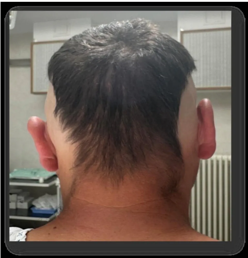
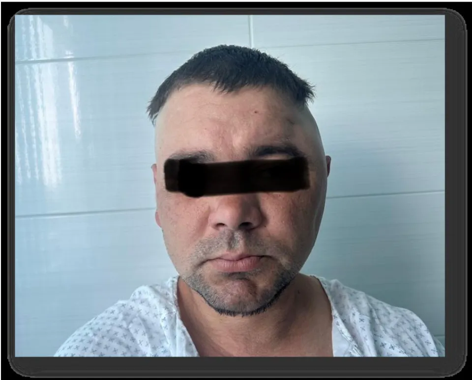
The patient considers himself ill for about 3-4 weeks, when he presented the symptoms of a chronic rhinosinusal pathology in exacerbation, after a few days the rhinosinusal symptoms subsided, but pronounced headache, vertigo, bilateral progressive otodynia began, which was followed by abundant right ear otorhea, insignificant in US, and postural instability. The patient did not go to the doctor. He administered topical outpatient treatment with pic. Floxadex, the condition with worsening in dynamics. For about 2 days, swelling and hyperimia have been installed in the projection of the zygomatic bone on the left. He called the 112 service, the patient was redirected to IMSP SCR "T.Moşneaga", for the consultation of the ENT doctor and specialized treatment.
Otoscopically, narrowed UD - CAE was highlighted, abundant suppurated eliminations, at aspiration of which the detachment of the posterior wall was highlighted, visible pulsation, TM is not visualized; US – narrowed, hyperemic CAE, MT is not visualized, the detachment of the posterior wall was highlighted. The suppurated eliminations were collected for bacteriological examination.
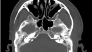
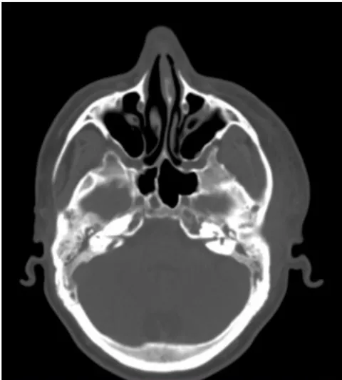
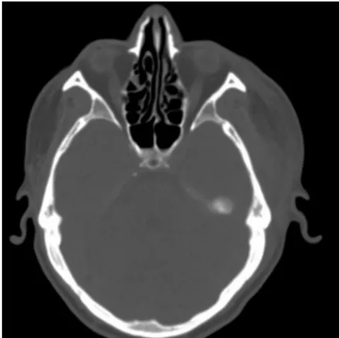
When performing computed tomography of the temporal bones (middle and inner ear), the mastoid cells and the bilateral tympanic house were totally opaque, the ossicular chain preserved, the intercellular septa preserved, bone defect in the squamous region of the temporal bone on the left, thickening of the tissues in the zygomatic region on the left, diffuse hypodense opacity of the subcutaneous layer with the deletion of the normal planes. (Figures 2, 3).
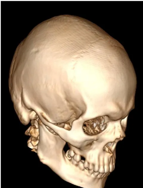
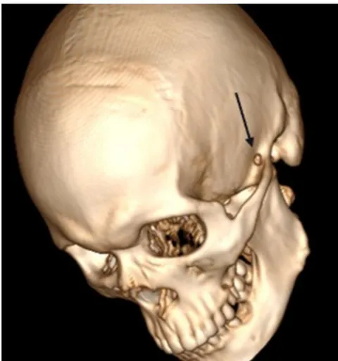
Subsequently, the patient was evaluated by the neurologist, excluding intracranial complications, after which the consilium was carried out jointly with the head of the ENT department, the head of the department with the decision to intervene urgently, by performing bilateral petromastoid clearance surgery with incision and drainage of the zygomatic collection on the left. Postoperatively, the patient was transferred to intensive care for monitoring, with the establishment of specific treatment - double antibiotic therapy according to the antibioti-cogram, anti-inflammatory, corticosteroid, etc.
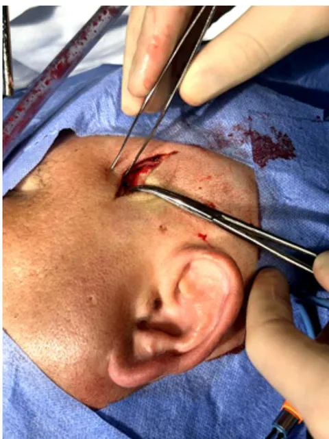
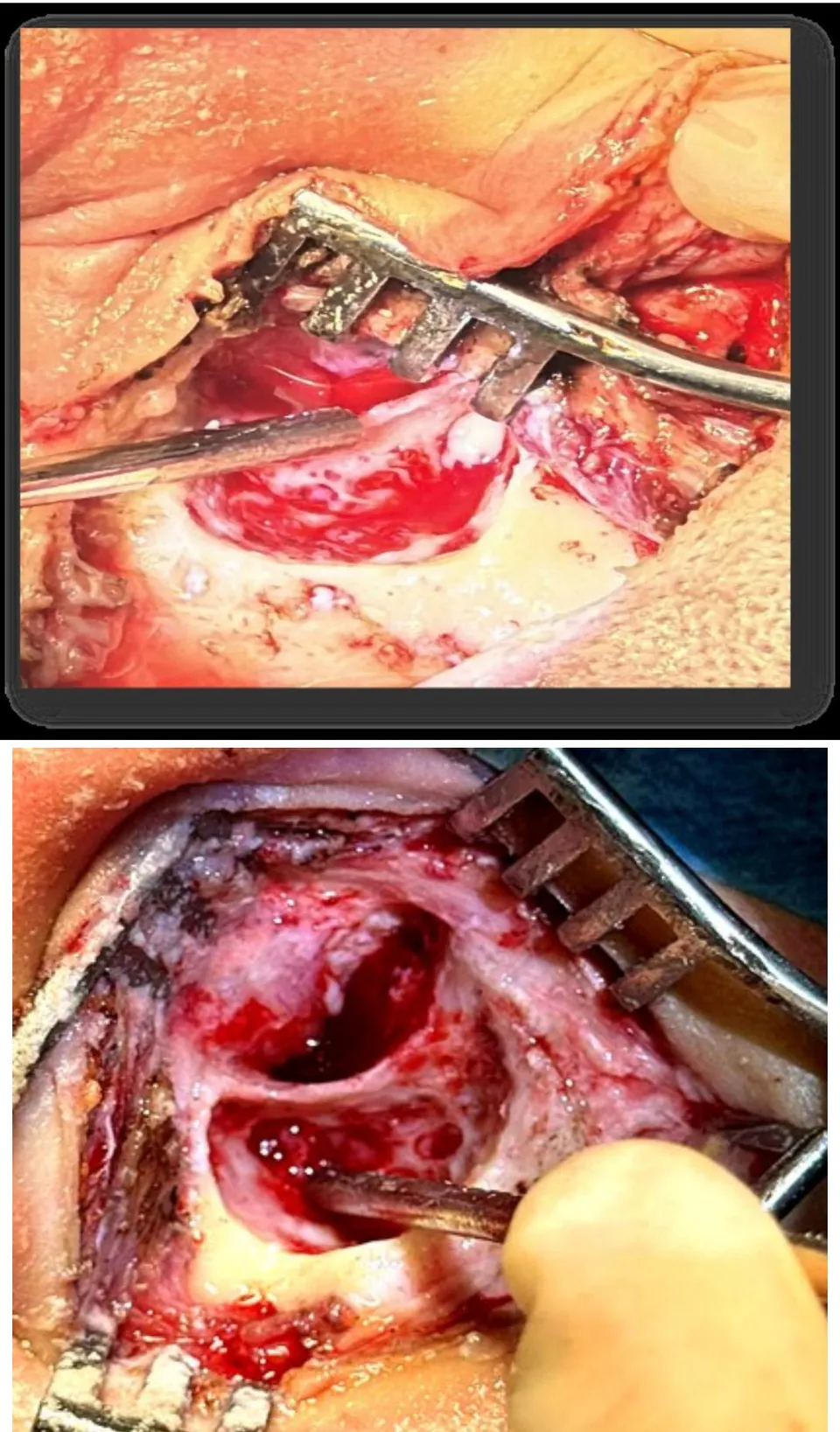
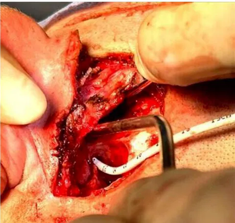
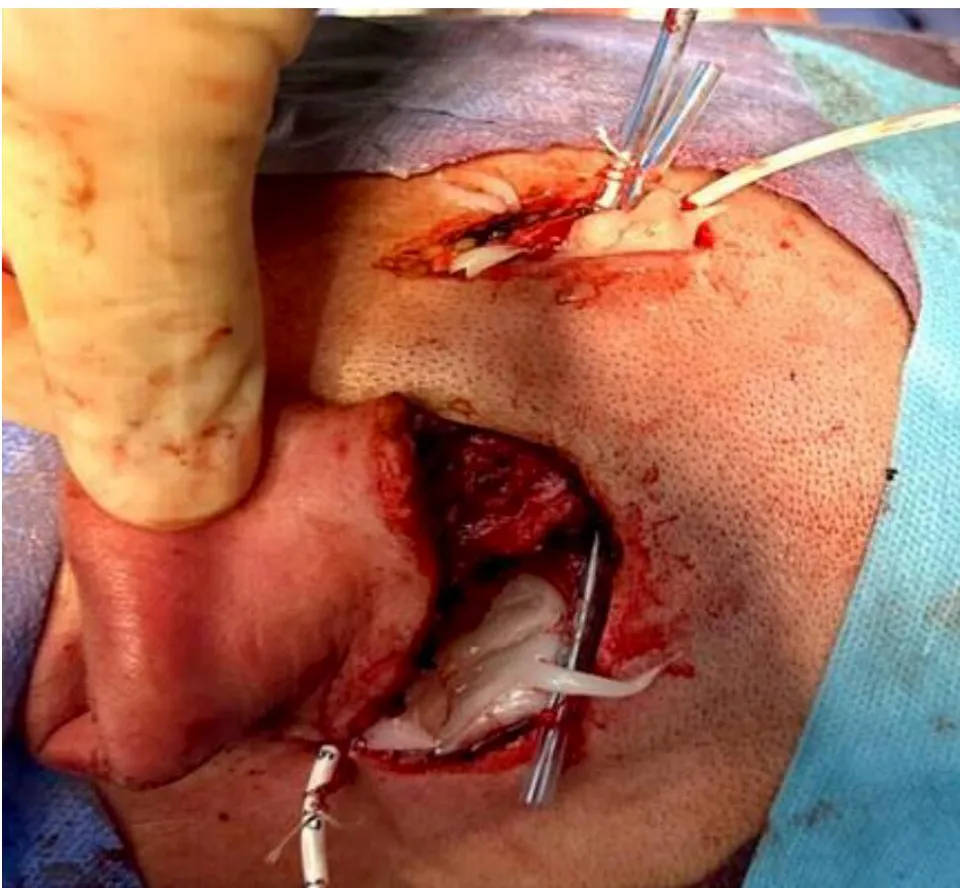
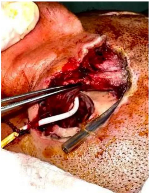
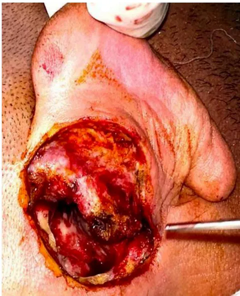
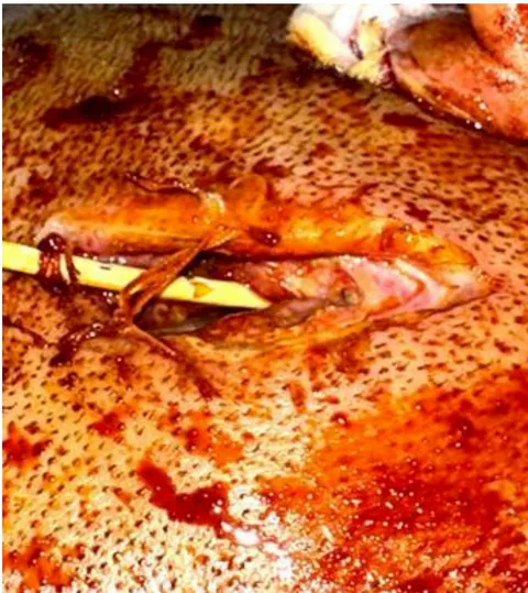
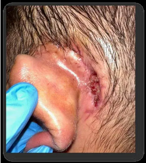
Daily aseptic dressing of the open wound was performed, with aspiration of the contents through the drainage tube, with subsequent wound washing. Subsequently, on the 7th day, secondary suturing of the retroauricular wound was performed.
III. DISCUSSION
Bilateral mastoiditis is an extremely rare condition, especially in adults, being most commonly seen as a complication of untreated acute otitis media. In the literature, cases of bilateral mastoiditis are reported sporadically, most of which are associated with compromised immunity, craniofacial malformations or persistent severe infections. Bilateral forms are considered challenging diagnoses due to the often non-specific symptomatology and initial subclinical evolution, which can delay early identification and appropriate treatment.
The complication of unilateral zygomaticitis, as observed in the case presented, is extremely rare and indicates the extension of the inflammatory process to the perimastoid and facial tissues. Zygomaticitis occurs as a result of the spread of infection through continuity or through the cellular spaces of the face, which can lead to localized facial edema, pain and, in severe cases, risk of compromised nerve function or deep suppuration. This clinical peculiarity gives the diagnosis a high degree of difficulty, requiring a detailed imaging evaluation, preferably CT, to identify the extent of inflammation and any purulent collections.
From a microbiological point of view, bilateral mastoiditis and associated complications tend to be caused by resistant pathogens or polymicrobial infections, which requires the prompt initiation of broad-spectrum antibiotic treatment, subsequently adjusted according to the results of the cultures. Surgery, whether in the form of mastoidectomy or drainage of the zygomatic collections, remains an essential component in the management of complicated cases, especially when there is a risk of dissemination to the deep or intracranial spaces.
The case presentation emphasizes the importance of maintaining a high degree of suspicion in the face of bilateral symptoms and signs of facial extension, even when the initial clinical signs are discreet. It also highlights the need for a multidisciplinary approach for optimal diagnosis and treatment.
In conclusion, bilateral mastoiditis complicated by unilateral zygomaticitis is a rare and provocative form of otological condition, with the potential for severe complications if not identified and treated early. Reporting these cases helps to raise clinical awareness and develop appropriate therapeutic strategies.
IV. CONCLUSIONS
Acute mastoiditis, although rare in the age of antibiotics, continues to be an otological emergency, with significant potential for endocranial and extracranial complications, especially in cases not properly treated.
Early diagnosis, based on thorough clinical evaluation and imaging investigations (CT, MRI), is essential to prevent complications and establish the indication for surgical treatment when necessary.
In the current context, marked by increased bacterial resistance and self-medication, atypical forms may become more and more frequent, requiring clinical vigilance and updating treatment protocols.
Interdisciplinary collaboration between ENT doctors, neurologists, imagingists, reanimatologists and surgeons is essential for the effective management of these complex cases.
Continuous medical education and correct information of patients about the complications of otitis media are essential steps in reducing the incidence of mastoiditis and its atypical forms.
Conflict of Interest
The authors declare no conflict of interest.
Ethical Approval
Not applicable
Data Availability
The datasets used in this study are openly available at [repository link] and the source code is available on GitHub at [GitHub link].
Funding
This work did not receive any external funding.
References
Cite this article
Related Research

Special Issue
Launch a focused special issue to highlight research, emerging trends, and expert insights in your academic field.