


IntelliPaper
Abstract
A study on Chronic Inflammatory Enteropathy (CIE) in dogs was conducted at Madras Veterinary College Teaching Hospital. Out of 40 dogs with persistent gastrointestinal signs, 16 were diagnosed with idiopathic CIE. The condition was more prevalent in 2-5 year-old male dogs, particularly in mongrel breeds. Clinical signs included chronic diarrhoea, vomiting, and weight loss, with disease duration of 1-3 months. Gastroduodenoscopy revealed hyperemia, friability, and granularity in the stomach and duodenum, while colonoscopy showed friability, hyperemia, ulceration, and discoloration of colonic mucosa. Histopathology of duodenal biopsies showed villous stunting, mucosal fibrosis, and lymphoplasmacytic infiltration. Immunohistochemistry indicated upregulation of CD3+ cells and Ki67 antigen. A positive correlation was found between the Canine Chronic Enteropathy Activity Index (CCECAI) and endoscopic and histopathological scores.
Explore Digital Article Text
I. INTRODUCTION
Chronic inflammatory enteropathy (CIE) is a term describing persistent gastrointestinal signs lasting over three weeks, with histological evidence of inflammation in the small or large intestine. Diagnosis requires exclusion of extra-intestinal, infectious, and parasitic diseases, as well as intestinal diseases of known etiology (Dandrieux,
2016). CIE involves complex interactions between the host genome, intestinal barrier function, microbiota, dietary antigens, and immune system (Allenspach & Mochel, 2020). In idiopathic CIE, T cell hypersensitivity and proliferation play a key role, with CD3 and Ki-67 antigens serving as markers for T lymphocytes and cell proliferation, respectively. Immunohistochemical analysis of CD3 and Ki-67 can help determine idiopathic CIE (Karlovits et al., 2019), potentially linking increased cytokine release to clinical symptoms.
II. MATERIALS AND METHODS
Sixteen client-owned dogs presented to the Referral Medicine unit of Madras Veterinary College Teaching Hospital, with the history and clinical signs of persistent or recurrent gastrointestinal signs for three weeks or longer and refractory to conventional therapies were selected for the study. These animals were subjected to detailed clinico-pathological, radiological, ultrasonographic, and special examination procedures. Gastro-duodenoscopy and colonoscopy were performed under general anesthesia by a trained endoscopist following standard operating procedures using a Veterinary Video endoscope (Karl Storz model No. 60914 PKS, Germany) with an outer diameter of 9.8 mm, biopsy channel diameter of 2.8 mm, and working length of 1400 mm.
Biopsies were taken with fenestrated, long oval cup biopsy forceps (Karl Storz) with a 2.2 mm diameter, and endoscope-aided mucosal biopsy samples were collected. Histopathology and immunohistochemistry were performed by a pathologist following standard operating procedures to confirm the diagnosis and assess immunological changes in the intestine by identifying specific markers (CD3/Ki67). The characterization of histologic changes in the endoscopic biopsy samples was performed according to the histopathological standards established by the WSAVA Gastrointestinal Standardization Group (Day et al., 2008). Immunohistochemistry was performed following a standard procedure (Carrasco et al., 2011), and the results were interpreted using the Immunoreactive Score (IRS) (Fedchenko et al., 2014).
III. RESULTS
The study revealed a high proportion of Chronic Inflammatory Enteropathy (CIE) in dogs aged 2-5 years, with males being more commonly affected than females. Mongrel dogs had the highest incidence, followed by German Shepherds and Retrievers. The duration of clinical signs prior to diagnosis was 1-3 months, with chronic diarrhoea being the primary complaint, followed by vomiting, mixed signs of vomiting and diarrhoea, and weight loss (Fig.1). All dogs were fed a homemade diet and had failed previous drug therapy. Body condition scoring revealed emaciation and thin body condition (BCS 1 and 2) in 10 dogs (Table 1). Fecal scoring showed scores of 4.5 (very soft feces) and 5 (diarrhoea) in the affected dogs.
The Canine Chronic Enteropathy Clinical Activity Index (CCECAI) was used to evaluate the severity of the disease condition. The CCECAI score of the CIE dogs expressed normal attitude and appetite with no vomiting in 9 out of 16 dogs to severe vomiting in 5 out of 16 dogs. Stool consistency was watery in 43.75% (7/16), very soft feces in 25% (4/16), slightly soft feces in 12.5% (2/16), and normal in 18.75% (3/16) dogs. Stool frequency was normal in 18.75% (3/16), slightly increased (2-3/day) in 25%, moderately increased (4-5/day) in 12.5%, and severely increased (>5/day) in 43.75% of dogs.
Mild, moderate, and severe weight loss were reported in 6.25%, 31.25%, and 43.75% of dogs, respectively. Albumin levels were normal (>20g/L) in 81.25% of cases, with mild (15-19.9g/L) and moderate (12-14.9g/L) hypoalbuminemia observed in 12.5% and 6.25% of dogs, respectively. Moderate peripheral edema was seen in 6.25% (1/16) of dogs.
Dogs with CIE were classified based on the summation of individual parameters as mild (4-5) 18.75% (3/16), moderate (6-8) 31.25% (5/16), severe (9-11) 31.25% (5/16), and very severe (>12) 18.75% (3/16) of dogs.
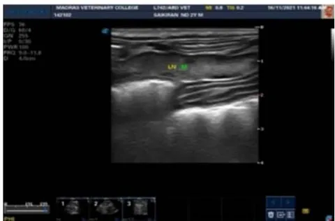
Hematology revealed leukocytosis, neutrophilia, lymphocytosis, and monocytosis in the CIE dogs. No remarkable changes were observed in serum biochemical analysis in the CIE dogs (Table 2). Radiography revealed gas-filled intestinal loops in one dog (Fig. 2). Ultrasonography was used to measure the wall thickness of the gastrointestinal tract in the CIE dogs, which was found to be normal in all CIE dogs except one. The wall layering and echogenicity were normal in all CIE dogs. Secondary changes observed in CIE dogs included mesenteric lymphadenopathy (Fig. 3), free abdominal fluid, and distended intestinal loops in few dogs.
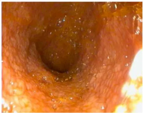
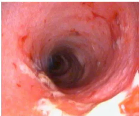
Gastro-duodenoscopic examination of the stomach and duodenum predominantly revealed hyperemia, increased friability, and increased granularity. Less common lesions observed included discoloration, thickening and corrugation, and increased mucus. In the colonic mucosa, the major changes observed were friability, hyperemia, ulceration, and discoloration (Fig. 4, 5, 6).
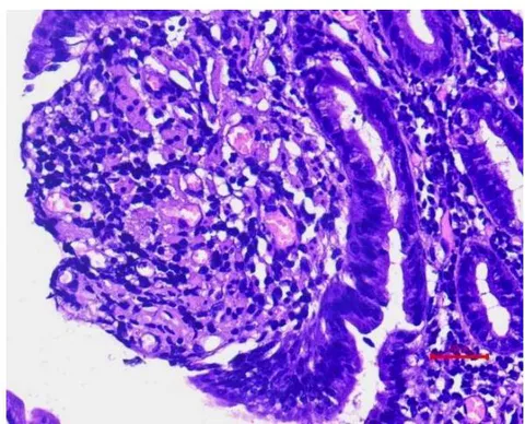
Histopathological examination of the gastric mucosa revealed inflammatory changes (lymphocyte and plasma cell infiltration), vacuolation or separation of focal areas of superficial epithelium, ulceration of surface epithelium and moderate form of mucosal fibrosis/glandular atrophy. Duodenal mucosal biopsy samples revealed severe morphological changes, including villous stunting and fusion, villous stricture, mucosal fibrosis, crypt epithelial hyperplasia, and inflammatory changes characterized by lymphoplasmacytic cell infiltration, which were recorded in all CIE dogs (Fig.7). In three Colonic biopsy samples inflammatory changes with mild surface epithelial injuries like attenuation, degeneration or vacuolation and loss of focal areas of superficial epithelium, crypt hyperplasia, dilation and distortion, mucosal fibrosis were noticed.
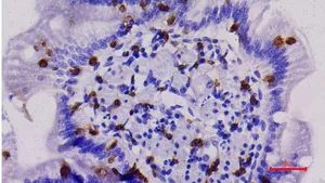
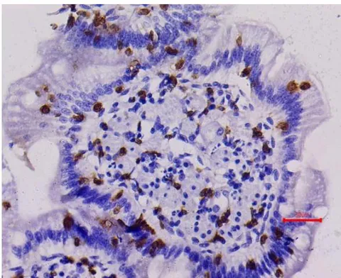
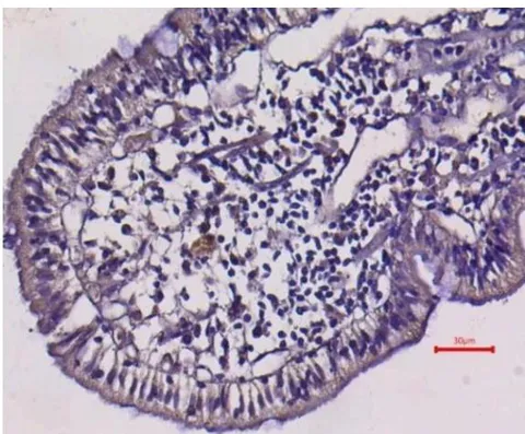
Immunohistochemical analysis of mucosal biopsy samples revealed varying degrees of CD3+ positive cell expression, with an Immunoreactive Score (IRS) of mild (2-3) in 37.5% (6/16), moderate (4-8) in 25% (4/16), and strongly positive (9-12) in 37.5% (6/16) of cases (Fig. 8). Similarly, the IRS for Ki67 positive nuclei was recorded as mild (2-3) in 43.75% (7/16), moderate (4-8) in 25% (4/16), and strongly positive (9-12) in 18.75% (3/16) of cases (Fig. 9). The results indicated up regulation of CD3+ positive cells and Ki67 antigen in the CIE-affected dogs.
A positive correlation was observed between the Canine Chronic Enteropathy Clinical Activity Index (CCECAI) and endoscopic score, as well as between CCECAI and histopathological score. However, no correlation was found between the endoscopic score and histopathological score.
IV. DISCUSSION
Chronic Inflammatory Enteropathy was mostly reported in middle-aged dogs. Volkmann et al. (2017) reported that dogs between 2 to 9 years old were commonly affected, while Allenspach et al. (2020) noted that CIE typically presents in middle-aged dogs due to environmental influences. The current study supports these findings, with a high incidence of CIE observed in dogs aged 2-5 years.
The breed distribution in this study, with Mongrel dogs, German Shepherds, and Retrievers being commonly affected, is also consistent with previous reports. Other breeds, such as Rottweiler's and Boxers, were also represented, which is in line with the findings of Kathrani, et al. (2011) and Jergens et al. (2012). The genetic predisposition of certain breeds, such as German Shepherds, to CIE has been attributed to single nucleotide polymorphisms (SNPs) in Toll-like receptor (TLR) genes (Kathrani et al., 2010).
The role of modifiable exposure factors, such as feeding practices and deworming protocols, in the development of Inflammatory Bowel Disease (IBD) has been highlighted by Hemida et al. (2021). Further studies are needed to investigate the impact of these factors on the development of CIE in dogs.
The findings of this study are consistent with previous reports that mixed breeds, particularly Mongrel dogs, are overrepresented in cases of Chronic Inflammatory Enteropathy (CIE) (Volkmann et al., 2017; SandhyaBhavani, 2018). The clinical signs observed in this study, including chronic diarrhea, vomiting, weight loss, and decreased body condition score, are similar to those reported by Jergens et al. (2003) and Sattasatuchana et al. (2017).
The Canine Chronic Enteropathy Clinical Activity Index (CCECAI) scores revealed that the majority of dogs had moderate to very severe forms of CIE, with increased stool frequency and weight loss being the predominant changes. These findings are consistent with Volkmann et al. (2017), who also reported moderate to severe forms of CIE in their study.
Gastro-duodenoscopy and colonoscopy, in combination with endoscopically guided biopsy, proved to be valuable diagnostic tools in this study, as previously reported by Jergens et al. (2012). The use of these techniques allowed for the visualization of gastrointestinal lesions and the collection of biopsy samples for histopathological examination, which aided in the diagnosis of CIE.
The endoscopic findings in this study are consistent with previous reports that describe various lesions in the gastrointestinal tract of dogs with Chronic Inflammatory Enteropathy (CIE). Slovak et al. (2014) noted that endoscopy provides a direct assessment of intestinal mucosal damage and can be used to measure disease activity index, with common lesions including erythema, friability, erosions/ulcers, and granularity. Similarly, Garcia-Sancho et al. (2007) reported gastric and duodenal lesions, such as mucosal erythema, granularity, friability, and erosions, in dogs with lymphocytic plasmacytic enteritis.
In the present study, the endoscopic findings revealed hyperaemia, discoloration, and friability in the gastric mucosa, while the duodenum showed predominant changes of friability, followed by hyperaemia, mucosal thickening, and corrugation, increased granularity, erosion/ulcers, discoloration, and increased mucus. The colonic mucosa exhibited friability, hyperaemia, ulceration, and discoloration. These findings are in accordance with the aforementioned studies.
Histopathology of endoscopic mucosal biopsy remains the gold standard for diagnosing CIE, providing valuable information on the extent and severity of mucosal inflammation and damage. The combination of endoscopy and histopathology allows for a comprehensive evaluation of gastrointestinal disease in dogs with CIE.
The histopathological findings in this study are consistent with the standards established by the World Small Animal Veterinary Association (WSAVA) International Gastrointestinal Standardization Group (Day et al., 2008). The morphological changes observed in the CIE dogs, including villous stunting, fusion, epithelial injury, and fibrosis, are similar to those described by Washabau et al. (2010) as characteristic of small intestinal inflammation.
The inflammatory cell infiltrate in the CIE dogs was predominantly lymphoplasmacytic, which is in agreement with previous studies (Jergens et al., 2010; Suchodolski et al., 2012; SandhyaBhavani, 2018). The presence of lymphocytes and plasma cells as the primary inflammatory cells is consistent with the diagnosis of CIE.
The histopathological changes observed in this study, including gastric pit epithelial hyperplasia, glandular or mucosal fibrosis, villous epithelial injury, and villous stunting and fusion, are also consistent with the findings of Jergens and Simpson (2012), who described minimal to pronounced inflammatory cell infiltration and mucosal architectural disruption in dogs with IBD. The consistency of these findings with previous studies highlights the importance of histopathology in diagnosing CIE and understanding the underlying inflammatory processes.
The pathogenesis of Chronic Inflammatory Enteropathy (CIE) involves a complex interplay of immune cells and cytokines. T lymphocytes, particularly Th1 cells, play a major role in the development of intestinal inflammation through the secretion of pro-inflammatory cytokines such as TNF and IFNγ (Maeda et al., 2013; Heilmann and Steiner, 2018). The imbalance between pro-inflammatory and anti-inflammatory responses, mediated by Th1, Th2, and Th17 cells, can lead to intestinal inflammation (Eissa et al., 2019).
The immunohistochemical findings in this study revealed up regulation of CD3 and Ki67 positive cells in the intestinal mucosa of CIE-affected dogs, indicating an underlying immunological reaction against unknown etiology and luminal antigens. The presence of CD3 and Ki67 positive cells in all CIE dogs with lymphoplasmacytic cell infiltration supports the role of immune-mediated mechanisms in the pathogenesis of CIE.
The study also found a highly significant positive correlation between the clinical activity index (CCECAI) and endoscopic score, which is in contrast to the findings of Allenspach et al. (2007). However, the positive correlation between CCECAI and histopathological score observed in this study is consistent with the report by Allenspach et al. (2019). The lack of correlation between endoscopic score and histopathological score is in agreement with the findings of SandhyaBhavani (2018). These results highlight the complex relationships between clinical, endoscopic, and histopathological findings in CIE.
V. CONCLUSION
This study provides valuable insights into the clinical, endoscopic, and histopathological features of Chronic Inflammatory Enteropathy (CIE) in dogs. The findings suggest that CIE is characterized by a complex interplay of immune cells and cytokines, with upregulation of CD3 and Ki67 positive cells indicating an underlying immunological reaction. The positive correlation between clinical activity index and endoscopic score, as well as histopathological score, highlights the importance of a comprehensive diagnostic approach in evaluating CIE. The study's findings contribute to the understanding of CIE pathogenesis and may aid in the development of effective diagnostic and therapeutic strategies for managing this condition in dogs.

| S. No | Clinical Signs | No. of Animals (%) |
| 1. | Diarrhea | 56.25% (9/16) |
| Melena | 66.67(6/9) | |
| Hematochezia | 33.33(3/9) | |
| 2. | Vomiting | 18.75%(3/16) |
| 3. | Vomiting with diarrhea | 25%(4/16) |
| 4. | Weight loss | 75%(12/16) |
| Emaciation (BCS 1) | 31.25% (5/16) | |
| Thin (BCS 3) | 31.25%(5/16) |
| S. No | Parameters | Control (n=6) | CIE affected dogs (n=16) | T value |
| 1. | Haemoglobin(g/d1) | 14.83+0.76 | 13.41 0.84 | |
| 2. | PCV(percent) | 41+1.72 | 37.81+2.18 | |
| 3. | Total erythrocyte(x10 /cu mm) | 6.55z0.27 | 5.8+0.35 | |
| 4. | Total leucocyte (x10 / cu mm) | 8.2z0.54 | 17.26+2.1 | 2.586* |
| 5. | Neutrophils (x10 / cu mm) | 6.042+0.59 | 14.25a1.97 | 2.499* |
| 6. | Lymphocytes (x10 /cu mm) | 1.02+0.1 | 2.32+0.31 | 2.507* |
| 7. | Monocytes (x10 / cu mm) | 0.27+0.04 | 0.75+0.08 | 3.226** |
| 8. | Eosinophils (x10 /cu mm) | 0.05+0.02 | 0.18+0.08 | |
| 9. | Basophils (x103/ cu mm) | 0.01+0.02 | 0.00+0.00 | |
| 10. | Serum Alkaline Phosphatase(IU/L) | 110.16+11.38 | 254.25+69.86 | |
| 11. | Total protein(g/dl) | 6.4+0.30 | 6.58+0.27 | |
| 12. | Albumin(g/dl) | 2.8+0.14 | 2.8+0.17 | |
| 13. | SerumCholesterol(mg/dl) | 156.50+7.52 | 129.62+18.2 |
NS-Statistically nonsignificant(P>0.05)
Conflict of Interest
The authors declare no conflict of interest.
Ethical Approval
Not applicable
Data Availability
The datasets used in this study are openly available at [repository link] and the source code is available on GitHub at [GitHub link].
Funding
This work did not receive any external funding.
References
Cite this article
Related Research


Special Issue
Launch a focused special issue to highlight research, emerging trends, and expert insights in your academic field.