


IntelliPaper
Abstract
RNase P ribozymes, derived from the M1 RNA of Escherichiacoli, have shown great promise as a novel nucleic acid-based gene interference approach to modulate gene expression. When the M1 RNA component of RNase P is covalently linked with a guide sequence (GS), it can be engineered into a sequence-specific endonuclease M1GS ribozyme. As the GS base-pairs with target mRNAs, it forms a structure that mimics a pre-tRNA-like substrate, allowing for M1 RNA’s structure-based recognition mechanism. These M1GS constructs function catalytically and irreversibly, capable of cleaving target mRNA substrates without relying on host proteins. M1GS activity enhancement has been achieved through an in vitro selection process that introduced mutations in the catalytic core of M1 RNA. This process generated ribozyme variants with greatly improved cleavage efficiency and substrate affinity. M1GS ribozymes have been successful in inhibiting herpes simplex virus 1 (HSV-1) by targeting genes critical for viral infection. HSV-1 is the causative agent of cold sores and may lead to severe morbidity and mortality in neonates and immunocompromised individuals. HSV-1 establishes lifelong latent infection, and novel anti-HSV-1 strategies are needed to block and eliminate viral latency and reactivation. Using HSV-1 infection as an example, this review will summarize the function of RNase P and its catalytic RNA, the enhancement and engineering of M1GS ribozymes, and their potential as a gene-targeting agent for therapeutic applications against HSV-1.
Explore Digital Article Text
I. INTRODUCTION
Nucleic acid-based gene targeting strategies have been at the forefront of major advancements in the field of molecular biology and therapeutic development. Among these strategies, antisense oligonucleotides, ribozymes, RNA interference (RNAi), and the genome editing technology based on the Clustered Regularly Interspaced Short Palindromic Repeats (CRISPR)/CRISPR-associated protein (Cas) RNA-guided nuclease systems have shown significant promise as tools for targeted gene regulation . Although the mechanisms vary greatly, ranging from passively blocking translation through complementary base pairing to introducing double-stranded breaks in DNA with site-specific endonuclease activity, each strategy provides a powerful and distinct approach to gene regulation. For example, hairpin and hammerhead ribozymes have demonstrated their potential in therapeutic applications such as the treatment of AIDS . These ribozymes contain a substrate-binding domain that, through Watson-Crick interactions, guides them to the target mRNA sequence, where the catalytic RNA domain can then hydrolyze a specific sequence .
RNA interference (RNAi) is a nucleic acid-based gene interference strategy that offers multi-turnover potential . Through its recruitment of cellular machinery, small interfering RNA (siRNA) directs the degradation of target mRNA substrates . However, at high siRNA concentrations, host protein complexes can be overwhelmed, potentially leading to off-target effects . CRISPR-Cas9 systems can be programmed to target DNA or RNA, but they may carry the risk of off-target cleavage and permanent genomic alterations .
Among the various nucleic acid-based gene interference strategies that interact with RNAs, the M1GS ribozyme offers a unique advantage of its endogenous catalytic activity guided by structural recognition . The M1GS ribozyme is derived from the catalytic RNA subunit (M1RNA) found in Escherichia coli RNase P, and is covalently linked to a guide sequence (GS) (Figures 1 and 2) . This guide sequence (GS) binds to target mRNAs in a sequence-specific manner and reshapes them into a pre-tRNA-like structure that M1 RNA can recognize and cleave . M1GS acts catalytically and irreversibly, allowing one M1GS to cleave multiple copies of the target mRNA. M1GS functions exclusively at the mRNA level, avoiding direct genomic alterations . Recent progress on the structures and functions of RNase P and M1 RNA has been summarized in recently published reviews and is outside the purview of this article . In this review, we will discuss the potential of RNase P-derived gene-targeting ribozymes for treating herpes simplex virus 1 (HSV-1), highlighting their design, optimization, and application in targeting HSV-1-encoded mRNAs.
II. HERPES SIMPLEX VIRUS 1
HSV-1 is a large, double-stranded DNA virus that establishes lifelong latency within sensory neurons . HSV-1 enters through mucosal epithelial tissue, where it replicates productively before traveling through the nerve termini to reach the sensory neurons, where latent infection is established and provides a viral reservoir for periodic reactivation . HSV-1 can cause symptoms such as cold sores and genital lesions, and in severe cases, can lead to severe morbidity or mortality . The viral life cycle for HSV-1 depends on the expression of conserved genes that encode proteins such as ICP4, a major transcription activator required for the expression of early and late genes during lytic infections, and thymidine kinase (TK), an enzyme important for nucleotide metabolism during replication in vivo .
Acyclovir is a widely used antiviral for HSV-1 [19]. This compound is structurally similar to guanosine but lacks the 3' hydroxyl group required for DNA elongation and is used to disrupt the viral replication process. Acyclovir is phosphorylated by thymidine kinase (TK), converting it into acyclovir monophosphate [19]. Host kinases convert it to its active triphosphate form. This active form competes with deoxyguanosine triphosphate and is preferentially incorporated by HSV-1 DNA polymerase, stalling elongation and terminating replication [19]. In immunocompromised individuals, resistance to acyclovir has emerged, as some HSV-1 strains carry TK-deficient mutations, and other strains express TK with structural mutations that prevent acyclovir from binding and getting phosphorylated [21, 22]. While acyclovir is considered safe due to its dependence on TK, high systemic concentrations of acyclovir in patients have been associated with renal impairments and symptoms such as confusion, agitation, or seizures [19]. These risk factors are elevated in elderly patients with underlying renal disease. These limitations highlight the need for novel strategies that can act through alternative mechanisms. The use of ribozymes, catalytic RNA molecules, may show promise and can be designed to degrade viral mRNA, inhibiting replication by targeting the mRNA level.
HSV-1 encodes immediate early (IE) genes that initiate reactivation, early (E) genes that encode TK and other proteins involved in viral DNA replication, and late (L) genes that encode viral capsids, envelopes, and proteins involved in lytic infection [17, 18]. The inherently heterochromatic and immunologically silent environment of neurons, combined with the IE promoter's affinity for repressive chromatin marks, facilitates the rapid chromatinization of HSV-1 DNA and the establishment of viral latency [23]. HSV-1 further promotes latency by expressing Latency-Associated Transcripts (LATs), non-coding RNAs that recruit host chromatin-modifying proteins to deposit repressive histone marks, thereby facilitating the formation of heterochromatin at the viral IE promoters responsible for reactivation [17, 24].
Current antiviral treatments, such as acyclovir, only act on the lytic phases of HSV-1 infection . This is clinically problematic, as reactivation is when HSV-1 is contagious and when symptoms that can be potentially severe or life-threatening take place. Unlike the latency reversal strategies used in HIV treatments, where the virus is forced into a treatable phase , similar approaches are not viable for HSV-1 infections, as cell destruction could result in irreversible neurological damage .
Efforts to treat HSV-1 latency through CRISPR-Cas9-targeted IE genes have not been very effective as the tightly packed HSV-1 DNA is inaccessible . However, meganucleases have been effective in depleting HSV-1 reservoirs by targeting essential genes during latency . Unlike the CRISPR-Cas9 systems, which rely on complementary base pairing, meganucleases can recognize specific DNA sequences through protein-DNA interactions in the major groove, a region potentially more accessible during latent repression . Further studies are needed to investigate the effectiveness of the meganucleases and the CRISPR-Cas9-based method for anti-HSV-1 applications.
HSV-1 mutants lacking LATs have been shown to establish latency at a lower level, indicating that latency arises through multiple mechanisms . HSV-1 also utilizes the host stress response to reverse its chromatin silencing . HSV-1's utilization of host machinery to establish latency and reactivation makes it therapeutically challenging to distinguish viral processes from normal cellular pathways. M1GS ribozymes can achieve high specificity by acting at the mRNA level, enabling them to degrade viral transcripts without affecting host gene expression. M1GS can target reactivation at its earliest step by targeting IE mRNAs that encode essential replication proteins, preventing HSV-1 progression to subsequent steps of the lytic cycle.
III. RNASE P AND ITS CATALYTIC RNA
RNase P is a ribonucleoprotein enzyme that cleaves the 5' leader sequence from tRNA precursors [11, 29, 30] and other small RNAs such as bacterial operon RNAs [31, 32], riboswitches [33], and signal recognition particle RNAs [34]. RNase P is found in all domains of life, indicating its essential, conserved function. The catalytic activity in RNase P is in the RNA subunit rather than the protein component [35]. RNase P has one protein component in bacteria, typically 4 in Archaea, and up to 10 protein subunits in Eukaryotes [11, 14]. RNase P from Escherichia coli is composed of an M1 RNA subunit, which is 377 nucleotides in length, and a 14 kDa, 119 amino acid-long C5 protein subunit that assists in M1 RNA's structural stability [12]. Both the RNA and protein subunits are necessary in vivo; however, M1 RNA has been shown to cleave pre-tRNA in vitro in the absence of the protein subunit under high divalent ion concentrations (i.e., ~100 mM) [35]. This phenomenon results from the positively charged ions shielding the negatively charged phosphate backbone of the M1 RNA, thereby reducing electrostatic repulsion and allowing it to fold into its catalytically active structure without the need for the C5 protein [35-43]. Under in vitro conditions where Mg concentrations are low, C5 protein is necessary for bacterial survival and works by stabilizing M1 RNA's structure into its catalytically active conformation, allowing M1 RNA to favor pre-tRNA over mature tRNA by improving its binding affinity to its substrates, and possibly pre-organizing metal ion binding sites [13, 35-43]. These roles have been supported by mutational and phylogenetic studies, as well as structural analysis of M1 RNA interaction with C5, using crystallography and cryo-EM [11, 12, 29, 44, 45]. Human RNase P consists of H1 RNA and at least 10 protein subunits, which are functionally analogous to the C5 subunit, but the precise functions of each subunit are not fully understood [14, 46].
IV. RNASE P SUBSTRATE RECOGNITION AND ENGINEERING OF GENE-TARGETING RIBOZYMES FROM RNASE P RNA
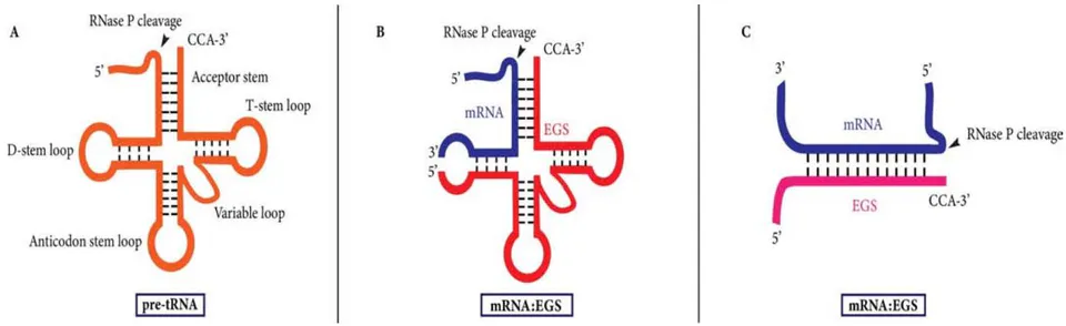
In E. coli, RNase P acts on substrates such as pre-tRNAs, 4.5S RNA precursors, and several other small RNAs [12]. These RNA substrates share structural motifs that include elements resembling the acceptor stem, T-stem/loop, and unpaired 5' leader regions (Figure 1A). These structures mimic the architecture of pre-tRNA and are what allows recognition by RNase P, reflecting RNase P's reliance on structure, rather than a specific sequence [47-49]. Early studies have shown that a shortened substrate containing the acceptor stem, T stem/loop, the 3' CCA sequence, and the 5' leader sequence can still be efficiently recognized and cleaved by RNase P and M1 RNA [48, 49], demonstrating that the full-length tRNA is not required as long as these structures are preserved (Figure 1). This study led to the development of the external guide sequence (EGS), which are short RNAs that bind to target mRNAs through Watson-Crick base pairing, reshaping them to mimic the structure of pre-tRNA (Figure 1B). This strategy leverages RNase P's conserved structure-based cleavage mechanism by converting it into a programmable, sequence-specific cleavage tool. EGSs expressed in both bacterial and human cells have been shown to inhibit the expression of the EGS-targeted mRNAs [50-56]. In one such study, EGSs were used to lower the levels of common reporter enzymes, β-galactosidase and alkaline phosphatase [51]. They suppressed the activity to 50-60% in E. coli expressing EGSs targeting these genes. In another study, drug sensitivity was restored in resistant strains of E. coli by targeting the drug resistance gene with EGS and RNase P [57]. Recent investigations indicated that EGS expressed in cultured mammalian cells inhibited the expression of the EGS-targeted mRNAs and infection of human viruses, including human cytomegalovirus (HCMV), HIV, hepatitis B virus (HBV), influenza virus, and Kaposi sarcoma-associated herpesvirus (KSHV) [58-63]. One of the main advantages of using the RNase P-EGS technology is that RNase P is expressed and active at all stages of the cell cycle, as it is responsible for processing all tRNA molecules . Due to the high specificity of RNase P cleavage, RNase P appears not to exhibit the off-target effects associated with other antisense strategies utilizing RNase H . The limitations of EGS, including delivery and efficacy, have led to the development of M1GS RNA.
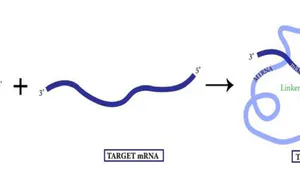
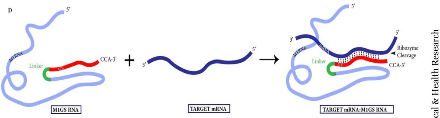
M1GS RNA addresses the issue of EGS's reliance on host RNase P by covalently linking the EGS to the catalytic M1 RNA (Figure 1C, Figure 2). The guide sequence contains an unpaired 3'-NCAA tail present in tRNA substrates that helps form the correct configuration. By tethering the GS to M1 RNA, the ribozyme forces the catalytic M1 RNA into proximity to the target site, thereby increasing cleavage efficiency and enhancing substrate binding [4, 13, 65, 66]. Researchers have shown that M1GS RNA allows for more efficient cleavage compared to unlinked M1 RNA and EGS under low Mg conditions, which better reflect in vivo conditions [4, 13, 42, 65]. M1GS has successfully targeted HSV-1 essential genes encoding TK and ICP4 in both in vitro and in culture cell settings [13, 67]. Although the C5 protein, the cofactor of M1 RNA in E. coli, is absent in human cells, its role seems to be compensated for by the protein subunits of human RNase P [14]. It is believed that the C5 analogous human proteins bind to regions of M1GS RNA that are recognized by C5. Studies have shown that C5 proteins enhance M1GS activity by 30-fold compared to purified human RNase P proteins, which enhance activity by 5-fold [13, 68, 69]. Further studies on developing M1GS variants that interact more effectively with human RNase proteins may be promising for future research.
V. ENHANCEMENT OF M1GS THROUGH IN VITRO SELECTION
Through an in vitro selection process, the efficacy of RNase P ribozymes has been further improved, making it more suitable for use in clinical applications (Figure 3) [70, 71]. M1GS variants that slice an mRNA substrate more efficiently were selected by first generating a randomized M1 ribozyme pool with mutations in the M1 RNA catalytic and conserved regions [70]. They were then annealed to 5'-biotinylated mRNA substrate, such as tk46, a model mRNA substrate derived from HSV-1 TK mRNA (Figure 3). Once annealed, these RNA-RNA complexes were passed over a streptavidin column, which binds tightly to the biotinylated mRNA substrate. This step removed any M1GS ribozymes that were improperly folded or failed to bind to the mRNA substrate. After the annealed complexes were allowed to bind, ions were introduced, allowing for RNase P-mediated hydrolysis, separating the complex from the column (Figure 3). The active ribozyme complex was captured from the column and purified using gel electrophoresis. Ribozyme RNAs were reverse transcribed and amplified by RT-PCR to regenerate DNA templates for recloning in subsequent, more stringent rounds with a shortened annealing and incubation period. This selection process was continued until the cleavage rate ceased to increase, and the most active ribozymes were cloned and sequenced to identify the mutations responsible for the enhanced activity [70]. The high-performing variants isolated in this selection exhibited many novel mutations near the ribozyme's catalytic core [70]. For example, one selected variant, R29, exhibited a 20-fold increase in catalytic efficiency and a more than 50-fold increase in binding affinity for an HSV-1 TK mRNA substrate compared to an M1GS ribozyme (M1-TK) derived from the wild-type M1 RNA sequence [70]. In a control mutant, where the point mutation was reverted to the wild-type sequence, the binding affinity reverted to the wild-type's value, confirming that this mutation was responsible for enhanced substrate affinity. UV crosslinking showed that R29 and similar variants had enhanced crosslinking with the 3' tail sequence of the TK mRNA substrate, suggesting that these changed nucleotides strengthened the ribozyme-substrate interactions through direct interactions with the substrate [70, 72]. This study was expanded in a subsequent investigation, leading to the isolation and characterization of variants V41, M95, and M200 [71]. Among these highly active variants, V41 exhibited the greatest catalytic efficiency with a 30-fold increase in cleavage efficiency and a 25-fold increase in binding affinity compared to
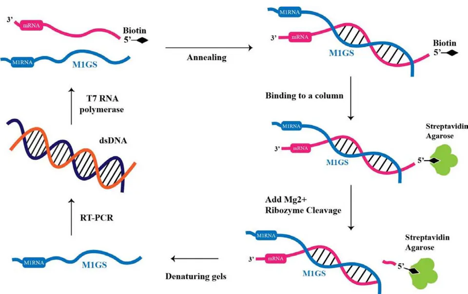
the wild-type M1-TK. V41, M95, and M200 carried point mutations within the M1 RNA, which were further shown to enhance substrate binding and were responsible for the increased activity . UV cross-linking and nuclease footprint analysis suggested that the point mutation was near the 3' tail sequence. These studies confirmed that the 3' tail sequence is a key interaction site for M1GS binding. Through this study, they demonstrated that catalytic activity and binding affinity can be significantly enhanced by optimizing this region to strengthen ribozyme-substrate interactions . The in vitro selection process represents an excellent approach for further M1GS optimization through targeted engineering.
VI. EFFECTIVE INHIBITION OF HSV-1 GENE EXPRESSION AND GROWTH BY M1GS RIBOZYMES
HSV-1 has a large DNA genome that encodes numerous essential proteins controlling replication, latency, and reactivation [17, 18]. TK and ICP4 are two important viral proteins that have been used as therapeutic targets in RNase P-based strategies. TK is involved in viral replication and has been used as a model gene. In contrast, ICP4 is a major transcriptional activator that is required for the expression of viral early and late genes .
6.1 Targeting HSV-1 TK
The first study applying the anti-HSV-1 RNase P ribozyme approach utilized an M1GS ribozyme targeting HSV-1 TK mRNA [13]. Researchers began by cloning M1GS constructs M1TK13 and M1TK13 (catalytically inactive M1 region) into retroviral vectors. HSV-1-infected human cells were then transfected by these vectors, and TK mRNA and protein levels were reduced by 80% in those expressing M1TK13 [13]. Cells expressing M1TK13 only exhibited a reduction of 9% in TK mRNA and protein levels, confirming that the catalytic activity of M1RNA was necessary for effective targeting of the TK mRNA in cultured cells. The M1 RNA in this study retained the wild-type sequence and its catalytic ability, setting the groundwork for future studies that introduced mutations in this region to enhance cleavage activity.
Follow-up studies investigated whether mutations introduced into the M1 RNA of the M1GS ribozyme could enhance its catalytic efficiency . These ribozyme variants were generated through mutagenesis of conserved regions in M1 RNA and selected through an in vitro process. One of the selected variants, R29 exhibited a 20-fold increase in catalytic efficiency and a more than 50-fold increase in binding efficiency compared to wild-type ribozyme (called M1-TK) activity . The R29 variant was then tested in HSV-1-infected cells. Cells expressing R29 exhibited a 99% reduction in TK mRNA levels and a 98% reduction in TK protein levels . These results confirmed that enhanced catalytic activity shown in vitro was also observed in a cellular context, validating the M1GS ribozyme's antiviral potential.
6.2 Targeting HSV-1 ICP4
The ability of RNase P ribozyme to inhibit HSV-1 gene expression and infection was further investigated using M1GS ribozymes that targeted the ICP4 gene . By targeting ICP4, researchers sought to determine whether knocking down the expression of an upstream regulatory gene could suppress the expression of the associated downstream genes. To test this, researchers used the V6-ICP4 ribozyme, which contains mutations in the M1 RNA covalently linked to a 13-nt-long GS targeting ICP4 mRNA . Two controls, M1-ICP4 (wild-type M1 RNA) and C-ICP4 (no M1 RNA catalytic ability), were tested alongside the V6-ICP4 construct. V6-ICP4 was 15-fold more active than M1-ICP4 and orders of magnitude greater than C-ICP4 . These ribozymes were tested in a cellular context where cells were transduced with a retroviral vector encoding V6-ICP4, M1-ICP4, and C-ICP4. These cells were then infected with HSV-1, and the levels of viral mRNAs and proteins were measured. V6-ICP4 expression resulted in a 93% reduction in ICP4 mRNA levels and a 92% reduction in ICP4 protein levels . M1-ICP4 also reduced ICP4 mRNA and ICP4 protein levels by 82% and 77%, respectively. C-ICP4 had minimal inhibition of around 6-8%, solidifying that antisense base pairing alone was insufficient for suppression. Suppression of downstream gene expression was shown with V6-ICP4, which reduced TK mRNA (an early gene) by 90% and late gene products ICP35 and gB by 90% and 91% [67]. M1-ICP4 also reduced expression, but at a lower level. These findings confirmed that the inhibition of ICP4 expression also reduced the expression of viral early ( ) and late genes ( ). The inhibition of viral growth in ribozyme-expressing cells was then tested and measured quantitatively through the number of plaques formed. Cells expressing V6-ICP4 had a ~4,000-fold reduction compared to the ~1,000-fold reduction in cells expressing M1-ICP4 ribozymes [67]. These results, by showcasing ICP4-targeted knockdown through M1GS ribozymes, demonstrated the viability of targeting upstream regulatory genes to suppress HSV-1 gene expression and replication [67, 73].
VII. ADVANTAGES AND DISADVANTAGES OF M1GS RIBOZYMES
M1GS ribozymes offer unique advantages over some of the other therapeutic tools. Classical antisense oligonucleotides rely on host RNase H to cleave RNA-DNA hybrids to degrade the mRNA target, but carry the risk of non-specific cleavage at non-target sites due to their tolerance of mismatches . In contrast, M1GS ribozymes require strict Watson-Crick base pairing to form a pre-tRNA-like structure, enabling their structure-based cleavage mechanism . Unlike other ribozymes, such as hairpin and hammerhead ribozymes, M1GS ribozymes do not require a specific nucleotide sequence (-GUX-) in the target mRNA . This allows M1GS to be capable of hydrolyzing almost any RNA target as long as it is accessible. M1GS ribozymes and M1RNA's catalytic activity have been shown to increase in the presence of human proteins, such as the cofactors of human RNase P, and also act irreversibly . These characteristics, along with the ribozyme's ability to hydrolyze multiple substrate molecules, make it a promising candidate for gene-targeted antiviral therapy.
Despite the many unique advantages offered by M1GS ribozymes, several concerns may need to be considered when using RNase P ribozymes for specific applications. One concern with M1GS ribozymes is the potential side effects on cellular physiology and viability resulting from the overexpression of M1GS RNA. Under high concentrations of M1GS, human RNase P and its associated pathways could be potentially disrupted . Additionally, M1GS RNA is prone to cellular degradation. While stability can often be enhanced through chemical modifications such as the addition of 2' hydroxyl or phosphorothioate linkages to resist endonuclease activity, these modifications could compromise M1GS's functionality . Due to M1GS's reliance on secondary and tertiary structure for its catalytic function, chemical modification could disrupt the precise folding required for its catalytic ability. Finally, the relatively large size of M1GS ribozymes, ~400 nucleotides, makes it challenging to deliver and synthesize. Compared to RNAi-based therapies, where delivery methods such as lipid nanoparticles and polymeric matrices are already established, M1GS RNA's large size may limit the use of these delivery mechanisms . This constrains M1GS delivery to alternative delivery strategies such as viral vectors or through the development of novel delivery mechanisms.
VIII. FUTURE DIRECTIONS AND CHALLENGES
Studies have shown that E. coli-derived M1 RNA can function in human cells . However, its catalytic efficiency is limited to the human RNase P protein's ability to compensate for the absence of the bacterial C5 protein . Several issues may need to be addressed to develop M1GS ribozymes for clinical applications in treating HSV-1 infection.
First, future directions could involve enhancing the compatibility of M1GS ribozymes with human RNase P proteins. One way this could be done is by mutagenizing contact regions of the M1 RNA domain that interact with RNase P proteins. These variants could be selected through an in vitro process involving human RNase P proteins to generate constructs better adapted to the human RNase P complex. Although M1GSs are highly specific, the sustained expression of M1GS and its effects on cellular physiology are not fully understood. Overexpression may lead to unintended side effects; therefore, future studies evaluating M1GS's potential off-target effects, long-term effects, and optimal dosage will be essential in determining its safe use in therapeutic applications.
Second, the efficient and tissue-specific delivery of M1GS ribozymes to the site of HSV-1 infection in vivo presents a challenge. M1GS ribozymes' relatively large size makes them incompatible with established delivery methods for shorter RNA molecules. As a result, viral vectors remain the most practical choice for M1GS expression and delivery. HSV-1 establishes a latent infection in sensory neurons ; this tissue specificity could be applied to the delivery mechanism of M1GS. Past studies have shown that Salmonella can be used as vectors for macrophage-targeted M1GS delivery in human cytomegalovirus infection . Future studies could explore neuron-specific vectors for non-invasive, sustained expression of M1GS in clinically relevant tissues and cells that are known to be infected by HSV-1 in vivo.
Finally, future studies could investigate the numerous important HSV-1 genes beyond TK and ICP4 as potential targets for M1GS. As discussed in this review, targeting ICP4, an IE gene, was highly effective in suppressing downstream gene expression and replication. Naturally, other IE genes, such as ICPo and ICP27 [18, 20], that are critical for reactivation from the latent stage, represent promising candidates for M1GS ribozymes. Beyond IE genes, early genes such as UL5 and UL9, responsible for origin recognition and replication initiation during genomic DNA replication, are also promising M1GS targets. By disrupting the expression of these genes, viral replication can be prevented even after reactivation. Another strategy could involve M1GS-mediated destruction of latency-associated transcripts (LATs). Studies have shown that HSV-1 mutants lacking LATs still establish latency at a lower level and remain capable of reactivation . Targeting LATs could induce the virus to enter the reactivation stage, albeit in a weakened state, allowing M1GS to target IE or early gene mRNAs, or with antivirals such as acyclovir. This approach could potentially address a major clinical challenge: current treatments are only effective during symptomatic reactivation . M1GS-mediated controlled reactivation may provide a safer therapeutic strategy that could minimize neuronal damage and reduce the severity of symptoms, which is beneficial for immunocompromised individuals and others at a higher risk of severe HSV-1 complications. Developing M1GS ribozymes against these genes could provide a broader and layered antiviral approach. By identifying and disrupting essential proteins involved in viral reactivation, replication, or immune evasion, a combination of M1GS-mediated knockdowns may circumvent the need for a singular, universal target.
IX. CONCLUSION
In this review, we have highlighted the gene-targeting activity of the engineered RNase P ribozyme, known as M1GS RNA, and how it can be optimized through in vitro selection. We have discussed the advantages of M1GS and summarized its efficacy in targeting HSV-1 by cleaving viral mRNAs encoding the TK and ICP4 protein. We outlined the potential of M1GS as an HSV-1 antiviral by discussing its ability to intercept reactivation by targeting immediate-early (IE) genes, as well as its potential to target latency-associated transcripts (LATs) and upstream regulatory proteins. M1GS's unique specificity due to its structure-based recognition and mRNA level of attack makes it a promising candidate for future therapeutic strategies targeting both HSV-1 and other viral infections characterized by latent or transcriptionally regulated gene expression.
ACKNOWLEDGEMENT
We are grateful to Phong Trang and Isadora Zhang for critical comments, insight discussions, and editorial assistance. We would also like to thank Robert and Colleen Haas for their generous support. T.S. is a Haas Scholars Fellow. The study has been supported by a Start-Up Fund (University of California).
Conflict of Interest
The authors declare no conflict of interest.
Ethical Approval
Not applicable
Data Availability
The datasets used in this study are openly available at [repository link] and the source code is available on GitHub at [GitHub link].
Funding
This work did not receive any external funding.
References
Cite this article
Related Research


Special Issue
Launch a focused special issue to highlight research, emerging trends, and expert insights in your academic field.