



IntelliPaper
Abstract
We have already presented our concept to close more extensive defects after tumor resection of basal cell carcinoma in the head and neck region in scientific journals [1, 2]. Essentially, the procedure consists of suturing a defect wound by approximation with an overlocking hem suture. over a period of about 3 months. The granulating wound is additionally covered by a collagen membrane and protected with a custom-made, stitched-on dressing plate. It seems to us to be logical to do this also for resection defects of other resection defects of other entities.
Explore Digital Article Text
I. INTRODUCTION
We have already presented our concept to close more extensive defects after tumor resection of basal cell carcinoma in the head and neck region in scientific journals . Essentially, the procedure consists of suturing a defect wound by approximation with an overlocking hem suture. over a period of about 3 months. The granulating wound is additionally covered by a collagen membrane and protected with a custom-made, stitched-on dressing plate. It seems to us to be logical to do this also for resection defects of other resection defects of other entities.
1.1 Anamnesis and Diagnosis
A 55-year-old male patient presented to our office in October 2021 for treatment of a suspicious dark skin tumor of the right cheek.
Anamnestically, a melanoma was known in the patient's father. Alio loco, a "lentigo maligna" had
1.2 Ttherapy and Course of Treatment
Given the well-documented clinical effectiveness of this methodology, we found it relatively easy to convince patients of this off-label use approach.
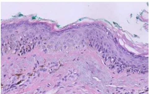
already been suspected. Therefore, a dermatological evaluation was obtained in consultation with a specialist. Since the findings there did not exclude the possibility of a benign finding, we initially decided on a close excision biopsy, which was then performed shortly thereafter. The histological evaluation by the Pathological Institute of the Marienhospital Stuttgart and the reference by the Laboratory for Special Dermatology, Microscopy and Molecular Dermatology of the University Dermatological Clinic Tübingen resulted in the diagnosis: "Lentigo maligna (melanoma in situ, Clark Level I),...marginal to the sides" (Fig. 1) Thereupon, resection was performed with a safety margin of 10 mm according to the guidelines [3, 4, 5].
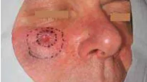
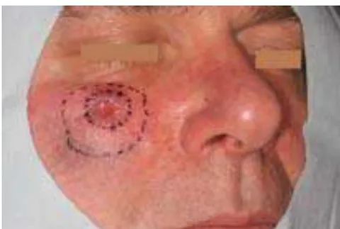
According to the image of a clock, four circular marginal incisions 0 - 3 o'clock, 3 - 6 o'clock, 6 - 9 o'clock, 9 -12 o'clock were taken and cranial, medial and caudal several from the tumor base. In the 6 - 9 o'clock region, a tumor spur required recutting until an Ro resection could be achieved (Figs. 2 - 5).
The main advantage of this procedure is that it avoids the need to elevate costly suture flaps with extensive incisions. These procedures can each be performed under local anesthesia with minimal surgical effort. The system consists of 3 partial steps:
Contraction of the wound edges already from the histographically confirmed excision via a circular overlocked hem suture (Maprolen 5/0 non-absorbable, Cutgut, Markneu- kirchen).
Covering of the open wound by suturing a 15 x 20 mm Mucograft® membrane (Geistlich
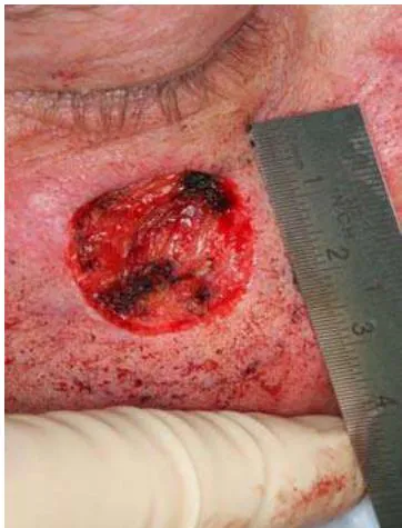
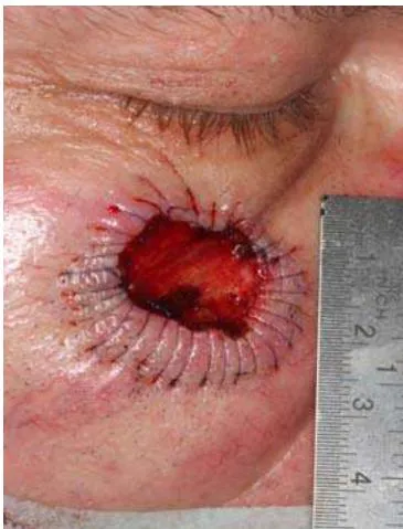
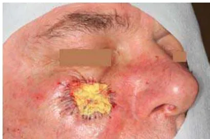
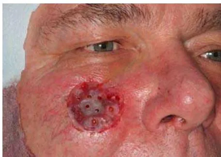
The contraction of the wound edges already in the first session of histographically confirmed tumor excision with up to 5 mm wound reduction in diameter is always amazing (Fig. 4). Distortion of the wound margins is not an obstacle due to the uniform reduction for the eventually due resection (second session) as in this case in the 6-9 o'clock section. The covering of open granulating wounds by absorbable membranes (Fig. 6) has been sufficiently described, so that this could be easily implemented in the technique of wound contraction [6, 7]. Overknot dressings are required to press the membranes or split skin grafts onto the support [Fig. 5]. In the facial region, however, these overtie dressings are impractical because they are cosmetically disturbing, and the disinfecting pastes and ointments, such as those used for the iodoform aureomycin tamponades commonly used in our field, in turn impede wound healing.
Furthermore, the odor is usually perceived as unpleasant by patients. Therefore, we have
Biomaterials, Baden-Baden, Germany) into the developing defect.
- Wound dressing by means of a customized pad (HS-Thermo bleach, clear, flexible foil 1.0 x 120mm ∅, Henry Schein, Langen).
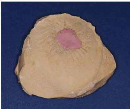
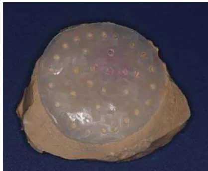
transferred the dressing plates used in oral surgery to the extraoral area. An impression of the surgical site is taken preoperatively with alginate compound, from which a plaster model is made (Fig. 7). A dressing plate is pulled over this using the thermoforming method and provided with perforations (Fig. 8). The perforations serve on the one hand to allow the wound secretion to drain off or to be cleaned with saline/water from the outside and ensure normal air circulation even under the cover. Fixation sutures can additionally be pulled through the perforations or they can be used again for anchoring "tension belt sutures". The transparency of the plastic allows continuous observation of the wound without the need for constant dressing changes.
After the resection margins were tumor-free, the mucograft membrane was stitched onto the wound with absorbable Marlin 6-o sutures in the third session. The first wound contraction already showed a significant reduction of the open wound area. At the same time, the second overlocked suture was applied. The dressing plate fixed with 5-o-Maprolen sutures remained in situ for 10 days. The patient was encouraged to moisten the wound with water, which has a cleansing effect and promotes epithelialization.
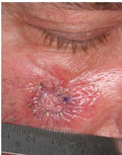
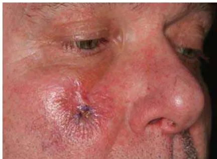
Three weeks later, another wound contraction was performed in the fourth session (Fig. 10).
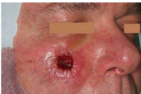
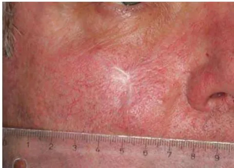
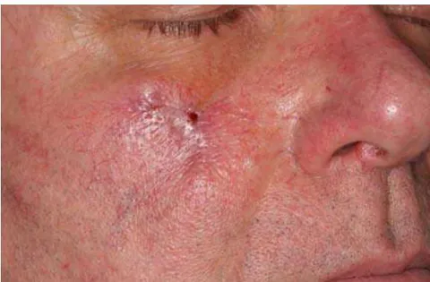
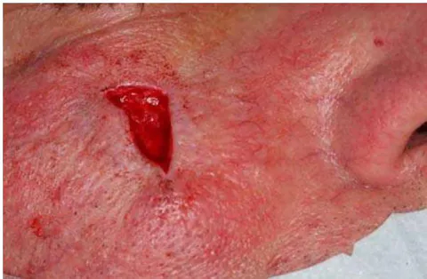
Since we had made the experience during longer observation over two years that the scar plate expands again somewhat due to traction from the wound edges (Figs. 11 - 12), we performed another scar excision with multilayer wound closure 10 months later - earlier was not possible due to scheduling reasons. The subcutaneous sutures should compensate for the centrifugal scar traction (Figs. 13 -14). Histology of the excised scar tissue showed no evidence of residual tumor.
II. DISCUSSION
Defects in the facial region of more than 1 cm in diameter are usually be treated with elaborate regional flap techniques which, depending on the type of incision, are associated with long scarring, depending on the type of incision. The procedure presented by us flap elevation and pulls the wound edges in usually 3 - 4 sessions by means of a circular, overlocked suture. suture. We see the advantages in the fact that larger defects can be defects can be treated. The procedure is comparatively low risk in terms of morbidity and is associated with much less scarring. scarring.
The sessions are performed on an outpatient basis under local anesthesia, with comparatively low effort and cost. No patient to whom we offered this procedure [2] opted for the classical procedures of plastic surgery in the head and neck area. head and neck region. Compliance was good in all patients and satisfaction with satisfaction with the course and outcome was high, as in this case. this case. The duration of 3 - 4 months until wound closure was interestingly not was not perceived by any patient as a major disadvantage. perceived as a major disadvantage.
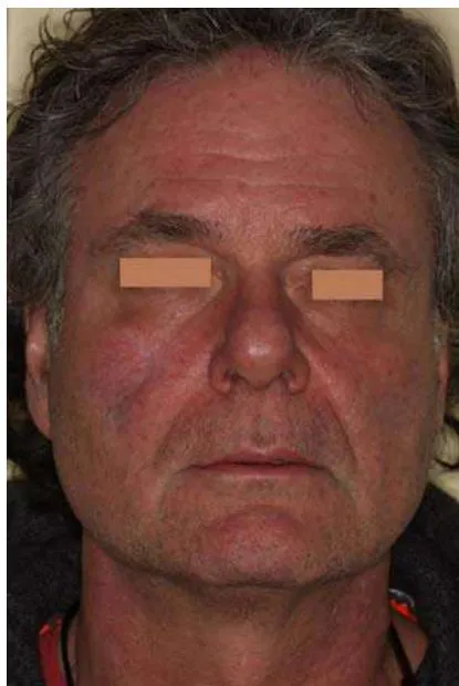
From our point of view, when used on younger patients, the operative patients, the surgical correction with multilayer wound closure to prevent to prevent the scar plate from pulling apart as a 4th step. This is a certain shortcoming.
However, even with more extensive flap plasty, surgical correction may be necessary.
III. CONCLUSION FOR THE PRACTICE
In our opinion, the concept of wound contraction of facial defects after tumor resection presented by us represents a real alternative for patients.
Compliance ethical guidelines
The patient, who could be identified from images or other information within the or other information within the manuscript, has given his or her written written consent.
Conflict of Interest
A. Born and U. Markmann declare that there is no conflict of interest exists.
Conflict of Interest
The authors declare no conflict of interest.
Ethical Approval
Not applicable
Data Availability
The datasets used in this study are openly available at [repository link] and the source code is available on GitHub at [GitHub link].
Funding
This work did not receive any external funding.
References
Cite this article
Related Research

Special Issue
Launch a focused special issue to highlight research, emerging trends, and expert insights in your academic field.