


Abstract
The typical presentation of cobalamin deficiency is macrocytic anemia with or without neurologic symptoms, adding that the most frequent cause is Pernicious anemia, an autoimmune disease. We present a case of a 61year-old man with neurologic symptoms, pancytopenia, and laboratory findings consistent with hemolytic microangiopathic anemia (MAHA), like Thrombocytopenic Thrombotic Purpura (TTP). The lack of response to plasma exchange (PEX) therapy, concomitant with low plasma levels of cobalamin, low reticulocyte count, macrocytosis and remarkably high lactic dehydrogenase (LDH) levels made us suspect a pseudo-Thrombotic Microangiopathy (Pseudo TMA). The diagnosis was confirmed with a normal serum ADAMPTS13 activity and a rapid clinical and laboratory improvement after cobalamin supplementation. We think the internist must know the wide variety of presentations of Cobalamin deficiency, since it is a reversible cause of bone marrow failure, and its’ misdiagnosis may result in unnecessary costly procedures.
Explore Digital Article Text
I. BACKGROUND
Cobalamin participates in cell maturation. When deficiency appears, it can lead to macrocytosis, immature nuclei and hyper segmentation in granulocytes at peripheral blood . Typically, hematological findings in cobalamin deficiency are megaloblastic anemia and neurologic symptoms that could not be present . The leading cause of cobalamin deficiency is pernicious anemia, an autoimmune disease characterized by antibodies against parietal cells and intrinsic factor . However, there are other clinical pictures related to this vitamin deficiency mimicking other clinical entities such as TTP or myelodysplastic syndromes.
II. CASE PRESENTATION
A 61-year-old man, with medical record of self-limited pancytopenia interpreted as transient hypersplenism eight years before, was admitted at internal medicine ward, derived from primary care presenting a new episode of pancytopenia described as asymptomatic. At his arrival, the patient presented slight disorientation in time and space, meanwhile the rest of the neurological exam appeared to be without findings. Clinical features such as fever, jaundice, hemorrhage, renal dysfunction and paresthesia were not found. Complete blood count reported macrocytic anaemia (hemoglobin 6.32 gr/dL), medium corpuscular volume (MCV) 115 fL, hematocrit 18.5%, white blood cells count 3.470/mm3 and platelet count 84.000/mm3 (Table 1). Blood smear showed schistocytes, dacrocytes, and stomatocytes, and negative direct Coombs test. Lactate dehydrogenase was high (4194 IU/L, plasmatic values between 150-350). Total bilirubin was 2.54 mg/dl, 55% indirect reacting*, and serum creatinine was normal (Table 1). ADAMTS13 activity and the presence of inhibitors were searched. The patient was transferred to an intensive care unit for urgent PEX, performing three sessions, and after second session of PEX, we realized lacking bone marrow compensatory response with low reticulocyte count (1.6%), persistent thrombocytopenia, and lower than expected drop in LDH levels (Figure 1), making megaloblastic anaemia confirmed by low plasmatic levels of cobalamin (140 pg/mL, NV 187 pg/mL – 883 pg/mL) (Table 1), suspecting Pseudo-TMA and began cobalamin supplementation.
IV. INVESTIGATIONS
| Variable | Reference Range | Results on ER admission and IMD* | Results after PEX and Before Vitamin B12** | Outcome results (7 days // 30 days after first dose of Vitamin B12**) |
| WBC, x103/μL | 4.4 – 11.30 | 3.47 | 2.93 | 7.70 // 5.91 |
| Neutrophils,% | 40 – 75 | 57.02 | 59.20 | 71.76 // 60.95 |
| Lymphocytes,% | 25 – 40 | 31.32 | 31.68 | 12.33 // 25.93 |
| Monocytes,% | 2 – 8 | 6.01 | 6.29 | 9.30 // 7.59 |
| Band neutrophils,% | 0 – 5 | 0 | 0 | 0 |
| Metamyelocytes,% | 0 | 0 | 0 | 0 |
| Promyelocytes,% | 0 | 0 | 0 | 0 |
| Myelocytes,% | 0 | 0 | 0 | 0 |
| Eosinophils,% | 2 – 4 | 4.51 | 1.70 | 4.66 // 3.70 |
| Basophils,% | 0 – 1 | 1.12 | 1.12 | 1.194 // 1.82 |
| Hemoglobin, g/dL | 13 – 17.5 | 6.32 | 7.57 | 9.82 // 13.82 |
| Hematocrit,% | 40 – 52 | 18.51 | 21.34 | 30.48 // 42.59 |
| Reticulocyte count,% | 0.5 – 1.5 | 1.6 | 9 // 1.4 | |
| Platelet count, x103/μL | 140-400 | 84 | 58 | 146 // 176 |
| MCV, fL | 80 – 96 | 115 | 96 | 99 // 87 |
| Total Bilirubin, mg/dL | 0,2 – 1,2 | 2.54 | 2.68 | 0.96 // 0.3 |
| Indirect Bilirubin, mg/dL | 0 – 1,0 | 1.40 | 1.9 | 0.5 // 0.2 |
| LDH, UI/LESR, mm/h | 125 – 2430-20 | 4,1941 | 972 | 372 // 151 |
| Crea, mg/dL | 0.72 – 1.25 | 0.90 | 0.7 | 0.7 |
| Blood smear: | - | Anisocytosis, Macrocytosis, Schistocytes, Dacrocytes, Stomatocytes | - | Anisochromia *** |
| Myelogram | Hypercellularity, erythroid hyperplasia, scarce blasts, dysplasia of the three blood cell lineages. (•) | |||
| ADAMPTS 13 Activity,% | 41 – 180 | 92 | ||
| Presence of ADAMPTS 13 inhibitors | No inhibitors present | |||
| Upper GI endoscopy | Atrophic pangastritis, intestinal metaplasia. Urease test: negative. | |||
WBC: white blood cell count, MCV: Medium Corpuscular Volume, AST: Aspartate transaminase, ALT: Alanine transaminase. AP: Alkaline phosphatase. Crea: serum creatinine. ESR: Erythrocyte sedimentation rate. ER: Emergency room, IMD: Internal Medicine Department, PEX: Plasma Exchange. GI: Gastrointestinal.
(*): Initial laboratory findings.
(**): After initiating Vitamin B Complex administration.
(***): Blood smear after one week of treatment with Vitamin B Complex didn't inform any abnormalities in the red blood cells' or the platelets' morphology.
(•): Myelogram performed one week after Cobalamin supplement.
{"image_source":{"path":"images/f3c45ec2f5fe8b2549ba5c3eacec76b73ef678970003572c3a5020a9966ebad8.jpg"},"content":"","chart_caption":[{"type":"text","content":"Blood count follow up from the initial evaluation to 30 day ambulatory medical control."},{"type":"text","content":"WBC: white blood cell count., ER: Emergency room, IMD: Internal Medicine Department, PEX: Plasma Exchange. VB12: Vitamin B12"}],"chart_footnote":[]} {"image_source":{"path":"images/a156d4c7378e44169543c2e4bf3b74526f5d5945706a42766ed727ddf762e14a.jpg"},"content":"","chart_caption":[{"type":"text","content":"ER: Emergency room, IMD: Internal Medicine Department, PEX: Plasma Exchange. VB12: Vitamin B12"}],"chart_footnote":[]}
{"image_source":{"path":"images/efb2e810ebb4643da18e31f388809492fb05948fbf4a1ed088f9eac7f60a61d1.jpg"},"content":"","chart_caption":[{"type":"text","content":"MCV: Medium corpuscular volume, ER: Emergency room, IMD: Internal Medicine Department, PEX: Plasma Exchange. VB12: Vitamin B12"},{"type":"text","content":"Figure 1: Laboratory Tests Follow-up"}],"chart_footnote":[]}
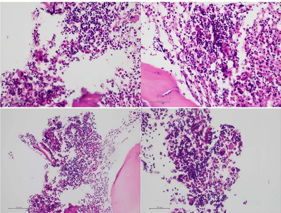
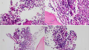
Bone marrow biopsy seen by optical microscopy, showing diffuse hyperplasia, megaloblasts and some scarce elements of myelodysplastic changes.
V. DIFFERENTIAL DIAGNOSIS
Pseudo-TMA due to cobalamin deficiency, in contrast to TTP, should be suspected when there is a deficient medullary response after acute hemolysis, verified by a low reticulocyte count and low intramedullary cell counts, without renal involvement . In TTP, there is a high reticulocyte count since it affects a normal bone marrow and diagnoses could be helped by the PLASMIC score when it is over five points (platelets count less than 30.000, hemolysis characterized by indirect bilirubin over 2 mg/dl or undetectable haptoglobin, plus no active cancer, and no history of stem cell transplant, INR less than 1.5, MCV less than 90fL and creatinine below 2 mg/dl, one point each other maximum seven points). However, this score is not definitive for ruling out, since our patient scored five points in PLASMIC diagnostic criteria except platelet count over 30.000 and MCV over 90fL. Moreover, other features have been described to suggest Pseudo-TMA, such as higher lactate dehydrogenase levels with slow reversion after PEX, higher mean platelet counts, and lower mean neutrophil count [3,4,8,9,10]. Going deeper in differential diagnostic workup, the finding of elevated homocysteine, methylmalonic acid, presence of hyper segmented neutrophils and megaloblasts in the peripheral blood smears are also useful to guide the diagnosis of cobalamin deficiency [1].
In addition, bone marrow study (myelogram and bone biopsy) may find 3 series dysplasia, encompassing any myelodysplastic syndrome as another differential diagnosis of cobalamin deficiency [1].
VI. TREATMENT
We performed three PEX sessions since we suspected TTP diagnosis, but after finding low medium-sized response with a reticulocyte index of 1.6 percent and low levels of cobalamin, procedure was withdrawn and it was decided to use intramuscular vitamin B complex (10.000 units daily for three doses), with excellent response. One week after Vitamin B12 supplementation, there was an impressive improvement in mental symptoms and laboratory tests, both in blood (Table 1, follow up tests) and bone marrow samples (myelogram and bone biopsy). The diagnostic study was negative for TTP, and the upper gastrointestinal endoscopy was consistent with pernicious anaemia (Table 1).
VII. OUTCOME AND FOLLOW-UP
The patient underwent a rapid, clinically significant improvement in his neurological condition and laboratory findings. Thus, one week after the initiation of vitamin B complex, a reticulocyte peak was evident. Afterwards, within two weeks of hospitalization and ten days of cobalamin supplementation, the patient was discharged without any neurological symptoms, mild normocytic anemia, and normalization of the other blood cell counts, with the indication of prolonged treatment with vitamin B12. Two weeks after discharge, with an almost normal blood count, ADAMTS 13 activity resulted normal (a sample taken before first PEX session), reflecting the absence of metalloprotease inhibitors. In the current context of contingency due to SARS-CoV2, when this case happened, a most comprehensive study of probable pernicious anaemia could not be performed, and the patient withdrew follow-up before pandemic isolation was stopped.
VIII. DISCUSSION
One common cause of cobalamin deficiency is pernicious anaemia, an autoimmune disease with plasmatic autoantibodies against the Intrinsic Factor . In our case, the gastric tissue biopsy result was consistent with pernicious anemia, but the antibodies could not be studied.
This cobalamin deficiency presentation, with clinical and laboratory findings consistent with TTP (neurological involvement, suggestive findings of MAHA encompassing anaemia, schistocytes in peripheral blood, thrombocytopenia, high levels of lactate dehydrogenase and indirect-reacting bilirubin) [6], is called Pseudo Thrombotic Microangiopathy (Pseudo TMA) [3,4,7,8,9,10]. Some case reports have described similar cases, postulating that laboratory findings were due to ineffective erythropoiesis. Other authors think there is a rise in homocysteine's blood levels, provoking endothelial dysfunction, fragmentation, and destruction of erythrocytes, the reason for appearing schistocytes in the blood smear [2]. The LDH levels appear to be higher in Pseudo TMA than in TTP, because of peripheral and intramedullary destruction of red blood cells; the presence of lower neutrophil mean count and less severe thrombocytopenia should help in differential diagnostic workup too [3,4, 8,9,10]. Another feature that guides us to cobalamin deficiency is the presence of lower reticulocyte count in Pseudo-TMA due to bone marrow failure, in contrast to being always elevated in TTP [1,4,5,7]. Moreover, elevated homocysteine and methylmalonic acid levels, hyper segmented neutrophils, and megaloblasts in peripheral blood smears in absence of acute kidney injury in Pseudo TMA, could be crucial for differentiation from TTP [1,5]. In addition, the response to cobalamin supplementation also suggested the etiology of hemolytic anemia due to the deep specific vitamin deficiency.
Pathophysiology of TTP consists in a quantitative or functional decrease of the protease ADAMTS13 on the endothelial surface, whose task is cleaving von Willebrand factor (vWf) from a macromolecule with a high capacity to recruit and activate platelets, into lower molecular weight segments with less activity [5]. Consequently, a decrease in ADAMTS13 activity promotes platelets activation and thrombus formation through every organ microcirculation [5].
Whereas, in our case, the correct diagnosis was made after significant medullary response after Vitamin B12 supplementation, meanwhile some elements of myelodysplastic syndromes were shown in the biopsy, but this data was not considered of importance because the fact that all cellular series responded to Vitamin B12 administration, including an early reticulocyte count peak.
Unlike TTP, a hematologic emergency that implies the urgent need of PEX, the Pseudo TMA does not need that therapy . The similarity of both clinical and laboratory pictures made PEX an initially appropriate indication, because the PLASMIC score had five points with only platelet count 84.000 and MCV over 90fL as negative findings .
The differential diagnosis between pseudo-TMA and TTP could be very subtle and requires a quick and profound analysis for correct diagnosis. Our opinion is that it is of utmost importance for internists to know the wide variety of presentations of cobalamin deficiency, since it is a reversible cause of bone marrow failure and misdiagnosis may lead to unnecessary costly and harmful procedures.
IX. LEARNING POINTS/TAKE HOME MESSAGES
Pseudo TMA and TTP are very similar in their clinical features.
TTP is a hematologic emergency that needs a quick PEX decision, whereas Pseudo TMA does not.
The presence of schistocytes in blood smear does not differentiate Pseudo TMA and TTP.
Clues to think in Pseudo TMA are a medullary response to hemolysis, finding low levels of reticulocytes and serum Vitamin B12, and normal levels of ADAMTS-13 activity without inhibitors.
X. INVESTIGATIONS
WBC: white blood cell count,
MCV: Medium Corpuscular Volume,
AST: Aspartate transaminase,
ALT: Alanine transaminase.
Crea: serum creatinine.
ESR: Erythrocyte sedimentation rate.
ER: Emergency room,
IMD: Internal Medicine Department,
PEX: Plasma Exchange.
GI: Gastrointestinal.
(*): Initial laboratory findings.
(**): After initiating Vitamin B Complex administration.
(***): Blood smear after one week of treatment with Vitamin B Complex didn't inform any abnormalities in the red blood cells' or the platelets' morphology.
(•): Myelogram performed one week after Cobalamin supplement.
Conflict of Interest
The authors declare no conflict of interest.
Ethical Approval
Not applicable
Data Availability
The datasets used in this study are openly available at [repository link] and the source code is available on GitHub at [GitHub link].
Funding
This work did not receive any external funding.
References
Cite this article
Related Research


Special Issue
Launch a focused special issue to highlight research, emerging trends, and expert insights in your academic field.