




IntelliPaper
Abstract
Drug delivery through the skin offers distinct advantages, including bypassing the liver's initial metabolism, maintaining consistent drug levels in the blood, and ensuring safety and adherence to treatment, which surpasses traditional oral or injectable methods. Microneedle technology has emerged as a revolutionary approach in this regard, offering enhanced delivery efficiency and patient acceptance. The main obstacle to transdermal administration, however, is the fact that only a small number of strong medications with optimal physicochemical characteristics can intercellularly and passively diffuse across skin barriers to reach therapeutic concentration using this method. Innovative strategies have been pursued to improve the drug's skin penetration. Microneedles, a microscale physical enhancement technique, have expanded the spectrum of medications that may be delivered transdermally and intradermally. These microneedles are usually between 0.1 and 1 mm in length, offering a minimally invasive yet highly effective method for drug administration. This review discusses the challenges associated with microneedle based TDDS, including formulation issues, skin permeability, and manufacturing complexities.We highlight recent advancements in microneedle design, materials, and fabrication techniques, shedding light on their potential to overcome these hurdles. One may create solid, coated, hollow, or dissolvable microneedles using a range of materials, including silicon, stainless steel, and polymers. Despite extensive research, several obstacles hinder the long-term, cost-effective production, and effectiveness of microneedles for transdermal drug delivery. This analysis identifies gaps in production technology and reviews characterization techniques. Microneedles show promise in various applications, including medicine delivery, vaccination administration, illness diagnosis, and cosmetics, indicating their versatility and wide-ranging potential.
Explore Digital Article Text
I. INTRODUCTION
For thousands of years, people have been using various chemical substances on their skin for a variety of reasons. These substances were utilized for medicinal purposes, to protect the skin, and even for cosmetic purposes . In ancient times, the Greeks developed a balm by combining water, olive oil, and lead (II) oxide. The olive oil served as a barrier, while the lead (II) oxide had a tightening effect . It wasn't until 1893 that Bourget proved the efficacy of salicylic acid in treating acute rheumatoid arthritis, which challenged the notion that the skin was impenetrable [3,4].
In the early 1900s, researchers made a significant discovery regarding lipophilic agents, which were found to enhance the permeability of the skin. Through Wolf's tape-stripping technique, Blank conducted a study and determined that the stratum corneum (SC) serves as the primary obstacle for the penetration and permeation of active pharmaceutical ingredients (APIs) [5,6].
The utilization of skin as a means of delivering drugs to systemic circulation was not adopted for commercial or scientific purposes until 1954. It was during this time that the effectiveness of 2% nitroglycerin ointment in managing angina pectoris was demonstrated. Consequently, this ointment became the initial commercially available formulation designed for transdermal delivery of active pharmaceutical ingredients into the systemic circulation .
1.1 Skin Structure
The skin, which accounts for approximately 15% of the body's mass and covers an area of 1.5 to 2.0 square meters, is widely recognized as the largest and most intricate organ in the human body. The first skin layer, i.e. the epidermis layer, is approximately 150–200 mm thick and is composed of viable cells. The structure consists of five layers based on the level of cell keratinization i.e. stratum corneum (SC, horny layer), stratum lucidum (clear layer), stratum granulosum (granular layer), stratum spinosum (spinous or prickle layer), and stratum germinativum (basal layer) [8]. Its vital role lies in safeguarding the body against detrimental environmental elements such as dehydration, disease-causing micro-organisms, and various forms of stress [9-12]. Moreover, the skin possesses distinctive attributes that render it suitable for the safe and efficient administration of medications. Specifically, topical, and transdermal delivery methods primarily target the skin for drug absorption. Nevertheless, the stratum corneum layer, the outermost lipophilic layer of the skin measuring 20 to 50 micrometers in thickness, often poses a challenge to the passive diffusion of drugs into the skin (Figure 1). The epidermis is composed of dead keratinocytes, the lipid matrix, and corneodesmosome, creating the well-known 'bricks and mortar' structure. The 'bricks' are keratinized corneocytes, while the 'mortar' is the continuous lipid matrix. Only moderately lipophilic compounds with a log P of 1.0–3.0 can penetrate the skin's lipid-enriched structure to reach the underlying skin layers. The covalent bonds between corneocytes and the lipid matrix form a strong bond, serving as the primary protective barrier function of the skin, which is also known as the main barrier limiting drug delivery rate [15].
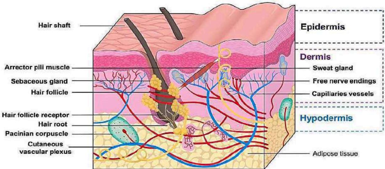 Figure 1: The Diagram Illustrates the Layers of Human Skin. Image Reproduced with Authorization from [16]
1.2 Transdermal Drug Delivery System (TDDS)
Transdermal drug delivery is gaining popularity as a preferred method of drug administration. This approach enables drugs to enter the bloodstream through the skin while retaining their effectiveness. As a result, it offers advantages such as increased bioavailability, sustained release, reduced side effects, and improved physiological and pharmacological responses . For example, in testosterone replacement therapy, transdermal delivery overcomes the limitations of oral and intramuscular methods by bypassing first-pass metabolism in the liver, thereby reducing the required dosage. Moreover, it eliminates the need for frequent injections and maintains a higher concentration of testosterone in the blood . However, the transdermal delivery of drugs is greatly influenced by the chemical characteristics of the drugs, impacting their absorption through the skin. As a result, only a limited number of drugs can be effectively delivered in therapeutic doses through this route . The transdermal drug delivery system necessitates the drugs to follow a complex path to penetrate through multiple layers of the skin, which consist of both aqueous and lipid domains, and ultimately enter the bloodstream . For a drug molecule to successfully traverse the stratum corneum (SC) layer, it must possess specific characteristics. These include a molecular weight below 600 Da, a Log P value ranging from 1 to 3, a well-balanced SC/vehicle partition coefficient, and a low melting point that correlates with good solubility, as predicted by the ideal solubility theory . Olanzapine is an example of a drug that possesses the necessary physicochemical properties for effective transdermal drug delivery. It is lipophilic (log P 2.8), has a low molecular weight of 312.4, and a low melting point (195 °C). The low bioavailability of olanzapine when taken orally and its vulnerability to loss during transportation result in only 40% of the intended dosage reaching the bloodstream. These attributes highlight olanzapine as a promising option for administration through transdermal drug patches . It is evident that numerous drugs do not meet the rigorous criteria for transdermal delivery .
The primary obstacle in transdermal delivery lies in the fact that only a limited range of medications can be effectively administered through this route. Currently, transdermal drugs that have proven successful possess molecular masses that do not exceed a few hundred Daltons. Additionally, these drugs exhibit octanol-water partition coefficients that strongly favor lipids and require daily doses of milligrams or less . Delivering hydrophilic drugs through the transdermal route has proven to be challenging. The transdermal delivery of peptides and macromolecules, including novel genetic treatments involving DNA or small-interfering RNA , has presented specific difficulties.
From a global standpoint, we propose that the progress in transdermal delivery systems can be classified into three generations of developmentFigure 2. The first generation involved the creation of many of today's patches through the careful selection of drugs that can penetrate the skin at therapeutic rates without requiring significant enhancement. The second generation brought about further advancements in delivering small-molecule drugs by enhancing skin permeability and the driving forces for transdermal transport. Moving forward, the third generation will facilitate the transdermal delivery of small-molecule drugs, macromolecules (such as proteins and DNA), as well as virus-based and other vaccines by specifically permeabilizing the skin's stratum corneum.
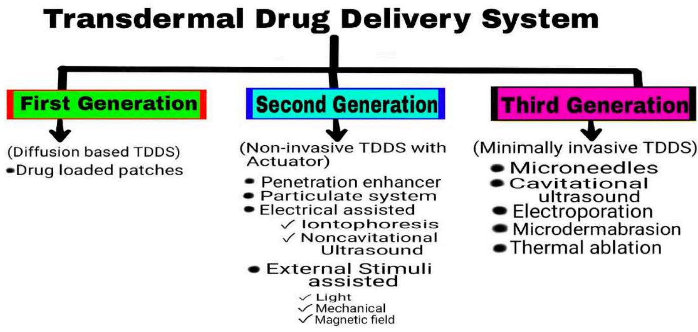 Figure 2: Generations of Transdermal Drug Delivery
1.3 Type of Transdermal Drug Delivery System
The transdermal drug delivery system (TDDS) is an innovative approach to delivering medication. It involves delivering a specific amount of medication through the outermost layer of the skin. This method ensures that the medication is released at a controlled rate over an extended period, allowing it to enter the bloodstream gradually. The TDDS can be classified as a unique type of drug delivery system that adheres to a zero-order drug release pattern . There are various methods of transdermal drug delivery available, which encompass:
1.4 Membrane Permeation Controlled TDD System
Within this particular transdermal drug delivery system, the drug reservoir is situated between the backing layer and the polymeric membrane. Illustrated in Figure 1(a), the drug reservoir region contains the drug that is distributed within the polymeric matrix solution to create a paste-like suspension. This suspension is subsequently released at a regulated pace through the rate controlling membrane, which can be either non-porous or micro porous [34]. Substances such as estradiol are administered through membrane penetration [35].
1.5 Matrix Diffusion Controlled Transdermal Drug Delivery System
In Figure 1(b), illustrates the presence of an occlusive base plate, drug reservoir, and polymeric membrane. The drug reservoir contains the drug, which is enclosed by the matrix. The matrix can consist of hydrophilic or lipophilic molecules. The drug and matrix together create a medicated disc-like structure, releasing the required amount of drug at a controlled rate into the systemic circulation. The drug is in a solution form within the matrix and is supported by an adhesive layer [36]. This method can be used to deliver drugs like nitroglycerine [37].
1.6 Reservoir Gradient Controlled Transdermal Drug Delivery System
This category is effective in addressing concerns associated with a non-zero release profile of the drug . The drug and polymer matrix are merged in a reservoir, which can transform into a reservoir gradient along the diffusional pathway across the layer. This process results in the controlled release of the drug, as illustrated in Figure 1(c). Glyceryl trinitrate is utilized as the drug in creating a deponit system for controlled delivery [34,39].
1.7 Microreservoir Dissolution Controlled Trans-Dermal Drug Delivery System.
The drug delivery system illustrated in Figure 1(d) combines matrix and reservoir dispersion types. It involves two main steps: creating the drug reservoir by suspending the drug in water-soluble or aqueous solutions, and then mixing/dispersing the suspension with a lipophilic polymer using strong mechanical force. This process leads to the formation of numerous microscopic reservoirs, ultimately resulting in the controlled release of the drug at a specific rate [34].
1.8 Microneedles- Innovating Drug Delivery Techniques.
A range of approaches can be employed to improve the delivery of drugs through the skin, such as the use of penetration enhancers, innovative formulation designs, and physical techniques . Among these methods, microneedles have recently emerged as the most efficient and dependable approach for transdermal drug delivery. This recommendation is supported by various research studies conducted in academic institutions and industrial companies . Microneedles, which are needles with sizes ranging from 25 to 2000 micrometers, have been found to puncture the layers of the skin in a precise and reversible manner, thereby disrupting the skin barrier function and creating numerous microchannels within the skin .
Microneedle technology has a rich history spanning over 40 years of development. The initial concept of microscale needles was introduced in a patent filed by Gerstel and Place, which was granted by the United States Patent and Trademark Office in 1976. The progress in the microfabrication industry has greatly facilitated the precise and controlled fabrication of microneedles. Subsequently, different types of microneedles, including solid, hollow, coated, dissolving, and swelling microneedles, have been developed in chronological order. Among these, the most recent design of microneedles for skin delivery is the hydrogel-forming swelling microneedle, which was developed in 2012 by Donnelly and his colleagues. In recent times, dissolving microneedles has garnered significant attention from researchers, leading to the invention of superior materials, novel designs, and optimized scalable production techniques. The extensive research conducted in this field has contributed to the growing popularity of microneedles. Furthermore, this evolving field has expanded to encompass cosmetic and diagnostic applications, as well as drug delivery to various tissues such as the eye, buccal mucosa, and gastrointestinal tract.
The initial microneedle design was patented in 1976, and a patent for a hollow microneedle device for intradermal drug delivery followed in 1996. A skin-piercing device was created in 1997, while silicon solid microneedles were first utilized for transdermal delivery of calcein in 1998. In 2000, researchers developed hollow microneedles for injecting a drug solution into the skin. The first coated microneedles were produced in 2004 to improve the transdermal delivery of desmopressin. Subsequently, in 2006, drug-loaded dissolving microneedles were manufactured to deliver bovine serum albumin and calceintransdermally. Finally, hydrogel-forming swelling microneedles were introduced in 2012 as the most recent type of microneedle.
Extensive research has delved into various aspects of microneedles, such as manufacturing processes , designs , drug delivery applications, safety measures , clinical studies , modeling, simulation , and more. Studies have shown that microneedles can penetrate the skin without reaching the dermis, where nerve fibers and blood vessels are located, to prevent pain or bleeding. Nguyen and colleagues recently explored the strategies for transdermal hormone delivery using microneedles, discussing trends, advancements, and challenges in transitioning from the lab to clinical settings . Ali and coworkers also examined the skin's anatomy and biomechanical properties concerning microneedle insertion and drug permeation, along with drug permeation modeling and clinical implementation of microneedles [52].
Transdermal delivery has seen significant improvement through the use of microneedles, expanding the possibilities for delivering small molecules , macromolecules , cosmeceuticals , and particulate systems . Microneedles are versatile in transporting molecules of varying sizes and molecular weights. Different microneedle systems have been developed, each with their distinct characteristics including geometry, size, design, layout, density, composition, and materials. Microneedles can be made from a range of materials such as glass, sugar, metal, silicon, ceramics, and polymers. These materials must meet specific requirements for microneedle production, such as mechanical strength, biocompatibility, and safety. Among these materials, biodegradable, biocompatible polymers have emerged as promising options and have garnered significant attention and interest . Various polymers can be utilized to create a variety of microneedles, such as dissolving, swelling, solid, coated, and hollow microneedles. Commonly used polymers for this purpose are SU-8 photoresist, cyclic-olefin copolymer, polycarbonate, poly (methyl Metha-acrylate), poly-lactic-co-glycolic acid (PLGA), polyglycolic acid, polystyrene, polylactic acid, poly (vinyl pyrrolidone), polyvinyl alcohol, and sodium carboxy methyl cellulose. PLGA, chitosan, and hyaluronic acid are among the frequently employed polymers in microneedle production . Recent research has also investigated a wide array of materials for microneedle fabrication, including natural, synthetic, and semisynthetic polymers, and particle composites . Microneedles composed of natural materials have garnered significant attention due to their exceptional compatibility and minimal skin irritation . Dabholkar provided a comprehensive overview of the utilization of natural materials, such as polysaccharides, polypeptides, and proteins, in the production of biodegradable microneedles . These natural materials include cellulose and its derivatives, starch, and complex carbohydrate polymers like chitosan, alginates, pullulan, chondroitin sulfate, chitin, xanthan gum, and hyaluronic acid. Protein polymers such as gelatin, zein, fish scale, collagen, and silk fibroin are also examples of materials used. Damiri et al. further delved into the discussion of various carbohydrates for microneedle fabrication .
Various types of microneedles have been utilized in transdermal drug delivery. These include solid, hollow, coated, dissolving, and swelling microneedles (Figure 4). Figure 5 displays microscopic images of dissolving microneedles. Both academic institutions and industrial companies have made significant advancements in the fabrication of microneedles on different scales. The scientific literature contains several reviews that discuss the various fabrication techniques for microneedles, such as microelectromechanical systems, micro molding techniques, additive manufacturing (including fused deposition modeling, stereolithography, digital light processing, and photon polymerization), atomized spraying technique, X-ray technique, laser technique (including laser cutting and laser ablation), droplet-born air blowing, drawing lithography, pulling pipettes, and micro-injection molding. These methods have been extensively explored and documented in the scientific community [46,68,69,74]. Among the various methods available, micro-molding stands out as the most commonly utilized technique for manufacturing microneedles in both academic and industrial environments [58,67,75]. Different microneedle-coating methods such as immersion coating, dip-coating, layer-by-layer coating, drop-coating, spray coating, electrohydrodynamic atomization, gas-jet drying, and piezoelectric inkjet printing are also employed. Ali et al. recently provided an overview of common techniques for producing dissolving microneedles, which include micro molding, drawing lithography (such as thermal drawing, electro-drawing, and magnetorheological drawing lithography), and additive manufacturing (3D printing) [68]. It is worth noting that 3D printing has gained significant attention as a promising method for microneedle production [76-78].
The fabricated microneedles underwent comprehensive characterization in multiple studies. Researchers analyzed various aspects including microneedle formulations such as drug solubility, drug-excipient compatibility, and rheological and interfacial properties. They also examined the geometry and morphology of the microneedles before and after insertion, as well as their mechanical properties including axial force, transverse force, base strength, and skin penetration force. Additionally, investigations were conducted on microneedle dissolution, drug release, drug-loading capacity, drug distribution, skin penetration efficiency, and safety aspects such as biological safety, skin irritation, and skin recovery. Furthermore, the physicochemical stability of the microneedles was assessed in terms of hygroscopicity, swelling behavior, stability, water content, and solid state.
Microneedles insertion creates multiple microchannels in the skin [79,80]. The successful microporation of the skin by microneedles is confirmed through various characterization studies. These studies involve evaluating the morphology, measuring skin resistance and transepidermal water loss, conducting histological analysis, dye binding studies, assessing microchannel depth using confocal laser scanning microscopy and optical coherence tomography, examining pore uniformity, and studying pore closure kinetics. Following microneedle insertion, the pores gradually close due to the skin's viscoelasticity and natural healing process. Several research groups have investigated the kinetics of pore closure, as it can impact skin irritation and the risk of infection. Researchers have reported that pore closure significantly influences microneedle-mediated drug delivery [81-83]. The time it took for pores to close varied from a few hours to 72 hours, depending on factors such as skin types (animal and human skin), experiment design (in vitro, in vivo, and clinical studies), occlusion, microneedle dimensions, and formulation pH [81,83,84]. Haridass and the team found that pores created by microneedle insertion (Nanopatch®) closed by
25% within 30 minutes and completely within 6 hours. As a result, microneedle-induced pores are temporary and reversible, resulting in rapid skin recovery within 1-2 days out of 39 [85].
Various advantages and disadvantages of microneedles have been explored in scientific literature. Microneedles address issues associated with hypodermic needles such as needlestick injuries, needle phobia, sharp waste, and the transmission of blood-borne pathogens. The painless and noninvasive nature of microneedle treatment enhances patient acceptance and compliance. Additionally, microneedles enhance drug bioavailability by bypassing first-pass hepatic metabolism and avoiding enzymatic degradation. Microneedles have the potential to offer a dose-sparing effect and a strong immunological response to vaccines. The temporary and reversible skin disruption caused by microneedle insertion helps reduce the risk of skin irritation and infection. There are several drawbacks associated with microneedles. One of the main concerns is that they can only hold a small amount of drugs. Additionally, polymeric microneedles may lack the necessary strength and mechanical properties to effectively penetrate the skin. Moreover, the viscoelasticity of the skin can limit the depth to which microneedles can penetrate. The variability in skin thickness, hydration level, and viscoelastic properties further complicates the task of achieving consistent penetration depth.
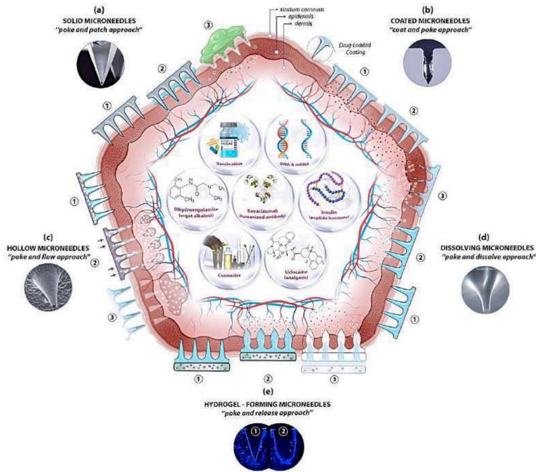
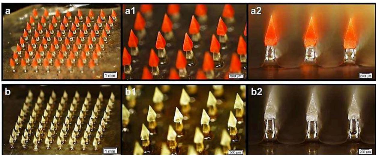 Figure 4: Diagram illustrating microneedle-facilitated transdermal drug delivery: (a) Solid microneedles enhance drug permeation by forming temporary hydrophilic microchannels in the skin. (b) Drugs are applied onto the microneedle surface and rapidly dissolve upon skin insertion. (c) Hollow microneedles pierce the skin, enabling drug solution injection. (d) Dissolving microneedles disintegrate upon skin entry, releasing the drug payload into the skin layers. (e) Swelling microneedles absorb skin fluid and expand to promote drug diffusion through the porous structure. Images reproduced with authorization from [86].
II. TYPE OF MICRONEEDLES FOR DRUG DELIVERY
2.1 Solid Microneedles
Solid microneedles typically involve a two-step process for drug administration. Initially, the solid microneedles are inserted into the skin and then removed, creating temporary hydrophilic microchannels. Following this, a drug-loaded topical formulation such as a gel, cream, lotion, ointment, or transdermal patch, is applied over the microchannels to facilitate drug delivery (Table 1) [80,88-90]. The microchannels formed by the insertion of solid microneedles enable passive diffusion of the applied drugs into the layers of the skin (Figure 2a). Upon the administration of the drug formulation to the affected area, the drug can be continuously delivered through microchannels created by microneedles until either the drug supply is exhausted, or the channels are sealed. The effectiveness of drug transportation into and across the skin is influenced by various factors such as the size, shape, sharpness, and density of the microneedles used for skin pretreatment [91,92]. Furthermore, the efficiency of microneedle-assisted delivery is greatly influenced by the physicochemical properties and molecular weight of the drugs [60].
Solid microneedles can be made from various materials such as glass, metal, silicon, and polymers. The common designs for solid microneedles include solid arrays, flexible patches, and roller types. The disruption of the skin caused by solid microneedles has proven beneficial for many biomolecules. Numerous studies have focused on using microneedles to administer substances like fluorescein isothiocyanate-labeled ovalbumin and insulin, ovalbumin-conjugated nanoparticles, human immunoglobulin G, calcein, bovine serum albumin, fluorescein isothiocyanate-coupled dextran, melanostatin, rigin, palmitoyl-pentapeptide, and genes [60,93,94]. In general, microneedle treatment significantly improves the delivery of most large molecules into the skin. Furthermore, the effectiveness of drug delivery is inversely related to the molecular weight of the drug [60,95].
Several research groups have explored the efficacy of solid microneedle therapy in delivering insulin intradermally, both in vitro and in vivo, resulting in a significant reduction in blood glucose levels . Martanto et al. reported an 80% decrease in blood glucose levels in diabetic rats after the insertion of solid microneedles. Moreover, these microneedles improved insulin delivery to a level comparable to 0.05–0.5 units of insulin administered via subcutaneous injection . Noteworthy, Qiu and team devised an insulin-loaded lyophilized hydrogel patch to achieve sustained and continuous drug delivery through microneedle-formed channels in the skin for up to eight hours. This novel formulation demonstrated a considerably longer duration of action compared to conventional subcutaneous injections. Additionally, insulin retained 90% of its bioactivity after being stored for six months at 4 °C .
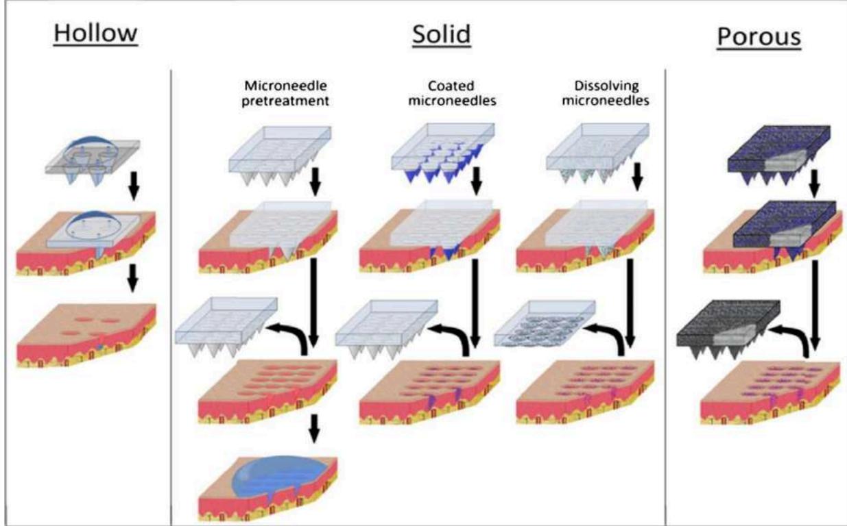 Figure 6: This Source Shows that Microneedles can be used for Drug Delivery with Various Structures Like Hollow, Solid, or Porous. Image Adapted from [114]
2.2 Hollow Microneedles
Hollow microneedles, which are downscaled hypodermic needles in micron size with a similar configuration (Table 1), enable the controlled injection or infusion of a drug solution into the skin layers (i.e., epidermis or dermis) in a non-invasive manner (Figure 2c). Passive diffusion is the simplest method of drug transport through hollow microneedles. However, due to the slow permeation of drugs into the dense skin tissue, researchers have applied pressure to enhance drug delivery [101]. One notable advantage of hollow microneedles is their ability to accurately deliver a large quantity of drugs into the skin [102,103]. A properly engineered microneedle must possess ample mechanical durability to avoid fracturing while penetrating the skin and minimize the risk of bore obstruction, a prevalent issue with hollow microneedles. Scientists have created hollow microneedles with off-center bores on the side of the tip to prevent blockages and enable the medication to access the adjacent skin tissue.
Scientific studies illustrate the effective application of hollow microneedles in improving the transdermal administration of various macromolecules, such as proteins, peptides, oligonucleotides, and vaccines.
Researchers have explored the use of hollow microneedles for painless and noninvasive insulin delivery, known as the "poke and flow" technique[104,96,105]. Studies have shown that intradermal delivery of insulin via hollow microneedles offers faster absorption and better treatment outcomes compared to traditional subcutaneous injections [106]. Additionally, retracting the microneedles partially has allowed for the injection of larger drug volumes. A study on children with type 1 diabetes demonstrated that insulin delivery through hollow microneedles led to quicker healing and reduced pain levels when compared to conventional injection methods[107]. McAllister and colleagues found that applying a pressure of 10 psi allowed a glass microneedle to deliver of insulin solution into the skin of a hairless rat over a period of 30 minutes [108]. Xenikakis and team, on the other hand, created two types of hollow microneedles through 3D printing and liquid crystal display techniques. The researchers analyzed the needle dimensions using scanning electron microscopy, the volumetric properties of microneedles and microchannels using microfocus computed tomography, and the mechanical properties and skin penetration efficiency using finite element analysis simulation. The resulting hollow microneedles successfully facilitated the delivery of insulin across human skin in laboratory settings [109]. In addition, hollow microneedles have the potential to improve the transdermal delivery of various macromolecules. These include -galactosidase, formaldehyde-inactivated botulinum toxoid [110], synthetic mRNA [111], cascade blue, dextran-cascade blue, FITC-dextran [112], human growth hormone, equine tetanus antitoxin [113], and ovalbumin-loaded PLGA nanoparticles [62]. To facilitate the injection of liquid formulations into the skin, 3M has developed a hollow microstructured transdermal system (hMTS). This system consists of hollow microneedles that are connected to a glass cartridge. By utilizing a spring-controlled mechanism, the hMTS device allows for self-injection of up to of drug solution. Notably, the hMTS device has demonstrated successful delivery of equine tetanus antitoxin and human growth hormone into the skin in vivo. Researchers have observed comparable pharmacokinetic profiles of these drugs in domestic swine when administered via hMTS or subcutaneous injection [113].
2.3 Coated Microneedles
An enhanced approach for enhancing transdermal drug delivery using solid microneedles involves applying drug formulations onto the needle surface (Table 1). Various coating methods (such as dip coating, casting, and deposition [115,116]) have been created and assessed for this purpose. Once the microneedles are inserted into the skin, the coating layer breaks down quickly, releasing the drug into the targeted skin layers (Figure 2b). In comparison to the solid microneedles' two-step application process, this single-step technique (coated microneedles) is significantly more effective, precise, and convenient. It is important to note that most in vivo studies on transdermal macromolecule delivery have utilized coated microneedles. However, it is worth mentioning that coated microneedles can only accommodate a small amount of drugs due to their limited surface area. Furthermore, an overabundant coating could lead to a decrease in the mechanical strength and sharpness of microneedles. Therefore, microneedles that are coated are advantageous for highly potent molecules that necessitate a lower therapeutic dosage, like desmopressin, human growth hormone, interferon alpha, and various macromolecules[118,119]. Scientists should strive to enhance the coating procedure and formulation to attain a precise, dependable, and consistent amount of drugs coated onto the needles.
Coated microneedles have proven to be effective in facilitating the penetration of various macromolecules into the skin. These macromolecules include desmopressin, bovine serum albumin, interferon-alpha, parathyroid hormone, peptide A, insulin, recombinant human erythropoietin alfa, bovine pancreatic ribonuclease A, antisense oligonucleotides, erythropoietin, ovalbumin, and human growth hormone . It is worth noting that coated microneedles have demonstrated the ability to deliver hydrophobic peptides into human skin in vitro and mouse skin in vivo . In a study conducted by Li and colleagues, metal microneedles were coated with various molecules such as proteins, immiscible molecules, and nanoparticles, enabling the delivery of multiple therapies using a single microneedle array . The bioavailability of human growth hormone and peptide A, when coated on solid microneedles, was found to be comparable to that of subcutaneous injections, thus highlighting the efficiency of coated microneedles in transdermal drug delivery .
Two prominent coated microneedle systems include the Macroflux® microneedle array (titanium microneedles) and the 3M solid microstructured transdermal system (sMTS). The Macroflux® system has the capability to coat a variety of biomolecules (such as biologics, peptides, proteins, and vaccines) onto the surface of solid microneedles. Notably, parathyroid hormone 1-34 (PTH 1-34), a medication used for treating postmenopausal osteoporosis, has garnered significant attention in both preclinical and clinical trials [121]. It is worth mentioning that PTH remained stable in the final product even after being stored for two years at 25 °C, thereby eliminating the need for any cold-chain or special storage requirements. The PTH-coated microneedles caused a sudden surge in drug plasma levels, reaching Tmax three times faster than the control FORTEO® subcutaneous injection [128]. Similarly, Macroflux® desmopressin-coated microneedles facilitated swift drug delivery in vivo, providing an effective dose for antidiuretic effects without causing pain or skin irritation [129]. Moreover, the 3M sMTS (coated microneedles) demonstrated the ability to transport a drug payload of up to 0.3 mg. Peptide A exhibited significantly enhanced stability when coated on the sMTS [122].
2.4 DissolvingMicroneedles
Dissolving microneedles, a unique design of microneedles, -has attracted considerable interest from both academic and industrial fields(Table 1). Ali and his team recently conducted a comprehensive review on dissolving microneedles, focusing on their designs and materials for delivering macromolecules through the skin [68]. These microneedles contain therapeutic substances within their polymer structure[44]. Once inserted into the skin, the drug-filled microneedles break down and dissolve in the skin fluid, releasing the drug(Figure 2d). This innovative system provides either immediate or prolonged drug release, depending on how quickly the polymer materials dissolve and how long the microneedles are applied [130–134]. The primary challenge with dissolving microneedles is their strength, which decreases as the amount of drug they can hold increases. Furthermore, the physical and chemical properties of materials and design factors greatly impact the strength and drug release rate of these microneedles[135]. The robustness of microneedles is directly influenced by the aspect ratio of microneedle length to base dimensions [136]. Recent studies on dissolving microneedles have focused on the design and geometries of these microneedles [41,42,44, 137,138]. Various innovative microneedle designs have been proposed and assessed for their effectiveness in skin penetration and drug delivery [139]. Biodegradable and water-soluble polymers such as carboxymethylcellulose [140], maltose [141], chitosan [75, 142, 143], polyvinyl alcohol (PVA) [144], hyaluronic acid, and polyvinylpyrrolidone are suitable for developing dissolving microneedles. Additionally, it is preferable to use mild manufacturing conditions to enhance stability and preserve the bioactivity of biopharmaceutical drugs [68,145].
Researchers have demonstrated that dissolved microneedles may efficiently transfer insulin into the skin, lowering blood glucose levels in diabetic rats, dogs, and mice . Particularly, there was a lot of curiosity in an insulin transdermal delivery system mediated by glucose-responsive microneedles . Specifically, there was no significant difference in the pharmacokinetic profile of insulin between subcutaneous injection and microneedle therapy . Moreover, insulin's stability, bioactivity, and bioavailability were all enhanced by encapsulating the medication in dissolving microneedles . For example, after a month at 25 or 37 °C, the insulin that has been encapsulated into starch and gelatin microneedles may still have over 90% of its bioavailability . Insulin-loaded microneedles with a relative bioavailability of 96.6 percent were created by Jung and coworkers using a gentle droplet-born air-blowing approach . To decrease the blood glucose levels efficiently and quickly in diabetic mice, Yu and colleagues created a "smart insulin patch" using a crosslinked hyaluronic acid matrix containing glucose-responsive vesicles . In a similar vein, Yang and colleagues created a glucose-responsive closed-loop device for glucagon and insulin transdermal administration. Changes in blood glucose levels have the potential to automatically modify the release of insulin and glucagon. Using mice and minipigs that had been given type 1 diabetes, the researchers showed that their microneedle technology was successful over a prolonged period [154]. To create polymeric microneedles with the required drug release kinetics, Demir et al. used gelatin methacrylate, polyethylene glycol diacrylate, and MoS2 nanosheets. In both ex vivo and in vivo experiments, the MoS2 needles were able to pierce the skin of mice and pigs and release insulin. In addition, the amount of blood glucose decrease brought about by microneedles was comparable to subcutaneous injection in pigs and mice [155].
Numerous biopharmaceutical agents, including calcein, bovine serum albumin, immunoglobulin
G [1], cyclosporin A [157], fluorescein isothiocyanate-labeled dextran [158], interferon-α-2b [159], polymyxin B [160], lysozyme [161,162], FITC-BSA [163], glucagon [164], human parathyroid hormone [165], vascular endothelial growth factor [166], monoclonal IgG [156], rhGH, desmopressin [167], and leuprolide acetate [168] are among the biopharmaceutical agents that are transdermally delivered by dissolving microneedles. A new tissue-interlocking microneedle based on hyaluronic acid was created by FakhraeiLahiji et al. to increase transdermal distribution of different biomolecules by enhancing needle-to-skin adhesion (Figure 7) [169].
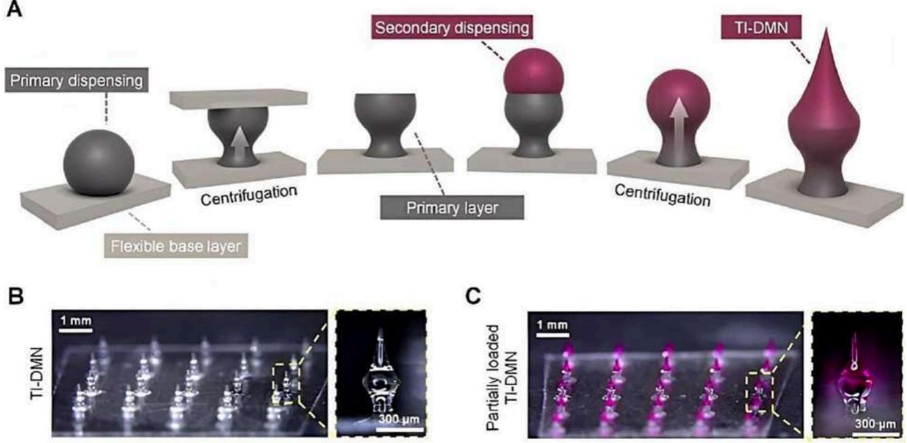 A Figure 7: Illustration showing the tissue-interlocking, dissolving microneedles based on hyaluronic acid. (A) the stages involved in fabrication; (B) microscopic pictures; and (C) A microneedle partly loaded with Rhodamine B. Pictures reprinted from [169] with permission
Table 1: An Overview of the Many Methods used to Administer Drugs using Microneedles, together with Information on the Kind of Needles that Correspond with each Method
| Drug delivery approach | Type of microneedle | Description | Reference |
| Poke and patch | Solid microneedle | medication is released via micropores created by microneedles. | [170] |
| Poke and flow | Hollow microneedle | The drug exits the bore. | [171] |
| Coat and poke | Drug-coated microneedle | Coating separation from the microneedle | [172] |
| Poke and release | Dissolving microneedle | Drug permeates the pores and dissolves there. | [173] |
III. MICRONEEDLE PRODUCTION
3.1 Materials
The production of MNs in research labs and pharmaceutical businesses has been made possible by the introduction of microfabrication manufacturing technologies in recent decades . As a result, the best materials for MN manufacture must be chosen using the following standards :
-
Delicate production without causing harm to delicate and unstable compounds.
-
Regulated or quick release of drugs; and
-
Enough mechanical strength to pierce the skin.
Silicon was used to create the first solid MNs since the creation of MNs was made possible by silicone's flexibility and industrial high-precision microelectronics instruments. However, because of their brittle nature, its primary drawback is the silicon MN breaking. These days, MNs are made of many different materials (Table 1), including titanium, nickel-iron, glass [177,178], ceramics [179], stainless steel [180,181,182], and titanium. Metal MNs are mechanically strong enough to pierce the skin, but one drawback is that they may produce biological waste [183,184]. It's interesting to note that nitinol is utilized in vascular surgery because of its benefits in terms of biocompatibility, flexibility, and shape memory [185]. Polymeric MNs offer improved solubility and are more practical when it comes to tip breakage [186]. MN production utilizes water-soluble polymers [84,187–189] and engineering plastics like CMC, polyglycolicacid (PGA), polylactic-co-glycolic acid (PLGA), polyvinyl alcohol(PVA), polyvinylpyrrolidone (PVP), polylactic acid (PLA), chondroitin sulfate, and polycarbonate. On the other hand, dissolving MNs consist of sugars such as maltose [190,191], dextran [81], or galactose [193,194–196]. Table 2 provides an overview of the materials that are appropriate for MN manufacture.
Table 2: Suitable Materials for Microneedle (MN) Manufacturing
| Material Type | Manufacturing Method | MN Type | Reference |
| Silicon | Etching, lithography | Solid, hollow, coated | [176,197,198,199] |
| Mesoporous silicon | Post-synthesis grafting method | [200] | |
| Nitinol | Multiple-pulse laser microhole drilling | Hollow | [185] |
| α-aluminum(III)oxide(α-Al2O3),zirconia | Lithography and ceramic sintering, micro molding, two-photon polymerization(2PP) | Ceramic(solid), hollow | [179,201] |
| Nickel/iron | Laser-ablation, micro molding, electroless plating, wet etching | Solid, hollow, coated | [202,203] |
| Stainless steel | Laser cutting, laser ablation, etching, electroplating, electropolishing, lithography, and micro stereolithography | Solid, hollow, coated | [204,205,206,181, 182,207-212] |
| Glass | Pulling pipettes | Hollow | [178,212,213] |
| PLGA | 2PP, micro molding | Hollow, solid, dissolving | [184,214] |
| Thermoplastic starch | Electro-discharge machining process | Dissolving | [215] |
| PLA | Fused deposition modelling (FDM), micro molding | Solid, dissolving | [105,216–218] |
| Titanium | Microelectromechanical systems (MEMS) | Solid,hollow, coated | [219,200] |
The manufacturing process for MN should be durable, reliable, repeatable, and exact according on the material that is used . MEMS, lithography techniques, laser cutting, laser ablation, metal electroplating, isotropic and anisotropic etching , injection molding , DAB method , surface/bulk micromachining, polysilicon micro molding , and additive manufacturing (AM) technologies (FDM , stereolithography (SLA) , digital light processing (DLP), and 2PP are some of the manufacturing methods for solid or hollow MNs and are covered in the following sections. Furthermore, a detailed description of coating MNs with an API-containing formulation is provided below.
3.2.1 Laser Cutting
3.2 Production Techniques for Microneedles
The processes of 3D laser cutting [204,205,206,182,207,208], laser ablation [209–211], and electroplating or electroless plating of metal onto positive or negative MN molds can all be used to create metal MNs.
Using an infrared laser, stainless steel or titanium sheets shaped like MNs are cut to create arrays of solid MNs (Figure 8). Some computer-aided design (CAD) software is used to produce the required form, geometry, and dimensions of MNs. After the laser beam conforms to the needle's predefined shape, MNs are cleaned in hot water and bent vertically at a 90-degree angle from the base plane. The next steps involve electropolishing, washing, and compressed air drying the MNs in order to remove, thin down, and sharpen the tips. A single row of MNs with various geometries and two-dimensional rows of metallic MNs may be produced using this manufacturing process [204, 205, 206,182,207,208].
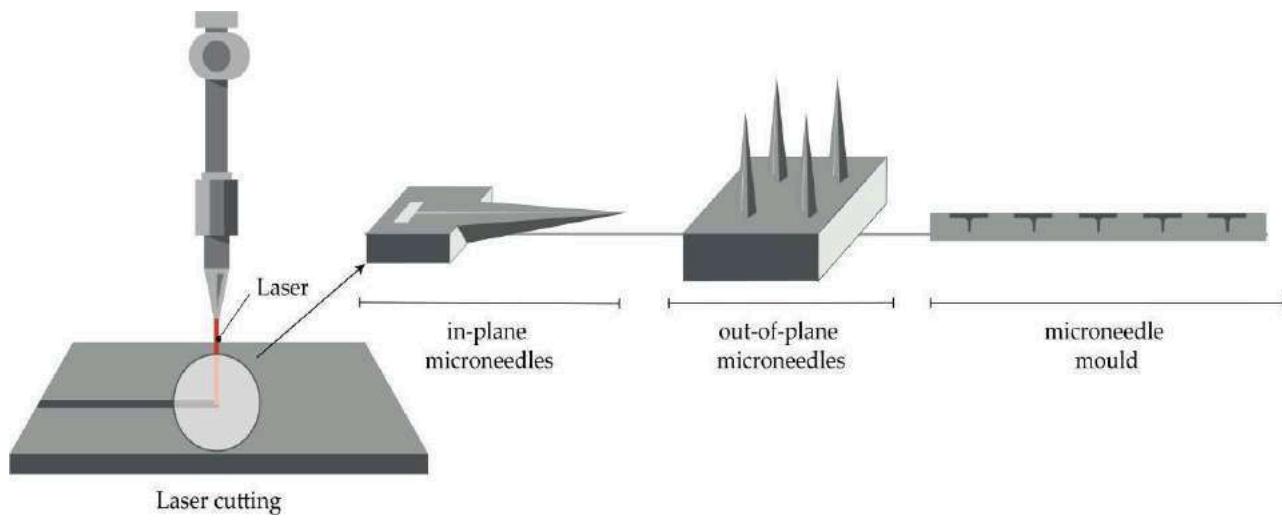 Figure 8: The Principle of Producing both in-Plane and out-of-Plane MNs and MN Molds by Laser Cutting [230]
Moreover, it is stated that molds for a been produced. In the first instance, PLA sheets dissolving MN patch and hollow MNs have that had previously been made using a micromolding process were made to have holes made from their sides using a KrF laser ( nm) [218]. The creation of MN patches on polymethylmethacrylate (PMMA) sheets using a CO laser was documented by Albarahmieh et al. Then, dissolving MNs containing terbinafine hydrochloride and methylhydroxy-4-benzoate were generated by pouring a chosen combination into PMMA molds [231].
3.2.2 Microelectro Mechanical Systems (MEMS)
Micro-electro-mechanical systems (MEMS) techniques have been used to directly manufacture solid and hollow micro-needles (MNs) as well as molds for dissolving MNs from a suitable material substrate . Three exact phases make up the production process: material deposition, patterning, and etching (Figure 1) . Because of the etchant's differing material selectivity, these processes lead to the development of intricate three-dimensional (3D) structures . In the initial stage, a thin layer is created on a substrate using chemical vapor deposition (CVD) or physical vapor deposition (PVD) that has a thickness ranging from a few nanometers to 100 m . Atoms are immediately transported from the source to the substrate during the gas phase of the PVD process, producing the film. Conversely, the CVD technique forms films as a result of a chemical reaction on the substrate surface .
During the second stage of the procedure, known as patterning, the required material is then transferred as a two-dimensional master pattern from the original photomask to the photosensitive-coated substrate. A silicon wafer is often used as the substrate, and one of the lithography processes—photolithography , ion beam lithography , or X-ray lithography is utilized to create the transfer process using a radiation source.
Photolithography is the most widely used kind of lithography. This procedure is based on the observation that some materials, including metals, become opaque when subjected to UV light ( nm), whereas glass remains clear. An opaque template called an optic mask is made during this operation to construct the required pattern in a wafer (Figure 9). The mask, made of a flat glass or quartz plate, only permits light to flow in a certain pattern [240]. The silicon substrate is first heated to around 900 °C with steam or humidified oxygen to form an oxide layer. It is then rotated and covered with an organic polymer known as photoresist material, which is UV-sensitive [236,139,241]. The solvent is eliminated, and the required photo-resistant pattern is formed when UV light and heat between 75 and 100 °C are applied [240]. Positive and negative resists can both be utilized in this stage. Compared to the negative resist, where the chemical bonds are reinforced, the positive resist exhibits photo-resistant polymer chains that break apart upon exposure to UV light, rendering the polymer more soluble in the developer's chemical solution (Figure 9) [241].
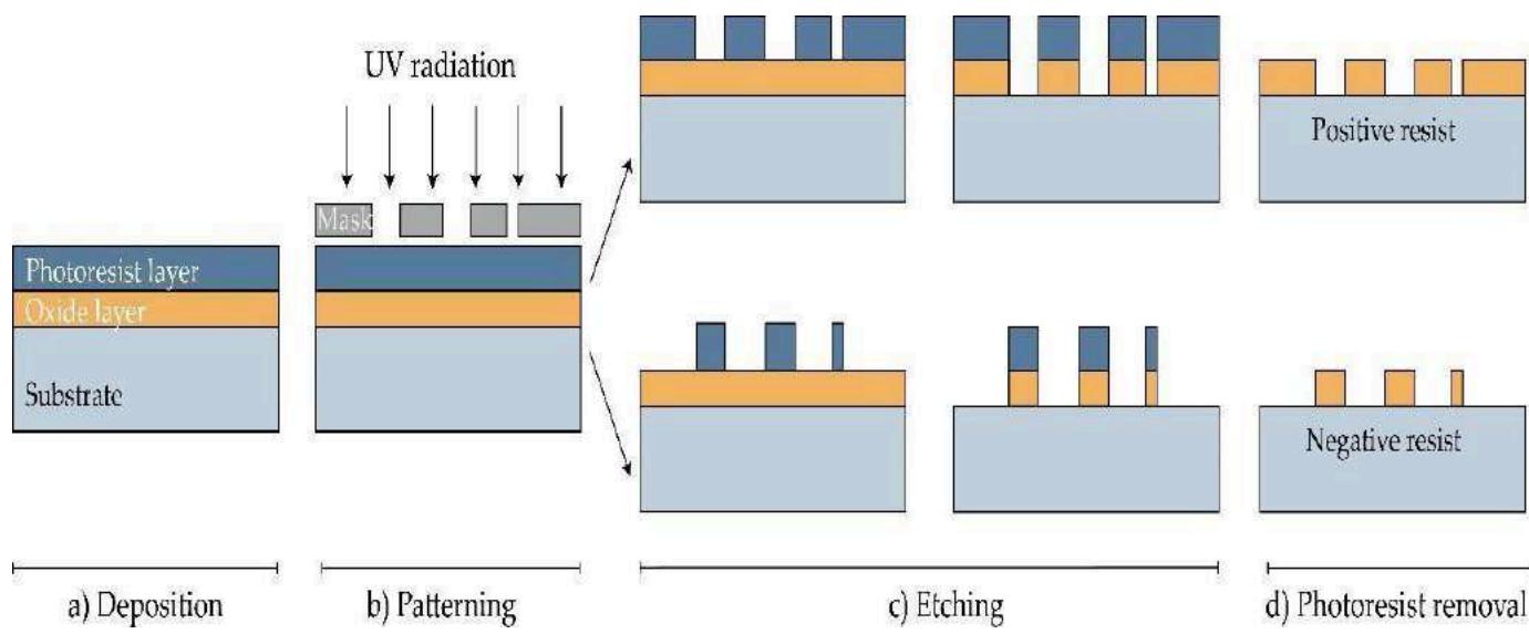 Figure 9: MNs are manufactured using photolithography [236]. (a) Deposition: To create an oxide layer on the Si wafer, it is either humidified or exposed to steam as a substrate. After that, a substrate is spin-coated with a photo-resistive substance. (b) Patterning: The photoresistive material is subjected to UV light guided by a mask. (c) Etching: SiO2 layer is etched after the soluble resist material is eliminated. (d) Photoresist removal: The photoresist layer is eliminated in this stage [230]
Moreover, photolithography makes it possible to manufacture MN molds. In this instance, a stiff silicone cast with a positive image is created, and the selected material is then applied after the creation of a negative mold from poly(dimethylsiloxane) (PDMS) [236]. In order to create a pattern on the material's surface, the exposed portions of the substrate are etched away using a powerful acid or caustic chemical. There are two different kinds of etching: wet and dry etching [233]. By immersing the substrate in the chemical liquid during the wet etching process, extra material is eliminated, resulting in metallic or silicon MN arrays. Anisotropic etching is the process of etching at a different rate from isotropic etching, which is done at the same rate [233,242]. However, the use of a vapor phase or plasma etcher is necessary to accomplish the dry etching procedure. It is possible to distinguish between two primary forms of dry etching: ion-beam milling (IBM) and reactive ion etching (RIE). A reaction between the gas and the substrate is made possible in the RIE process by the gas's excitation into a reactive state. By adjusting the gas pressure, the amount of ions that affect the degree of isotropy may be changed. Ions can be accelerated by the electric field, which also increases the etching's direction. To physically remove the material to be etched, inert ions are accelerated from a source in the IBM method . While RIE can produce structures, it has a poor etching rate and finds it difficult to maintain a high width-to-height ratio. The procedure known as the Bosch process, or deep reactive ion etching (DRIE), works well for producing off-plane MNs. Hollow MNs with a lumen of several hundred micrometers (width-to-height ratio of 30:1) are created using this technique . The greatest results are obtained by combining isotropic dry and anisotropic wet etching to generate well-defined and sharp MN tips, even though wet etching can lower production costs compared to dry etching .
3.2.3 Micromolding Method (SolventCasting)
The process of producing dissolving MNs typically involves pouring the liquid formulation into an MN mold that has already been constructed . A silicon wafer is often used as the starting material for mold . The wafer is then oxidized at . CVD is utilized to cover a wafer and lithography techniques are employed to form a needle geometry, which is then subjected to RIE (see Section 3.1.2). After filling the molds with a liquid polymeric solution, air pockets are wafer and lithography techniques are employed to form a needle geometry, which is then subjected to RIE (see Section 3.1.2). After filling the molds with a liquid polymeric solution, air pockets are extracted using a vacuum or centrifuge [246,247]. The molds are then dried in the oven, and MNs are taken out after they have cooled (Figure 10) [224]. The benefits of this approach are found in the comparatively straightforward and economical manufacturing of MNs at room temperature [248]. Furthermore, it has been documented that MNs made from biodegradable polymers, comprising both natural and synthetic substances, possess the necessary shape and strength to effectively penetrate the skin [184, 248]. It is worth noting that micromolding has also been employed in the fabrication of ceramic MNs [208].
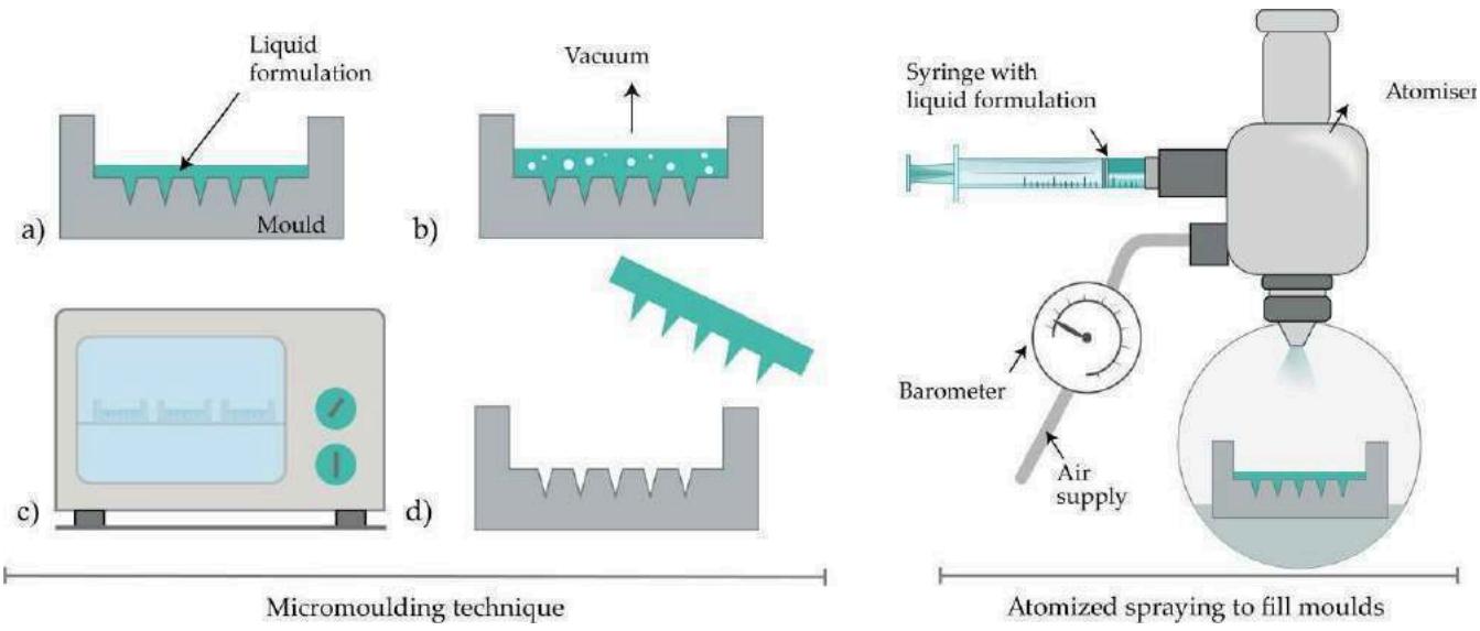 Figure 10: The process of producing MNs using micromolding involved many steps: (a) filling the mold with the liquid formulation; (b) vacuum degasification; (c) drying; and (d) extracting the MNs. Right: Filling molds with atomized spraying. Image from [230]
3.2.4 Laser Ablation
This approach to material processing, which includes metal processing, is top-down. Solid metal arrays are created when light pulses cause a desired shape to bulge on a metal plate . However, the creation of plasma ions and electrons is unsuitable for the creation of structured materials because of the high-intensity laser pulses. Consequently, as seen in Figure 11, Omatsu presented a brand-new, efficient, and time- and money-saving fabrication technique for producing metal MNs based on circularly polarized optical vortices with nonzero total angular momentum. Tantalum MNs with a vertical height of more than 10 m and noticeably tiny tip radii were fabricated, as the authors described . Evens et al. presented a brand-new technique in 2020 for creating solid polymer MNs with laser-ablated steel molds. Additionally, this mold was used in the injection molding procedure to create the polymer MNs. This low-cost manufacturing approach allows for the variation of MN height and the acquisition of acute tip radii [211].
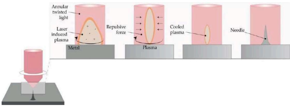 Figure 11: The Omatsu et al. (Modified from [210]) Principle of Metal MN Synthesis Utilizing Twisted Light with a Spin. Image from [230]
3.2.5 Atomized Spraying Method
This technique solves the issues related to the restricted ability to produce dissolving MNs in large quantities with the appropriate shape and physical properties. Additionally, it is possible to reduce the issues arising from the impacts of liquid viscosity and surface tension during the filling of the MN molds. Sugars (fructose, trehalose, and raffinose) or polymers (PVA, PVP, CMC, HPMC, and sodium alginate) can be used to make dissolving MN. In summary, an atomized spray is created via a nozzle that is attached to a liquid formulation and an air source (Figure 11). The mixture is poured into PDMS molds and allowed to air dry for two hours. This approach can also be used to make MN that dissolves in laminate-layered and horizontally layered structures [101].
3.2.6 Droplet - Born Air Blowing Method (DAB)
Conventional MN manufacturing procedures have resulted in drug inactivity owing to UV radiation and heat exposure. Among the drawing lithography techniques is the DAB approach, which was put out by Kim et al. [251]. This process, which uses air blowing to form polymer droplets into MNs, allows for manufacture in temperate climates without the need for heat or UV light [184].
To put it briefly, the procedure starts with the prepared solution being dispensed onto the upper and lower plates. Next, the higher plate is lowered to facilitate droplet contact. This causes the viscous solution to extend as the upper plate moves higher. Next, as shown in Figure 7 [251, 223,252,253], air blowing eliminates any remaining water and pulls the droplets off a substrate to solidify them in the appropriate form.
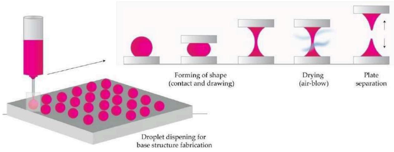 Figure 12: The Fundamental Idea Behind Droplet-Born Air Blowing (Dab) Techniques (As Amended by [253]). Image from [230]
Thus, direct control over drop size and API concentration is made possible by applying one drop of polymer per milliliter. Insulin-loaded dissolving MNs were created using this 10-minute procedure, and they were successful in lowering blood glucose levels in diabetic rats .
A new technique with a shadow mask made it possible to produce MN uniformly and solved droplet formation issues related to poor throughput. By using this technique, the authors reported optimized hole width and thickness of the shadow mask together with regulated medication dose .
3.3 Process of Additive Manufacturing (AM)
A recent area of study is additive manufacturing, sometimes referred to as 3D printing, which is used to make molds and MN arrays. Using computer-aided design software (CAD) to build a three-dimensional item is the initial stage in all AM technologies. To tessellate and slice the 3D shape into digital layers, the CAD model is first converted to an STL file. After that, the printer is configured with printing parameters, and the STL file is sent to it using specialized machine software. By layering appropriate material (such as liquids, ceramics, thermoplastic, plastic, photopolymer, powders, or even living cells), the printer creates the model .
MN arrays were effectively fabricated using additive manufacturing technologies, FDM [216,217], and photopolymerization-based methods such as SLA [226,227,228,260–263], DLP [135–138], and 2PP [214,229,264,265]. Compared to conventional manufacturing methods, these state-of-the-art technologies provide several benefits, such as affordability, ease of use, the capacity to construct intricate geometrical products with the flexibility to alter original designs at any point, and the ability to produce devices tailored to individual patients [259,266].
3.3.1 Two-Photon-Polymerization (2PP)
The 2PP approach makes it possible to fabricate 3D structures at minimal cost, layer by layer, from solid, liquid, or powder precursors in both microscale and nanoscale architectures. For the purpose of polymerizing resin into MN structure, a femtosecond or picosecond laser is directed within a liquid resin droplet . Photopolymerization is accomplished by a mechanism that relies on the temporal and spatial overlap of photons . The method has several benefits, including enhanced geometric control, scalable resolution, and a high degree of flexibility. It may also be carried out in traditional facilities .
The first report on employing 2PP to create MNs from Ormocer® (organically modified ceramic) materials was made by Doraiswamy et al. [201]. According to Trautmann et al., 2PP was used to create hollow MNs with internal laser-generated microchannels [264]. Moreover, another research team used 2PP to print ultra-sharp polymer MNs [265]. Using 2PP 3D, Cordeiro et al. reported a method for creating superior MN array master templates [229]. According to Gittard et al., 2PP may produce MNs with a variety of geometries, including rocket-shaped, mosquito fascicle-shaped, in-plane, and out-of-plane MNs [214].
3.3.2 Fused Deposition Modelling (FDM)
Using CAD software to design the MN and optimizing its shape in accordance with printer specifications is the first step in being ready to print it using a standard FDM printer .
The appropriate thermoplastic material is then fed into the printer via rollers in the form of a filament and heated by heating elements into a molten state to a temperature slightly over its softening point (glass transition temperature ). Gears guide the melted or softened material as it is pushed toward the head end of the printer, where it is extruded via a nozzle and placed layer by layer on a build plate. The material cools and solidifies in less than a second (Figure 8) [254–257]. 3D structures may be created by the printer's head moving in the x, y, and z axes while the platform can move in the z-axis [258].
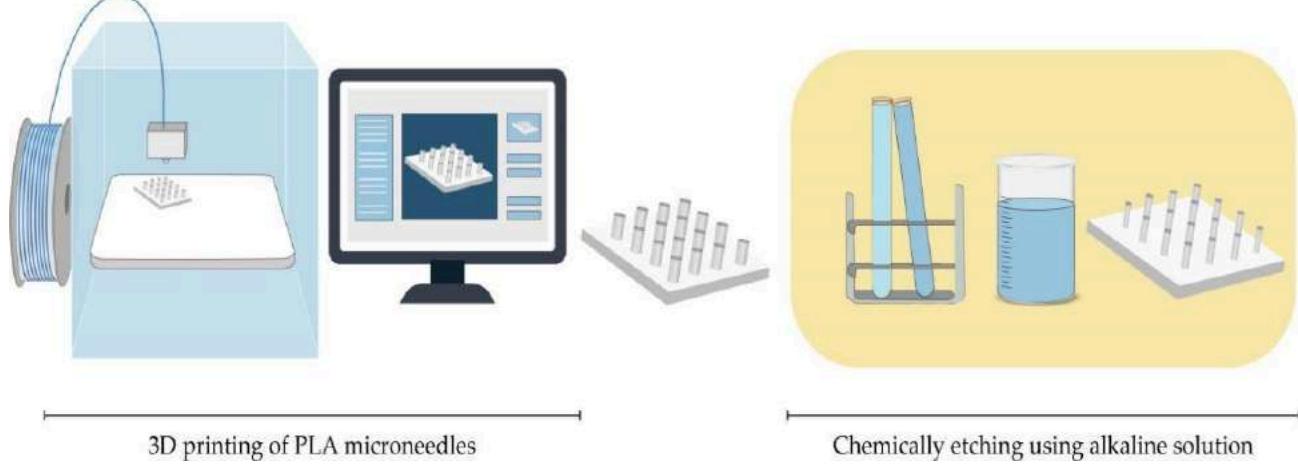 Figure 13: Mns Are Fabricated using Fused Deposition Modeling (Fdm) Techniques and Then Etched in an Alkalinesolution [216,217]
A variety of standard filaments are used in FDM printers, including nylon, acrylonitrile butadiene styrene (ABS), PLA, PVA, high-impact polystyrene (HIPS), and polyethylene terephthalate glycol-modified (PET-G). The dimensions of the filaments used in commercial print heads range from 1.75 mm to 2.85–3 mm [256].
Processing factors such as nozzle diameter, feed rate, building plate and nozzle temperatures, printing speed, layer heights, and part-created orientation should all be tuned during an FDM process . FDM is a flexible and affordable way to manufacture MNs, but its primary drawback is its poor print quality. For the first time, Luzuriaga et al. described combining FDM with a post-fabrication etching phase to produce needles with the perfect size and form . FDM was also effectively employed by
Camovic et al. to print MNs, which were then coated (Figure 13).
3.3.3 Digital Light Processing (DLP)
Another method based on photopolymerization is DLP, which uses light projections to polymerize photosensitive polymers. Using a high-resolution projector to flash the object's whole cross-section at once in the form of volumetric pixels, this approach is quicker than Stereolithography(SLA) [254]. Gittard et al. reported that DLP may be applied to the creation of MNs. In their work, they effectively used DLP to print solid MN array structures for wound healing applications in a variety of geometries using an acrylate-based polymer [267]. A desktop DLP 3D printer was also effectively utilized by El-Sayed et al. to create MN molds for the distribution of nanoparticles [268]. Using microstereolithographic (DLP) equipment,
Lu et al. created drug-loaded MN arrays for transdermal administration of a chemotherapeutic medication.
3.3.4 Stereolithography (SLA)
SLA is the most widely utilized method for printing MNs because of its high resolution, precision, and perfect surface finish. The first publication on the creation of MN arrays for transdermal medication distribution using the lithography-based multiphoton polymerization 3D printing approach came from Ovsianikov et al. [179]. The photopolymerization of liquid resin using photo-active monomers under UV light is the foundation of this technique. MNs are created by successive resin layers solidifying in the presence of intense light, such as a UV laser beam directed by scanner mirrors [256]. A laser beam applied to a resin's surface produces an MN pattern that gives the material a distinct depth. MNs are cured in the UV chamber after being cleaned in an alcohol bath to get rid of any unpolymerized resin residues [228,262].
SLA is a rather slow, costly, and constrained printing process due to its limited choice of printing materials (lack of biocompatibility) while producing high-quality components at a fine resolution (of up to 10 m) [269]. This photopolymerization-based method was reported to be used by several research groups to produce solid MNs, hollow MNs, and MN molds [226, 260]. Using a Class 1 biocompatible resin that had exceptional mechanical strength and was coated with insulin-sugar films, Pere et al. and Economidou et al. created MN arrays using SLA [228,262].
3.4 Methods of Microneedle Coating
3.4.1 Gas-Jet Drying
Using a gas-jet applicator, the medication suspended in a coating solution is dried using a process known as "gas-jet drying" (Figure 14) [270]. It works particularly well for curved MNs because dip-coating's lengthy drying time is inconvenient in these circumstances. The liquid used for wet coating on the MNs' surface may migrate and alter in thickness, which might affect the dosing accuracy. Additionally, short (less than 90 microns in length) and tightly spaced (less than 20,000 cm 2) MNs can be used with this approach [271].
A tiny coating of gold was applied to solid silicon microprojections. The whole length of the microprojection is coated with the solution, which contains methylcellulose, surfactant, and model medication and has the perfect surface tension and viscosity. In order to guide the coating liquid onto the MNs and away from the base, the drying process began with a gas jet operating at 6–8 m/s at an incidence angle of horizontally. The microprojections' coated layer had a thickness of 5 m and grew quickly to prevent the coated material from moving on the base substrate and to allow it to cure. The uniform distribution and quick drying of the coating solution, the ability to remove excess coating solution from the base substrate, and the relatively constant viscosity of the bottom layer of the solution are only a few benefits of this technology [272].
It is possible to enhance the transport of big vaccine molecules via the SC by altering the gas jet technique used to coat MNs. The homogeneity and displacement of the drug from the whole MNs to the tips were guaranteed by rotating the patches, eliminating the patch edge, and raising the incidence angle from 20 to 70 degrees. The protective immunological response was produced at far lower levels than with the intramuscular injection. Additionally, these MN patches enhanced and prolonged the stability of the vaccination [273].
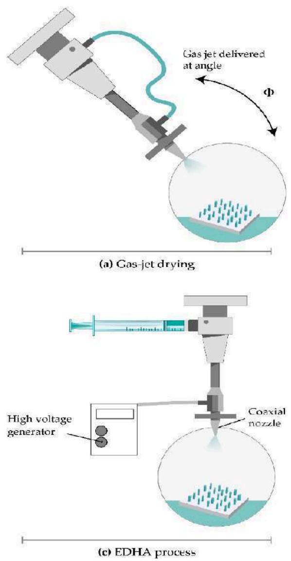 Figure 5: Images of chitosan-poly(L-lactide-co-D, L-lactide) microneedle array loaded with rhodamine B dextran (a, a1, a2) and ovalbumin (b, b1, b2) are depicted in the microscopic form. These images have been reprinted with permission from [87].
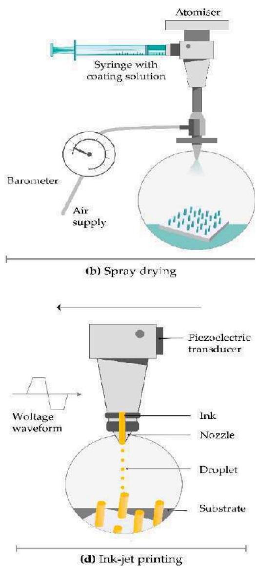 Figure 14: Strategies for Coating MNs. (a) Gas-jet Drying, (b) Spray Drying, (c) Electrohydrodynamic Atomization (EHDA) Procedures, and (d) ink-jet Printing are the Four Methods Mentioned above Image from [230]
3.4.2 Electrohydrodynamic Atomization (EDHA)
An electrical field creates charge inside the droplets during the process of electrohydrodynamic atomization (EHDA), which produces atomized droplets by moving a liquid. Droplets of liquid shoot out of a nozzle when the critical voltage is reached. Subsequently, it is placed onto a grounded collector situated beneath the tip of the nozzle (Figure 14) [274]. Drugs, polymers, and solvents are all present in the coating liquid. EHDA can produce fibers (electrospinning) and particles (electro spraying). Because of the insulating polymeric masks, this approach only coats the MN tips and leaves the base substrate uncovered. However, a significant amount of drug waste occurs on the mask over the basic substrate .
The EHDA technique is available in three different configurations: coaxial (two or more immiscible liquids are injected into separate nozzles), multiplexed (formulation is injected into a single or coaxial nozzle array), and single-needled (one nozzle is filled with formulation using a syringe pump). The coaxial method permits prolonged and regulated drug release while shielding the medication from direct environmental exposure . Particle size, size distribution, porosity, shape, and surface charge are significantly influenced by the following factors: solution viscosity, surface tension, flow velocity, voltage, and distance between the nozzle and collecting platform. Jet stability is affected by the properties of the material. The low electrical conductivity of the solvent is the primary need for the EHDA process .
This technique is used to the administration of delicate biomolecules such as proteins and peptides that become unstable when administered orally, as well as insulin, folic acid, gold utilized in gene transfer, and titanium dioxide as an antibacterial agent . Ali et al. used the EHDA method to coat MNs with fibers and particles. The researchers concluded that MNs coated with PVP in ethanol released more quickly than MNs coated with polycaprolactone in dichloromethane, which displayed a sustained release profile. The skin was effectively penetrated by both varieties of MN, and the electrospun MN covering released a significant quantity of the medication after 6 hours .
3.4.3 Spray Coating
Spray coating refers to the process of creating droplets using fluid pressure. Fine droplets (less than 280 m) are deposited onto an MN array, outspread, and combine to create an unbroken film coat. Atomization is the initial stage that produces tiny droplets (Figure 9). Subsequently, droplets cling to one another and deposit, crashing on the surface. Droplets coalesce on the substrate in the last stage to create an unbroken film covering [278].
The droplet size is determined by the nozzle design, concentration, input ray, viscosity, surface tension, and density of the coating solution, as well as processing parameters like the air-to-liquid mass ratio, spraying duration, atomization air pressure, gun-to-surface distance, and air cap setting. Spray density and spray velocity control the amount of droplets that land on the surface . In addition to producing dissolved MNs, the spray coating technique may effectively apply an entire, micron-sized film-coating on silicon MN arrays (see Section 3.2.5).
McGrath et al. produced an atomized spray by connecting a nozzle to a supply of compressed air and a coating solution. They employed tape to secure silicon MN patches to the adjustable stage, and a peristaltic pump and syringe driver were used to regulate the liquid input rate. Fast film-forming and improved droplet coalescence on the MN surface were achieved with a coating solution consisting of HPMC, CMC, and surfactant .
3.4.4 Dip-Coating
By dipping MNs in drug formulation, the technique known as "dip-coating" selectively coatings the MN shaft while preventing contamination of the MN array's base substrate. You can employ different molten liquids, organic solvent-based solutions, or aqueous solutions. On the MN surface, dipping causes a liquid film to develop; drying then turns the adhering liquid film into a solid coating . To stop the coating solution from rising when the MN shaft touches the base substrate, the viscosity and surface tension of the coating solution should be properly regulated. Either thin-film dip-coating or masked dip-coating can be used to selectively coat the MN shaft. In order to prevent the coating solution from flowing through to the base substrate, masked dip-coating requires the employment of a masking plate . By ensuring that the coating solution's thickness is less than the MN's height, thin-film dip-coating eliminates the possibility of coating solution and base substrate contact by ensuring the coating solution's little capillary rise .
The thickness of the coating on the MN shaft directly affects the amount of medication coated on the MNs. By increasing the rate at which MNs leave the coating solution, improving solution viscosity, and increasing the number of dips, it is possible to obtain a larger thickness and drug mass. Surfactants lower the solution's surface tension and provide a homogeneous, integrated coating. The thickness of the coating is also affected by the drying period in between dips .
Dip-coating is a particularly practical technology for MN production because of its low cost and simple fabrication procedure. Upgrading the procedure with a roller, a fixture, a limit, and a dam board can result in optimal medication delivery . This method's primary flaw is its long drying process, which might result in the loss of drug dispersion from MN surfaces. Also, if the MNs are tightly spaced apart, surface tension may prevent them from coating uniformly .
Stainless steel was used to create solid MNs by electropolishing and laser cutting. Then, with micron-scale control over the length of the coated shaft, appropriate quantities of CMC and Lutrol F-68 NF were utilized to improve viscosity, decrease surface tension of the coating solution, and prevent contamination of the base. Vitamin B, calcein, bovine serum albumin, plasmid DNA, and viruses were all coated on the MNs . When recombinant human growth hormone was dip-coated onto titanium MNs, the absolute bioavailability was comparable to that of commercial subcutaneous injections. The authors concluded that MN patches could eventually take the place of these injections due to their simplicity of use and absence of discomfort .
Vaccine-coated MNs should be directed toward skin immune cells, where maintaining protein integrity is crucial since alterations in protein structure might affect immunological response and vaccination efficiency . Heparin, a pH-responsive copolymer, and a combination of DNA vaccines were coated on MN bases. Electrostatic repulsion between the co-polymer and heparin allowed for the release of vaccinations . Intracellularly encoded antigens, given to necessary effector cells directly for cytolytic action, are the basis of DNA vaccines. MNs enable the delivery of DNA vaccines to the dermal antigen-presenting cells by penetrating the skin's epidermis. Additionally, by using polyelectrolyte multilayers with adjuvant components, they facilitate the intracellular co-delivery of DNA vaccines . The primary drawback of MNs coated with DNA vaccines is their low immunogenicity and coating efficiency. By increasing stainless steels affinity for plasmid DNA, nano-patterned MNs boosted vaccination effectiveness and immunological response. Because of their more hydrophilic surface, nano-patterned MNs demonstrated superior DNA vaccine loading capacity and dip-coating efficiency. Increased cell proliferation indicated improved cytocompatibility. The most significant difference was that their cellular immune responses were stronger .
When the influenza vaccine is coated on MNs, antigen activity may be reduced. In addition to increasing viscosity, CMC caused viral particle aggregation, which resulted in a reduction in vaccination activity. Trehalose was substituted for CMC to ensure that the antigen and its activity were protected since it prevented particle aggregation and had improved heat stability [287].
Antigen activity may be decreased on MNs coated with the influenza vaccination. CMC increased viscosity and also led to the agglomeration of virus particles, which decreased vaccination efficacy. Trehalose, which had better heat stability and inhibited particle aggregation, was used in place of CMC to preserve the antigen and its activity .
Another variation of the dip-coating technique is dropping coating. It means that the MN array should be dropped into the coating solution rather than dipped into it. A non-uniform coating of the MNs and the base, liquid segregation from the MN tip, and substrate buildup between MNs are caused by slow solvent evaporation. It proceeds to the point where the base is the region that is primarily covered. By heating the patch or vacuum-drying it, these disadvantages can be overcome .
IV. DELIVERY SYSTEM'S CHALLENGES USING MICRONEEDLES
In the foreseeable future, approving the transfer of MNs from research labs to pertinent businesses is a thrilling but challenging process. Some important concerns and obstacles should be taken into consideration as soon as possible in order to transition this revolutionary technology from the lab bench to viable goods in the relevant markets.
The future of the area and its commercial uses may be determined by the obstacles and proactive approaches to overcome them that we will examine in the following sections. The next sections address the primary challenges and concerns about the creation of a delivery system based on microneedles, which are summarized in Figure 15.
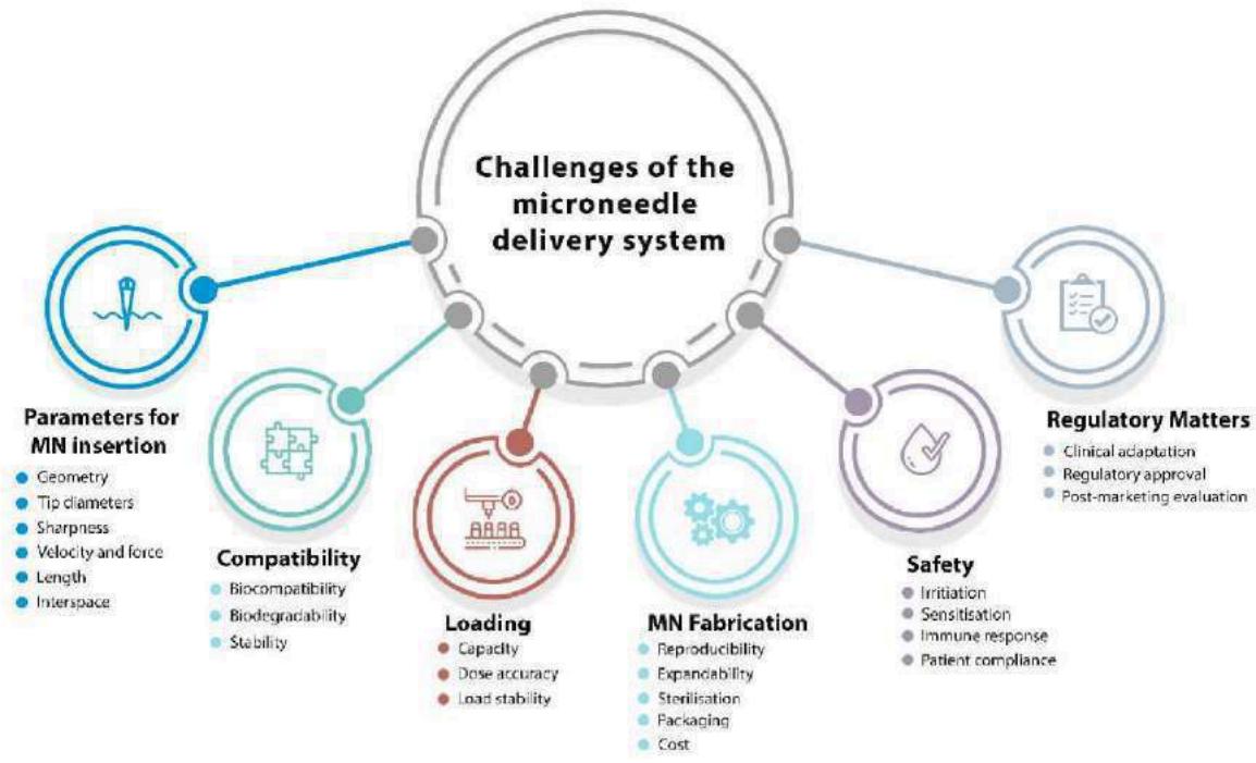 Figure 15: Factors Influencing the Delivery Technique based on Microneedles [365]
4.1 Parameters Affecting MN Insertion
One essential necessity for MN patches is that they must be able to pierce the skin enough. The properties of the skin, which might differ from person to person and across the body, should also be considered while handling this issue. A number of factors, including shape, base and tip diameters, length, and interspace (centre-to-centre spacing), significantly influence how MNs enter and penetrate the skin to overcome its elasticity . For any MN application, a "one-size-fits-all" strategy cannot be considered during the design or development phases. The shape of individual MNs and the array, MN materials, the MN management strategy, and the properties of skin tissue are all highly correlated with the infiltration and active delivery efficacy of MNs . By altering the microneedle's composition and form, the mechanical strength, insertion depth, and drug release profile may be precisely adjusted to suit certain medications and purposes.
4.2 Biocompatibility, Biodegradability, and Stability
Biocompatibility is one of the safety features of MN systems used in clinical settings. A number of studies are necessary to assess the biocompatibility of MN products based on contact durations of less than 24 hours, between 24 and 30 hours, and more than 30 hours in order to guarantee that they are safe for human exposure. [294]. The comparable tests for the first two periods are the intracutaneous reactivity, cytotoxicity, sensitization, and irritation tests. For the later stages of usage, genotoxicity and subacute/subchronic systematic toxicity testing are also advised. Since biodegradable materials can be safely broken down and eliminated from the body, it is preferable to employ them in microneedle construction. As a result, efforts to fabricate MN utilizing biodegradable polymeric materials have been made recently. The capacity to incorporate medicine into the microneedle matrix for skin discharge via biodegradation or dissolution in the skin's bodily fluid is the main advantage of polymeric microneedle systems.
4.3 Loading Capacity and Dosage Accuracy
Loading capacity: The maximum amount of medication that a coated microneedle device can bolus dose is around 1 mg. While hollow microneedles provide "asneeded/on-demand" dosage or continuous infusion, squeezed skin tissue following microneedle insertion may block central exits. MNs can penetrate the skin's barrier qualities, but their effectiveness is mostly reliant on the biological formulation's passive diffusion into the skin. Large doses may be challenging to deliver as a result, and a significant portion of the dose may be lost on the skin's surface. Because of this, there has been hesitation to employ this technology for some clinical applications due to the timing of application and the difficulty to monitor dosage administration. The delivery of vaccinations, for which dose consistency is essential, is one instance. Recent studies have shown that administering vaccines directly to the epidermis and dermis of the skin can generate immune responses with much smaller vaccine doses compared to the traditional method of injecting into the muscles. However, if only a tiny fraction of the given dose reaches the skin, the advantages of this approach may be reduced. While this obstacle can be overcome, vaccines require a minimum dosage to activate immunity, which may be more difficult to achieve through passive diffusion.
Dosage accuracy: It is important to pay careful attention to the dose accuracy of MN delivery systems in continuous medication administration. Various techniques utilizing separable microneedles have been suggested to reduce the duration of patch wear and expedite the extraction of formulation from the microneedles . Protein medications, such as insulin, erythropoietin, glucagon, growth hormones, and parathyroid hormones, are difficult to store and administer because bio-macromolecules quickly degrade and become inactive. In addition to using stabilisers, the ideal way to address these issues would be to consider all of the variables involved in the manufacturing of MN, including polymer concentration, sterilization, packaging, and temperatures for both production and storage. As was previously mentioned, MNs may be produced using a variety of materials and kinds. Accurate management of medication delivery efficiency using solid MNs presents certain challenges. Because of its narrow coating surface area, coated MNs can effectively administer precise dosages of a medicine, but their drug loading capacity is restricted. If dissolvable microneedles are made predominantly of hydrophilic, biocompatible, and biodegradable materials and if the cargo can be released completely inside the skin's interstitial fluid without producing undesired debris, then it is conceivable to encapsulate pharmaceuticals in the matrices of MNs. Reservoir leakage can be avoided when transferring relatively high dosages and different medications with regulated release (slow or quick delivery). Dissolvable microneedles may be a useful tool for stabilizing and maintaining nanoscale compositions while enhancing the penetration of nanoparticles through the stratum corneum barrier. Numerous methods have been extensively researched, and a number of analytical techniques have been established for both in vitro and in vivo tracing and tracking the trip of nanomaterials with their important payloads [297,298].
4.4 Skin Irritation and Recovery
The skin is an extremely receptive organ to the MN administration of any medicinal substance due to its immunogenic nature. As a side effect, mild and transient erythema may appear based on the medication's size, composition, and kind. During clinical trials, skin irritation, sensitization, and immunological response need to be assessed as part of MN product safety evaluations. Before doing any clinical studies on humans, this safety problem must be assessed by animal testing. On the other hand, if other challenges have been adequately handled as previously mentioned, the skin's high level of immunological responsiveness may offer a chance for MN-based vaccine administration.
4.5 Cost of Microneedle Fabrication
To fully implement microchip-based microneedles into therapeutic applications, current microneedle manufacturing procedures must be enhanced to large-scale production. Although comprehensive economic evaluations of the technology have not yet been completed, it is easy to assume that, similar to any new technology, the clinical use of MNs may be relatively costly because of the intricate fabrication and storage processes as well as the drawn-out approval process .
V. MECHANICAL PROPERTIES OF MN
The mechanical properties of the MNs must be considered during the MN design process since they will be exposed to an applied force during epidural implantation. The MNs must be strong enough to prevent failure in the MN array in order to do this . According to Lutton et al. , no one test can accurately replicate and monitor the mechanical characteristics of the needle and the implantation of the MN in vivo. As a result, the MN should be subjected to a variety of mechanical tests for characterization. Axial force, transverse force, base plate break, and insertion force are among the several mechanical test kinds performed on MNs. Furthermore, several studies have been conducted to examine the connection between MN production parameters and mechanical characterization .
5.1 Transverse Force
The transverse force test entails applying a force using the y-axis parallel to the MN base plate. The transverse fracture force measurement is crucial because the roughness of the skin surface may cause the MN to bend transversely . Furthermore, the transverse force, in addition to the axial force, completes the whole picture of the mechanical characteristic of the MN and hence forecasts the MN's bending behavior after insertion . This test's drawback is that it requires manual alignment of the metal probe with a predetermined MN length .
Using the TA.XT-plus Texture Analyzer (Stable Micro Systems, Surrey, UK), Donnelly, et al. determined the transverse failure force of MN arrays [307]. Park et al. conducted a second investigation using a force-displacement station and a microscope to quantify the transverse force [308]. A PDMS arrangement supported the MN vertically on a metal plate under perpendicular stress. After testing the transverse force until the MNs broke, it was shown that displacement rises linearly with an MN base diameter. A micromechanical tester (Instron® Model 5969; Instron, Norwood, MA, USA) was used by Demir et al. [306] to measure the transverse force of the MN (Figure 16B).
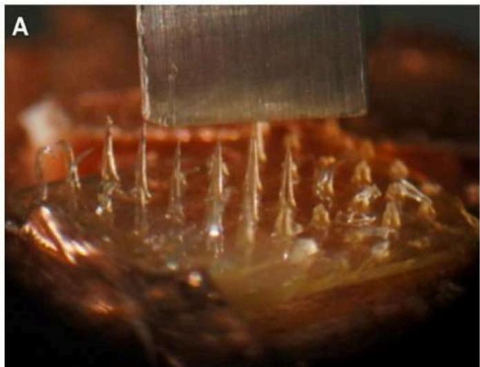
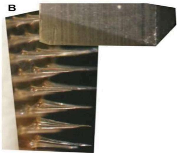 Figure 16: (A) Using the Instron® Model 5969 micromechanical tester, a digital image shows the SA MN placed up against the metal mill to detect the axial fracture force (Instron, Norwood, MA). (B) To evaluate the transverse fracture force using a micromechanical tester (Instron® Model 5969, Instron, Norwood, MA), MN shafts were transversely forced on the metal mill. [306]. Reproduced with permission from Characterization of Polymeric MN Arrays for Transdermal Drug Delivery by Demir et al., published in PLoS One (2013)
5.2 Axial Force
The most popular test is the axial force test, which involves exerting force on the MN array's base as well as the vertically oriented needle tips . This mechanical test is crucial since it establishes the needles' failure force. Since it provides a rough range (expectation) of needle insertion force, knowing the needle failure force measurement is the most significant information, also known as the safety point .
Various tools and computation techniques have been used in several axial force investigations to ascertain the MN failure force. Davis et al. computed the force and displacement data [310] to measure the failure (ScopeTest1, EnduraTEC, Minnetonka, MN, USA). Additionally, Demir et al. used a universal testing apparatus (Instron® Model 5969, Instron, Norwood, MA, USA) (Figure 16A) [306] to evaluate the fracture force. Additionally, Khanna et al. used motorized actuators (Thorlabs Motorized Actuators, Morganville, New Jersey, USA) and a compression load cell (LCFA-500gF sensing capacity, Omega Co., Norwalk, CT, USA) to study the axial fracture tests [311]. Using a TA-XT2 Texture Analyzer (Stable Microsystems, Haslemere, UK) and a light microscope (GXMGE-5 digital microscope, Laboratory Analysis Ltd., Devon, UK), Donnelly et al. conducted compression mechanical tests [312]. Using a displacement-force test station (Model 921A, Tricor System, Elgin, IL, USA), Park and Prausnitz assessed the failure test [313].
5.3 Insertion Test
The axial force does not provide an exact measurement like the insertion test, which makes the insertion test more significant and distinct from the axial force. Additionally, this test was conducted on a variety of skin subjects, including humans, rats, and pigs (Figures 17 and 18). The ability to load and apply the medication to the skin is one benefit of utilizing an MN. Even if the fracture force of the needle is simulated by several mechanical tests, it's crucial to confirm the findings using real skin.
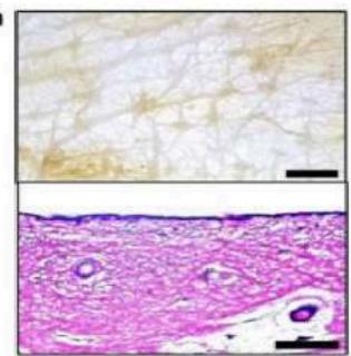 a
a
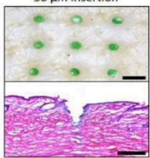 b 50 μm insertion
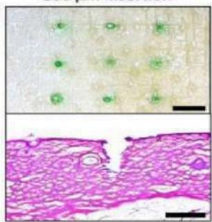 c 100 μm insertion
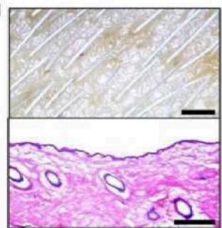 d
d
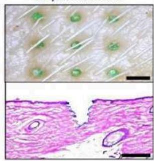 e 50 μm insertion
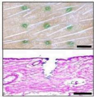 f 100 μm insertion Figure 17: Histological analyses and microscopy pictures of hairy and hairless pig cadaver skin: (a) Skin of hairless pig cadaver before DMN implantation. (b) A 600 m tall DMN is inserted every 50 m. The depth of the DMN insertion into the skin was 650 m. (c) Compared to DMNs implanted 50 m deep, the base area of those placed 100 m deep was less visible on the skin's surface. According to a histological analysis, the DMNs were implanted 700 m deep in the skin. (d) The skin of a hairy pig cadaver before DMN injection. (e) DMNs placed 50 m deep into hairy skin seemed identical to DMNs implanted into the skin of a hairless pig cadaver.(f) The hairy pig cadaver skin was pierced 700 m deep by DMNs that were placed 100 m deep. Scale bars: 500 m for histology pictures; and 2 mm for microscopy images [314].Source: Shayan F. Lahiji et al., A patchless dissolving MN delivery system providing quick and effective transdermal medication administration; Springer Nature, 2015. Reproduced with permission [300,301]
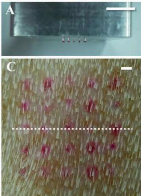
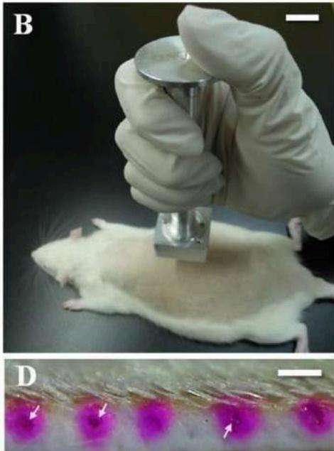 Figure 18: Investigation of in vivo skin penetration: (A) Troy MNs were arranged in an array using an applicator. (B) The applicator was put on rat dorsal skin vertically by hand. (C) Skin image with Troy MNs added. The vertically sliced line utilized to extract sectional tissue is represented by the white dotted line, and the array of red dots depicts the place where rhodamine B-encapsulated Troy MNs were pierced. (D) Sectional view of the skin. Delivered rhodamine B is shown by red dots in the skin, while undissolved DMNs are indicated by a white arrow. 10 mm (A, B) and 1.0 mm (C, D) scale bars [315]. The Troy MN: A Rapidly Separating, Dissolving MN Formed by Cyclic Contact and Drying on the Pillar (CCDP), by Kim et al., is reproduced here with permission from PLoS One (2015) [301,302]
VI. FROM CLINICAL TRIALS TO COMMERCIAL DEVELOPMENT
MNs have so far been created from several materials utilizing a range of manufacturing techniques, and with a wide range of shapes, either with or without an MN application device. While MNs have been the subject of much research concerning transdermal drug administration and vaccine distribution, these systems may also be tailored to target delivery to other tissues, including hair follicles, the mucosa of the mouth and vagina, and the muscles that control the anal sphincter [315]. These days, MNs are also being investigated for the delivery of drugs to the cornea, sclera, and suprachoroidal region in the eyes [316]. MNs (Dermaroller®, Dermapen®) are already in the advanced stages of research and are being sold for cosmetic skincare. MNs have been studied recently as a component of the monitoring/diagnosis system to enable entirely painless transdermal bodily fluid sampling . In order to demonstrate the effectiveness and safety of MN delivery systems over conventional administration methods, the bulk of these clinical trials employ MN injection systems and MN array-based patches.
For transdermal medication delivery to be effectively created and the finished product to have sufficient and repeatable penetration, the type of MNs and their shape must be carefully chosen. Typically, an MN application device—either impact-insertion or manual hand-held—is required to enable self-administration. A wide range of pharmaceutical firms and research facilities, such as 3M, Zosano Pharma, Alza Corporation, Becton-Dickinson Technologies, Valeritas, Vaxxas, Microneedle Therapy System, Nanopass Technologies Lohmann, Therapie-Systeme AG, and others, are involved in the development of Minnesota-based products.
Only a small number of MN devices are already on the market, and the majority are still in clinical studies. Becton-Dickinson Technologies created the first commercially available MN device, called Soluvia® (Figure 19A). However, some authors contend that this device actually uses very short hollow needles to successfully inject ID using a standard syringe barrel rather than true MN arrays [18]. 2009 saw the launch of Intanza® by Sanofi Pasteur, the first influenza vaccination to target the dermis [318]. The medicine Intanza is removed from use in the European Union in 2018 at the request of the holder of the marketing license, despite several clinical trials showing that the drug's advantages outweigh its hazards [319]. The FDA gave Nanopass Technologies' MicronJet® its approval in February 2010. This single-use MN device was used to administer insulin, lidocaine, and the influenza vaccination intradermally. It is made up of four hollow silicon needles that are less than in length and are linked to a plastic device. The business finished a Phase 1 clinical research in 2009 to compare the insulin delivery system's pharmacokinetics and pharmacodynamics while using a standard needle with the Micronjet® [320]. The business created MicronJet600®, a new device version, to enhance device performance, particularly the insertion approach [321]. A clinical trial was conducted in 2019 by Yonsei University to examine the immunogenicity and safety of delivering Bacillus Calmette-Guerin (BCG) using a standard needle vs using the Micronjet600® device (Figure 19B) [322].
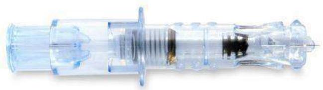 (A)
(A)
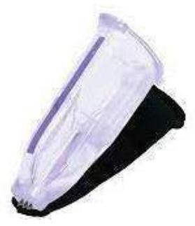
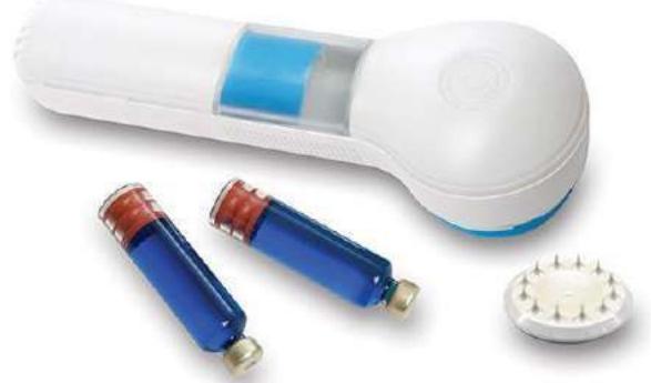 (C)
(C)
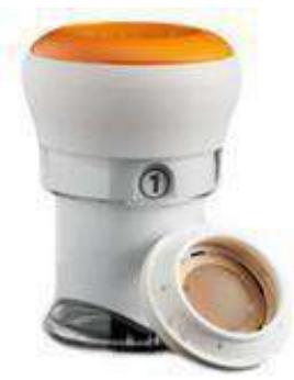 (B) (D)

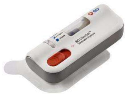
 (E) (G)
 (F) (H)
Figure 19: Current MN devices. (A) Soluvia , (B) Micron Jet 600, © Microstructured Transdermal System , (D) Qtrypta , (E) SCSMicroinjector , (F) Microinfusor , (G) MicroCor , (H) Bullfrog Micro-Infusion Device. Image from [230]
VII. MN APPLICATIONS
Researchers, scientists, and industry players are all very interested in MNs. Numerous research has shown that MN has the potential and capacity to be administered in many domains. These include administering medications, administering vaccines, diagnosing illnesses, and applying cosmetics.
7.1 Disease Diagnosis
Several well-established bioassays that collect bodily fluid samples in order to evaluate and track medical conditions can be used to monitor disease diagnosis and treatment efficacy. The existing approaches cause discomfort and call for specific tools, skilled medical staff, and specialized methodologies . On the other hand, bioassay solutions with straightforward installation and painless experience are provided by microneedle technology .
Numerous illnesses, including diabetes [324], Alzheimer's [325], and cancer [326] can be identified by a hollow MN. Another use for MNs is in patient health monitoring. To measure the glucose level, for instance, a hollow glass MN may be utilized [327]. Moreover, the MNs system for electrocardiography signal optimization was proposed by O'Mahony et al. [328]. To track alcohol levels in artificial interstitial fluid, an enzyme based on microneedles was functionalized [329]. Early-stage osteoarthritis biomarkers were identified using microneedles containing nanoparticles [208]. Hydrogen peroxide, lactate, dissolved oxygen, and glutamate were detected using microneedles [330].
7.2 Drug Delivery
In 1998, solid silicon MN was used for the first time to deliver drugs . Human growth hormone was applied transdermally to the skin of hairless rats using a dissolvent MN patch . An MN patch filled with caffeine that dissolves was able to regulate the weight of obese mice and function as an anti-obesity therapy . Salmon calcitonin was administered using a coated MN patch . A protein antigen (ovalbumin) was injected into the skin of hairless guinea pigs using a solid microneedle [335]. Insulin [336], BSA [337], and calcein [337] were all delivered using metal MNs and solid silicon MNs. Moreover, MNs have been employed in transdermal permeation of many medications, including paracetamol, ketoprofen, and ibuprofen [338]. Additional medications administered using microneedles include of glycerol, L-ascorbic acid, riboflavin, aspirin, docetaxel, pilocarpine, lidocaine, hydrochloride, and ketoprofen [330]. Some research has successfully achieved microneedle injection into chicken thigh [339], and brain tissue [340], even though the majority of studies employed microneedle array for drug delivery into mice, pigs, and human skin.
7.3 Cosmetic Application
MNs are frequently employed in cosmetic applications, including hair growth and skin therapy (Figure 20). A dissolvable MN patch based on hyaluronic acid was created by Kim et al. to provide ascorbic acid and retinyl retinoate intradermally [341]. Using a solid MN, Kumar et al. demonstrated an improvement in the local delivery of eflornithine (used to treat face hirsutism) both in vitro and in vivo [342]. Moreover, two alopecia areata patients were successfully treated with Minnesota technique [343]. Following therapy, hair growth was observed in these individuals. A MN has been used in successful clinical studies for hypertrophic burn scars [344], atrophic acne scars [345], and atrophic face scarring [346].
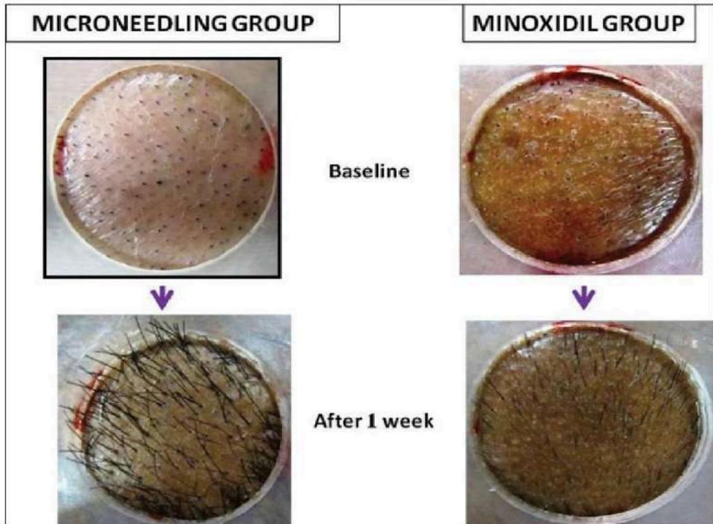 Figure 20: The Microneedling Treatment Group Showed Faster Hair Regrowth after One Week [215]. Image from [301,347]
7.4 Vaccine Delivery
One popular kind of MN used for vaccine administration is a dissolvable MN. In place of the conventional hypodermic injection needles used to provide vaccinations, the dissolvable MNs were employed. Dissolvable MNs are biocompatible, reliable, scalable, and do not produce biohazardous waste, in contrast to other forms of MN . Vaccines against malaria, diphtheria , influenza , hepatitis B , HIV , and polio were administered by soluble molecular nanoparticles (MNs).
Coated MNs arrays have been successfully employed for vaccination applications, while dissolvable MNs are the most often used form of MNs for vaccine administration . Pigs' immune systems were strengthened in a trial by using a coated MN and the Bacillus Calmette-Guérin (BCG) vaccine, which was easy, safe, and compliant to give . Hepatitis C virus protein was effectively encoded in a DNA vaccine coated on a microneedle in another investigation . In mice, the microneedle was successfully primed for certain cytotoxic T lymphocytes (CTLs). In addition, a coated microneedle containing influenza viral antigen was used to vaccinate mice .
Rather than administering the anthrax recombinant protective antigen vaccine by injection, hollow MNs have been employed to do so . In a mouse model, a hollow microneedle was tested as a vaccine against plaque . When compared to intramuscular injection, the immune system responded similarly in a human clinical study employing hollow microneedle influenza vaccination .
Table 3: List of A Few Clinically Effective Microneedle Drug Delivery Devices that Can be used to Introduce Medication into Tte Bloodstream [364]
| Drug | Microneedle type | Use | Reference |
| Donepezil HCL | Drug-coated microneedle | Alzheimer's disease | [360] |
| Sinomenine HCL | Dissolving microneedle | Analgesia, anti-cancer, anti-inflammatory | [361] |
| Meloxicam | Dissolving microneedle | Arthritis | [362] |
| 5-aminolevulinic acid | Coated microneedle | Skin tumors | [363] |
VIII. RESEARCH GAPS AND PROSPECTS FOR THE FUTURE
In contrast to alternative methods, this study presents the advantages of using MN for several applications. Furthermore, a number of studies recommend various materials, manufacturing processes, and needle kinds for the creation of an MN array. A large-scale clinical experiment was suggested for the use of MNs for various applications. Nonetheless, the field of MN array manufacturing still has deficiencies. We provide an overview for MNs in this part about the use of next-generation techniques, such as additive manufacturing, for COVID-19 testing and immunization, as well as the scaling up of manufacturing processes and predictive modeling of materials and manufacturing techniques.
8.1 Forecasting Model for MN Production
There is insufficient knowledge of the design parameters for the manufacture of MNs, which calls for more investigation. By examining the PDMS MN's dimensions with various laser power and scanning speed values, Chung and Tu expanded their research on producing the MN array to include merging CO2 laser processing and polymer molding [366]. The impact of hole width and pulse shot number on fabrication depth was investigated by Aoyagi et al. Additionally, they observed that sidewall smoothness was influenced by both hole diameter and repetition rate [367]. Nevertheless, no research has been done on methods of fabricating MNs arrays by process parameter adjustment. The performance of manufacturing processes has been improved in the literature using a variety of predictive model types [368–373], which may be expanded for MN creation. Previous research has shown that new manufacturing techniques combined with computer modeling may produce complex biomolecules for use in biomedical applications. Moreover, a prediction model that connects manufacturing factors to drug elution characteristics and sensing applications is needed.
8.2 MN and Additive Manufacturing
A cheap cost, shorter fabrication time, and high-quality resolution are all provided by the promising technique known as additive manufacturing . Compared to traditional MN manufacturing methods, the device size and formulation may be changed with few postprocessing steps, therefore fabricating an MN array with a 3D printer would be beneficial. Direct-write techniques can also be applied to coat MNs with various biomolecules for effective medication release. Johnson and Procopio recently completed the first investigation in which they manufactured an MN structure using a commercial 3D printer . To create tiny needles, they employed an Autodesk Ember printer with decreased layer height and enhanced antialiasing. An inexpensive SLA 3D printer was used in another recent work to manufacture MN arrays . This investigation was expanded to evaluate the insertion of MN into the skin using a 30N force.
8.3 Scaling Up of Manufacturing Process
The possibility for large-scale MN manufacture is highly sought after given the recent rise in MN applications and the dearth of commercial MN goods (there are now just 13 MN items available)
[393]. More understanding of chemistry and manufacturing materials, according to Bhatnagar et al., would enable enterprises to meet their financial targets and realize higher profits and large-scale production [394]. To create a single MN array, the majority of existing MN manufacturing techniques need many phases [353]. By addressing this constraint, new research opportunities of the decrease or integration of the number of processes necessary for the fabrication of an MN become available.
8.4 Covid19 and MN
The MN strategy is a strong contender to combat the coronavirus (COVID-19) pandemic, as its effects are being felt globally. MN-based oropharyngeal swabs were presented by Chen et al. [395] to lower the number of false negative results in COVID-19 testing. This idea enables highly efficient viral capture, allowing medical professionals to distinguish between positive and negative samples. Since the COVID-19 vaccination is now accessible, those who are capable of self-administering the shot may receive it from MNs.
8.5 Next MN Generation
Several investigations have used synthetic MN to provide medications and vaccinations in vivo. Making an MN capable of delivering macromolecules with large molecular weights and high hydrophilicity, however, is an impending problem . Irritation, skin allergies, and redness are other problems related to utilizing an MN for medication administration . There are several MN devices on the market, such as the Dermaroller, however, no biodegradable polymer MN has yet to be released for sale . Furthermore, no MN that may include protein products is available for purchase .
IX. CONCLUSION
Achieving effective MN mediates transdermal and intradermal distribution mostly depends on breaking through the stratum corneum barrier. MNs technology in the era of transdermal medication administration is summed up in this study. Because of its benefits, a lot of research and studies have been done on the creation of MNs. This study has provided illustrations of several MN design types, materials, and production techniques.
Many MN systems with unique delivery mechanisms have been created and deployed in the last few decades to deliver tiny or large compounds. As this thorough review highlights, recent studies have demonstrated that transdermal transport efficiency of small molecules, salt forms, excipients, and other formulation parameters is improved by transiently disrupting the skin microchannel lifespan. The synergistic impact of coupled enhancement in addition to MN therapy, vaccinations, and intradermal and transdermal distribution of macromolecules, including therapeutic peptides and proteins, were discussed. Additionally, the literature examines MN mechanical testing and their characterisation.
In conclusion, this work highlights the research deficit related to MN manufacturing. Despite the fact that MNs are mediating a number of novel transdermal products, they have not yet realized their full potential. It is becoming more and more clear that there is a gap in permitting cost-effective manufacturing for large-volume manufacture of MNs as our understanding of MN-mediated grows.
Funding
This research received no external funding.
Conflicts of Interest
Regarding this work, the writers have no conflicts of interest to disclose.
Conflict of Interest
The authors declare no conflict of interest.
Ethical Approval
Not applicable
Data Availability
The datasets used in this study are openly available at [repository link] and the source code is available on GitHub at [GitHub link].
Funding
This work did not receive any external funding.
References
Cite this article
Related Research




Special Issue
Launch a focused special issue to highlight research, emerging trends, and expert insights in your academic field.
