




IntelliPaper
Abstract
The therapeutic efficacy of medicinal plants is attributed to polyphenolic chemicals, which are essential bioactive components. The bark of the mango plant, Mangifera indica, is a rich source of polyphenols that have antibacterial, anti-inflammatory, and antioxidant properties. For plant-derived products to be consistent, effective, and quality controlled, however, thorough phytochemical standardisation is necessary. The purpose of this research is to provide a comprehensive phytochemical profile by utilising High-Performance Liquid Chromatography (HPLC) and Liquid Chromatography-Mass Spectrometry (LC-MS) to analyse and interpret the polyphenolic content of Mangifera indica bark powder.Mangifera indica bark powder methanolic extracts were made and then separated chromatographically using HPLC in conjunction with photodiode array (PDA) detection. Acetonitrile and aqueous formic acid were used as the mobile phase in a gradient elution technique using a reverse-phase C18 column. Major polyphenolic components, such as Mangifera, quercetin, gallic acid, catechin, and ellagic acid, were identified and quantified using standard reference compounds using UV detection at 280 nm. The structural identity and fragmentation patterns of the identified polyphenols were further confirmed by LC-MS analysis.According to the study,Mangifera is the main component of Mangifera indica bark, which is a strong source of polyphenolic chemicals. Researchers studied the pharmacognostic characteristics of Mangifera indica's bark. Organoleptic studies of the air-dried bark powdered rind revealed character like brown colour, sweet odour and unique flavour taste. The physiochemical characteristics, which include drying loss, values for total ash, acid-insoluble ash, water-soluble ash, swelling index, and foaming index, were analyzed. Investigate the solubility and extractive value of various solvents, such as methanol, ethanol, hydroalcohol, ethyl acetate, petroleum spirit, water and hexane. The ethanol solvent was found to have a higher extractive value. Flavonoids were found in the aqueous extract according to a preliminary phytochemical investigation.
Explore Digital Article Text
I. INTRODUCTION
A vital source of compounds used as food additives, agrochemicals, medications, flavourings, aromas, colours, and biopesticides, plants produce secondary metabolites, which are biosynthetically produced from primary metabolites. The medicinal plants shown a wide variety of pharmacological and therapeutic effects, according to recent papers (1-6). Protein, carbohydrates, glycosides, phenol, volatile oil, alkaloids, flavonoids, phlobatannins, terpenoids, tannins, and resins were all detected by phytochemical screening of Mangifera indica. Immunomodulatory, cardiovascular, hypolipidemic, anti-obesity, anti-cancer, antidiabetic, reproductive, dermatological, antioxidant, hepatoprotective, nephroprotective, CNS and neuro-protective, gastrointestinal, anti-anemic, and anti-snake venom activity were just a few of the many pharmacological actions of Mangifera indica. The purpose of this review was to highlight Mangifera indica's pharmacological action and chemical components (7). In terms of export volume, the three most traded tropical fruits worldwide in 2021 were pineapple, avocado, and mango, according to the Food and Agriculture Organisation of the United Nations (FAO) (8). With 2.3 million tonnes exported that year, mangoes made up of the total among mangos, mangosteen, and guava, representing a increase over the previous year (8). That indicates the economic significance of fruits in the market. Postharvest losses result in financial losses for farmers, distributors, and the agricultural sector because of the fruit's perishable nature, microbial deterioration, issues with transportation, marketing, and overall unfavourable handling circumstances (9,10,11,12). Based on FAO estimates, worldwide (13), "With the exception of the retail stage, of the food produced in 2016 was lost from the farm. Which fruits and vegetables make up nearly . Dehydration is used as a conservation technique because of the fruit's market value and inherent propensity to deteriorate (14). is significant because it makes it possible to produce fruit that keeps better for longer than fresh fruit (15). This reduces losses in terms of money and goods. Fruit preservation is difficult, though, because conventional methods of dehydration, like convective hot air drying, can change the fruit's physicochemical characteristics, nutritional value, and sensory qualities, such as colour, texture, flavour, and perfume(9). by causing the fruit to lose volatile and heat-sensitive substances due to exposure to high temperatures (16); additionally, airflow drag can transfer substances that dissolve in the media from the solid (17).
Hence, it is necessary to determine the distinctions between products dehydrated using convective hot air drying and alternatives such freeze-drying and the reactance window.
Freeze-drying process operates in ultra-vacuum with the use of pre-frozen substance (18), water from which is extracted through the sublimation of ice into steam and not through its liquid phase (19). According to the Spanish original, "it is based on the use of water as the main means for power transfer" (20). Reactance window technology uses radiating hot water through a transparent sheet to transfer heat that comes into interaction with the food that will be dried under atmospheric pressure. The film in question is a Mylar® film (21).
In light of this, the study's objective is to assess several methods of dehydrating mango (Mangifera indica) using a Systematic Literature Review (SLR) in order to ascertain how they affect the fruit's physicochemical, nutritional, and sensory qualities, which act as quality indicators (22). It also aims to analyse bibliographic information, such as pertinent keywords, authors, years of publication, products, fields of use, and mango varieties used.
Because they now have a broad guideline to assess which technology is best considering the process's operating conditions and the intended qualities of the final product this paper seeks to be beneficial to entrepreneurs and industrialists.
 A
A
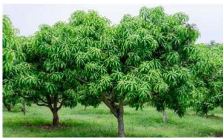
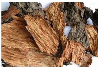 B Figure 2: (A) Whole plant of Mangifera indica (B) Mangifera indicaBark
II. ROLE OF MANGIFERA INDICA IN NEURODEGENERATIVE DISEASE
Neurodegenerative illnesses like Huntington's disease (HD), Parkinson's disease (PD), Alzheimer's disease (AD), multiple sclerosis (MS), and amyotrophic lateral sclerosis (ALS), Thus make a characterized of a pathologies group by separate etiologies with pathophysiological characteristics and unique morphological characteristic. There are many pieces of evidence that indicate the multifactorial conditions which encompass (a) aberrant protein dynamics with impaired protein aggregation and degradation, (b) oxidative stress and free radical production, (c) mitochondrial dysfunction and dysfunctional bioenergetics, and (d) metal toxicity and pesticide exposure as potential etiologies of these disorders (23).
Mangiferin, quercetin, and gallic acid are bioactive compounds found in mangos (Mangifera indica) that have neuroprotective properties. According to research, these substances may be able to stop oxidative stress, inflammation, and -amyloid aggregation—all of which are major contributors to neurodegenerative illnesses including Parkinson's and Alzheimer's. Mango extracts may be a natural treatment for neurodegenerative diseases since they have been demonstrated to enhance cognitive function and prevent neuronal damage(23).
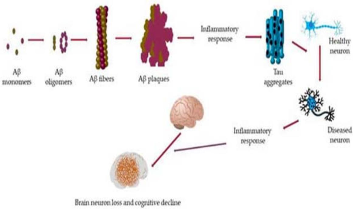 Figure 3: Role of Mangifera Indicain Neurodegenerative Disease
III. NEURODEGENERATIVE DISORDER TYPES
A. Alzheimer's Disease [AD]
A progressive neurological disease, Alzheimer's disease causes -amyloid plaque buildup, memory loss, and cognitive impairment. It results in impaired brain function with an impact on daily activities and quality of life.
B. Parkinson's Disease [PD]
A neurological condition that impairs mobility, Parkinson's disease causes bradykinesia (slow movement), stiffness, and tremors. Motor and cognitive impairment are brought on by the brain's dopamine-secreting neurones degenerating.
C. Huntington's Disease [HD]
Parkinson's disease is a neurodegenerative condition that impacts movement, resulting in tremors, stiffness, and bradykinesia (slow movement). It is caused by the degeneration of dopamine-secreting neurons in the brain, which produces motor and cognitive dysfunction.
D. Amyotrophic Lateral Sclerosis [ALS]
The long-term, steadily worsening neurodegenerative disease known as amyotrophic lateral sclerosis (ALS) damages nerve cells in the brain and spinal cord. Muscle weakening, paralysis, and ultimately the loss of voluntary movement are the results. Motor neurone degeneration causes the condition, which hinders the brain's capacity to connect with muscles. There is presently no cure for ALS, which over time significantly impairs breathing and mobility.
E. Multiple Sclerosis [MS]
Multiple sclerosis (MS), a chronic autoimmune disease, affects the central nervous system and causes damage. nerves and interferes with brain-body communication. Muscle weakness, exhaustion, eyesight impairments, and coordination problems are some of its symptoms. The illness affects everyday living and mobility and advances in an unpredictable manner with relapse and remission phases (24).
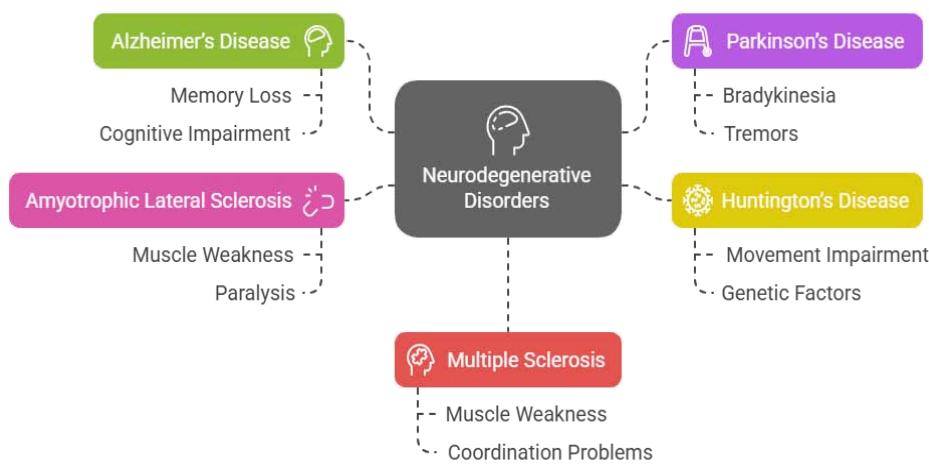 Figure 4: Neurodegenerative Disorder Types IV. MATERIALS AND METHODS
4.1 Apparatus and Instrument
The Camag HPTLC system consists of a Linomat 5 applicator, a Camag TLC scanner 3, and WinCATS software. The Shimadzu HPLC system consists of an LC AT10 VP pump, an SPD M10 AT VP detector, and CLASS M10A software. A Jasco V-560 UV/Vis spectrophotometer is also employed.
4.2 Chemical
Chemical such as ethanol, methanol, ethyl acetate, and petroleum ether purchased from Bio Liquid Research Pvt Ltd. We conducted the remaining phytochemical tests in our pharmacognosy lab at the MET Faculty of Pharmacy, which is part of the Moradabad Educational Trust Group of Institutions.
4.3 Plant Material
Mangifera indica Barkpowder, which is dark brown incolour, was obtained from the Herbal Garden MITGI, Faculty of Pharmacy, and identified Herbarium of Hindu college Moradabad.
4.4 Preparation of Mangifera indica Bark Powder
The plant material (bark) of Mangifera indica were taxonomically identified and authenticate with and collected at local market of Moradabad.
4.5 Yield Calculation
By computing the percentage weight (w/w) ratio between the produced extract and the simplicia utilised, the yield is determined (25).
V. CRUDE EXTRACT EXTRACTION FROM BARK POWDER
5.1 Crude extracts are extracted using the cold maceration process from bark powder
The extraction process for the selected plant parts is described below. Plants Active components were tracked using the cold extraction method (26).
| Method: | Cold Maceration |
| Principle: | Cold Percolation |
| Apparatus: | Iodine Flask |
| Temperature Maintained: | 25 – 30°C |
| Quality of Plant Bark Powder Used: | Fine Grand Powder |
| Volume of Each solvent used: | 500 ML |
| Period of Extraction with Each Solvent: | 3 – 4 days. |
{"image_source":{"path":"images/9646656ebc7bbd16debb6ca616a7307cc2e969ab7bddd41337f3753bba0bc73f.jpg"},"content":"","chart_caption":[{"type":"text","content":"Figure 5: Cold Maceration Extract Process"}],"chart_footnote":[]}
5.2 Extraction of Crude Extracts from bark powder by Soxhlet Extraction method
Collect Mangifera indica bark, wash it to remove any debris, and air-dry it. Grind the dried bark into a fine powder to maximize the surface area, which enhances extraction efficiency.
| Method: | Successive extraction |
| Principle: | Continuous hot percolation |
| Apparatus: | Soxhlet extractor |
| Temperature maintained: | 35 – 40°C |
| Quality of Plant Powder Used: | Fine Grand Powder |
| Volume of Each Solvent Used: | 500 ML |
| Period of Extraction with Each solvent: | 3 days |
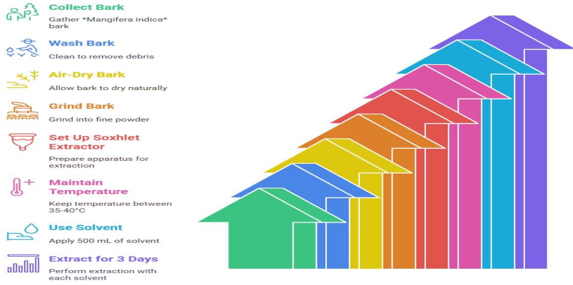 Figure 6: Soxhlet Extraction Process
Powdered crude drug was separately extracted by solvents of ascending polarity such as methanol, water, hydro-alcohol, ethanol, ethyl acetate, and petroleum spirit. The extraction yields were considerably different, the maximum being obtained by ethanol with a yield of 86.9%. The process of extraction was done step by step with continuous hot percolation using Soxhlet apparatus.
5.3 Macroscopic Studies of Mangifera indica bark
Macroscopic examination of Mangifera indica(mango) bark shows its scaly, fissured, and rough outer surface with dark brown to greyish-brown color. The inner bark is reddish-brown, fibrous, and emits a faint astringent smell when cut. It possesses a moderately hard texture with a bitter flavour. The bark is frequently lenticel-marked and ridged, which gives it a rugged look.
5.4 Microscopic study of Mangifera indica bark
Microscopic examination of Mangifera indica bark shows a distinct periderm, cortex, and phloem with clear mechanical and functional features. The occurrence of stone cells and resin ducts indicates its protective and defensive function. These anatomical features are responsible for the medicinal nature of the bark, which makes it a good source for pharmacological applications.
5.5 Physicochemical Parameters Mangifera indica bark Powder
Table 2 presents the physicochemical characterization of M. indica bark powder. The assessment of physical constants of medications is a crucial factor in identifying adulteration or poor management of pharmaceuticals. The leaf had a moisture content of merely 7.5%, hence inhibiting bacterial and fungal proliferation. Acid-insoluble ash, water-soluble ash, and totalash were the three different ways we measured the ash value. With 1.5% acid-insoluble ash and 1.5% water-soluble ash, we calculated that the total ash content was 9.6%. Calculate the 12% values for the foaming index and swelling index. Table 1 shows the bark of M. indica's extractive value. We observed the highest extraction value in methanol solvent, and the lowest in petroleum ether. We assessed the solubility of the methanol extract of M. Indicabark in six solvents of differing polarity. The extract exhibited high solubility in ethanol and methanol but reduced solubility in non-polar solvents (Table 3).
Phytochemical analysis of Mangifera indica bark powder detects the bioactive molecules like tannins, flavonoids, alkaloids, and saponins responsible for its medicinal activities. Phenolic molecules and glycosides are also present with antioxidant and antimicrobial activity. Secondary metabolites like steroids and terpenoids are found to be present in abundance in the bark, verifying its use in traditional medicine.
VI. RESULT AND DISCUSSION
Advanced Phytochemical Analysis of Mangifera indica Bark Powder
Table 1: Macroscopic Studies of Mangifera indica bark: The colour, texture, thickness, odour, taste, moisture content and bulk density of the powdered drug were studied (14).
| Colour | Greyish brown to dark powder |
| Texture | Smooth |
| Thickness | Around 0.1 to 1.5 cm |
| Odour | Slightly astringent |
| Taste | Bitter and astringents |
| Moisture Content | Low moisture content |
| Bulk density | Depending on an particle size and storage conditions. |
Table 2: Standardization Parameters: Mangifera indica bark Powder Table 3: Extractive Value of Mangifera indica bark Powder
| S. No | Specifications | %W/V |
| 1. | Moisture Content | 7.5% |
| 2. | Acid Insoluble Ash | 1.5% |
| 3. | Total Ash Value | 9.6% |
| 4. | Water Soluble Ash | 12% |
| 5. | Swelling Index | 5.5% |
| 6. | Foaming Index | 4mm |
| 7. | Foreign Index | No Foreign Index |
Solvent extraction yields vary significantly: Ethanol produces highest yield at 86.9%.
| S. No | Types of Solvent | % Yield (W/V) |
| 1. | Ethyl acetate | 67.2% |
| 2. | Petroleum Sprit | 0.8% |
| 3. | Methanol | 15.2% |
| 4. | Ethanol | 86.9% |
| 5. | Hydroalcohol | 9.6% |
| 6. | Water | 4.8% |
Table 4: Screening of Mangifera indica Bark Powder Phytochemically To identify the bioactive components, photochemical screening was performed on Mangifera indica bark powder.
VII. DETERMINATION OF MAXIMUM ABSORPTION WAVELENGTH
| Tests | Decoction |
| Flavonoids | + |
| Alkaloids | + |
| Saponins | + |
| Tannins | + |
| Terpenoids | - |
| Glycosides | + |
| Steroids | - |
| Phenols | + |
| Carbohydrates | + |
| Proteins | - |
The spectrophotometer UV-Vis is used to determine the maximum absorption wavelength (Figure 1). As a result, Mangiferin Indicahas an absorbance of 0.601 with a maximum absorption wavelength of 320 nm.
{"image_source":{"path":"images/377e143ec1572671de8e4af3f0122944b5d540badc803521eb7dcc48288333fa.jpg"},"content":"","chart_caption":[{"type":"text","content":"(A) Mangiferin Indica"}],"chart_footnote":[]} {"image_source":{"path":"images/41e595ec459a8825565ae7379236abf73ab069456fb5475f5d4004b5a2984d4a.jpg"},"content":"","chart_caption":[{"type":"text","content":"(B) Mixture of Qurectine and MI"}],"chart_footnote":[]}
{"image_source":{"path":"images/62e9189103fe93529fec06e3be28acfb3d726eacd12f9b85c362f599cd32e11c.jpg"},"content":"","chart_caption":[{"type":"text","content":"(C) Qurectinr Standard"},{"type":"text","content":"Figure 7: UV Spectrum of Mangiferin Indica 10 "},{"type":"equation_inline","content":"\mu"},{"type":"text","content":" g/mL Ethanol"}],"chart_footnote":[]}
VIII. ULTRAVIOLET SPECTROSCOPY
8.1 Preparation of Standard solution
Following carefully weighing 50 mg of pure Mangiferin Indica, it was put in a 50 mL volumetric flask and diluted with methanol to the appropriate level. It was then diluted until it created a solution with 100 g/mL, 200 g/mL, 300 g/mL, 400 g/mL, and 500 g/mL concentrations (27).
8.2 Determination of Maximum absorption wavelength
A 100 g/mL pure Mangiferin Indica solution is pipetted up to 1 mL, after which it was moved to a 10 mL volumetric flask and filled with the proper amount of methanol. The spectrophotometer UV-Vis is then used to identify the dilution's peak absorption wavelength, which is between 200 and 400 nm (28).
HPTLC: A reliable analytical technique used for the separation and identification of phytochemicals in powdered Mangifera indica bark is High-Performance Thin-Layer Chromatography (HPTLC). HPTLC yields high-resolution chromatographic fingerprints, which facilitate qualitative and quantitative evaluation of bioactive compounds. The technique guarantees accurate identification, reproducibility, and validation of plant-derived constituents.
8.3 Preparation of Standard Stock Solution of Marker
10 mg of Mangiferin Indica were transferred into a 10 ml standard flask and dissolved in 1 ml of methanol to achieve a concentration of . and then adding more methanol to get the volume up to 10 ml. Among the stock solutions mentioned above, one containing of the final concentrations were set at 200, 400... 1200 ng/spot once the marker was created. HPTLC and solutions ranging in concentration from 0.2 to 0.4 to 1 g/ml were made for HPLC examination.
8.4 Standardization by HPTLC
In the current study the HPTLC fingerprinting analysis and standardization was conducted on Mangifera indica extract. Different organic solvents such as petroleum ether, chloroform and methanol in order of their increasing polarity were employed in preparing the selected plant extract and methanol to extract the formulation. Optimized mobile phase system employed was ethyl acetate:methanol: Ethanol: water, 10:1:1:0.5% v/v/v/v. The stationary phase on which separation was done was pre-coated plates with silica gel on aluminium sheets. 310nm was the wavelength used for fingerprinting and standardization.
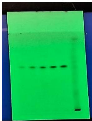 Figure 8: HPTLC Platecontaining optimized mobile phase system employed was ethyl acetate: methanol: Ethanol: wate
8.5 Recording of the HPTLC Chromatogram
50 l of each of the solution of the Mangifera indica extracts and its formulation were used with CAMAG semiautomatic applicator on to individual plates and chromatograms were developed. The plates were photo-metrically analyzed according to detection technique and chromatograms were taken. The fingerprinting of extracts was taken and the different. The Rf values achieved were recorded. The Rf values achieved in the respective extracts (plant as well as its formulation extracts) corresponding to respective marker is detected in the plant extracts and their formulations that correspond to the marker, the peak areas of the detected component (Mangiferin Indica) are recorded. The marker's calibration graph is used to determine the quantity of component present.
{"image_source":{"path":"images/2eba573dd47c4986a53305862ff1946cf40a691b912104c8c3328c77939031d3.jpg"},"content":"","chart_caption":[{"type":"text","content":"Figure 9: The calibration graph at different wavelengths in HPTLC"}],"chart_footnote":[]}
| S. No | Rf | Substance | Max @ |
|---|---|---|---|
| 1. | 0.58 | Quercetin | 309 nm |
| 2. | 0.57 | Quercetin | 311 nm |
| 3. | 0.57 | Quercetin | 383 nm |
| 4. | 0.57 | Quercetin | 381 nm |
| 5. | 0.57 | Quercetin | 380 nm |
| 6. | 0.55 | Quercetin | 300 nm |
| S. No | Rf | Substance | Max @ |
| 1. | 0.58 | Quercetin | 309 nm |
| 2. | 0.57 | Quercetin | 311 nm |
| 3. | 0.57 | Quercetin | 383 nm |
| 4. | 0.57 | Quercetin | 381 nm |
| 5. | 0.57 | Quercetin | 380 nm |
| 6. | 0.55 | Quercetin | 300 nm |
8.6 Validation
A number of factors, including linearity, accuracy, limit of detection (LOD), limit of quantification (LOQ), intraday and interday precision, sample application and measurement repeatability, stability evaluations, and selectivity, were taken into consideration when calibrating the HPTLC processes for the selected marker.
8.7 Range and Linearity
Markers were developed in different concentrations and tested using HPTLC methods. For HPTLC linear regression, data is observed to have a good linear relationship overa range of sdv = 11.96.
{"image_source":{"path":"images/6272ce8d7cccc41adc466f87cca99992b5740d3f4eeff6fe447d670a788e3091.jpg"},"content":"","chart_caption":[],"chart_footnote":[]}
Figure 10: Linearity and Range Different HPTLC Chromatogram at various wavelength of standard markers with sample Extract
| S. No | Vial | Rf | Amount | Area |
| 1. | 1 | 0.58 | 200.00 μg | 720.63 |
| 2. | 1 | 0.57 | 400.00 μg | 1711.34 |
| 3. | 1 | 0.57 | 600.00 μg | 2547.95 |
| 4. | 1 | 0.57 | 800.00 μg | 3083.15 |
| 5. | 1 | 0.57 | 1000.00 μg | 3343.94 |
| 6. | 2 | 0.55 | 389..03<180.00 μg MI Extract |
{"image_source":{"path":"images/c6ea81256aac97b63508b236ed27609ba8ca35564c4afe748d48d553ca49a967.jpg"},"content":"","chart_caption":[],"chart_footnote":[]} {"image_source":{"path":"images/73aba516425329e7537888c0698914ae77f7ef2d9adab0e8667bd4d169c27b81.jpg"},"content":"","chart_caption":[],"chart_footnote":[]}
| Peak | Start Rf | Start Height | Max Rf | Max Height | Max% | End Rf | End Height | Area | Area% | Assigned substance |
| 1 | 0.53 | 1.5 | 0.58 | 35.4 | 100.00 | 0.61 | 0.1 | 720.6 | 100.00 | QUERCETIN |
{"image_source":{"path":"images/6da500831c2f95d743ecd12533d31b3a4e235ee722c3bbb241efb825cc8757c0.jpg"},"content":"","chart_caption":[],"chart_footnote":[]} {"image_source":{"path":"images/60db15afa60e4a6185b1d25f4f0a9dacf4cb2d2c3e688229fa2c0c22471a223a.jpg"},"content":"","chart_caption":[],"chart_footnote":[]}
{"image_source":{"path":"images/4762b45f59a2495ef4db379d8fb7ac92893aefc54dac4c0f969092489a92698e.jpg"},"content":"","chart_caption":[],"chart_footnote":[]} {"image_source":{"path":"images/3da064186d9320f80aef4a0490cafc242bdfc791fce5da841b3a4ff94b49721e.jpg"},"content":"","chart_caption":[],"chart_footnote":[]}
{"image_source":{"path":"images/5b14716f726d5df6544500b3651aa15fe769459cfa78b9d306dbde4cab9783a4.jpg"},"content":"","chart_caption":[],"chart_footnote":[]} {"image_source":{"path":"images/1b52f981c15dd100f820e465533df4f93b86a413fe9ad70c7eebff6d0677f42e.jpg"},"content":"","chart_caption":[],"chart_footnote":[]}
{"image_source":{"path":"images/5f44d441f487df40d9e4e5854d012451cb2fe21ebd2ad61120f4b784a48e67f7.jpg"},"content":"","chart_caption":[],"chart_footnote":[]} {"image_source":{"path":"images/600f662d4d85d4a550e25236f62cc671d9f9b7b943d80f20c0e1389add398bc0.jpg"},"content":"","chart_caption":[{"type":"text","content":"Figure 11: Different HPTLC Fingerprint of Methanolic Extract"}],"chart_footnote":[]}
Figure 12: Overall Different HPTLC Fingerprint of Methanolic Extract
| S. No | Appl. Position | Appl. Volume | Viral# | Sample ID | Active |
| < 1. | 6.8 mm | 0.4 μl | 1 | Quercetin Std. | Yes |
| < 2. | 14.0 mm | 0.8 μl | 1 | Quercetin Std. | Yes |
| < 3. | 21.2 mm | 1.2 μl | 1 | Quercetin Std. | Yes |
| < 4. | 28.4 mm | 1.6 μl | 1 | Quercetin Std. | Yes |
| < 5. | 35.6 mm | 2.0 μl | 1 | Quercetin Std. | Yes |
| < 6. | 42.8 mm | 3.0 μl | 2 | MI extract | Yes |
Polydispersity Index and Particle size: Polydispersity Index (PDI) is an important parameter to assess the homogeneity of particle size distribution in Mangifera indica bark powder. It reflects the homogeneity of the sample, and lower values represent a more homogeneous dispersion. PDI is an important factor in deciding the stability and consistency of pharmaceutical and herbal products.
{"image_source":{"path":"images/1746b4dcb7f51a1a01a83706f0a9997be8c97d2d4b459efdf3a7b3ca52cf7316.jpg"},"content":"","chart_caption":[],"chart_footnote":[]}
Figure 13: Polydispersity Index and Particle size Mangifera indica
| Name | Mean | Standard Deviation | RSD | Minimum | Maximum | |
| Z – Average (nm) | 671.4 | - | - | 671.4 | 671.4 | |
| Polydispersity Index (PI) | 0.2835 | - | - | 0.2835 | 0.2835 | |
| Intercept | 0.8449 | - | - | 0.8449 | 0.8449 | |
IX. CONCLUSION
In summary, pharmacognostic standardization and quality profiling of Mangifera indica bark powder reveal crucial information regarding its prospective therapeutic use, particularly in neurological disorder management. Through a detailed examination of its macroscopic and microscopic characters and Physico-chemical attributes, it has been proven that Mangifera indica bark contains a multifaceted profile of bioactive constituents, which may contribute to its neuroprotective actions. The availability of vital phytochemicals like alkaloids, flavonoids, and terpenoids, along with its proven antioxidant and anti-inflammatory properties, indicates its potential application in the management of diseases like Alzheimer's disease, Parkinson's disease, and other neurodegenerative diseases. In addition, the development of strong standards for the quality of the bark powder guarantees its reliability and consistency for therapeutic purposes. The results highlight the necessity for additional in-depth research, such as clinical trials, to comprehensively assess the safety and efficacy of Mangifera indica bark in neurological use. As research evolves, this plant is promising as a valuable natural resource for integrative strategies in managing neurological health. The pharmacognostic profile further highlights the significance of quality control and standardization of herbal medicine to ensure that patients are administered safe and effective treatments from natural sources.
Future Aspects
The potential of the pharmacognostic standardization and quality profiling of Mangifera indica bark powder as an antidote against neurological disorders is immense. With further development in research, one of the primary regions of interest will be the complete isolation and characterization of bioactive molecules within the bark, employing novel methods such as high-throughput screening and liquid chromatography-mass spectrometry. These investigations will unveil new molecules with neuroprotective effects. Additionally, clinical trials are important to confirm the effectiveness, safety, and ideal dosage of Mangifera indica bark for neurological disorders to ensure that the plant can be made a part of mainstream treatment approaches. More studies on how it affects neurobiological pathways, especially on how it acts upon them, will also be important in determining its application in fighting neurodegenerative diseases.
Conflicting Interests
The authors acknowledge their individual contributions to the study and declare that they have no financial or other conflicts of interest.
Contribution of the Authors
All authors participate in the conception and design of the study. Shivani Agrwal and Rahul Chauhan conducted the material preparation, data collecting, and analysis. Shivani Agarwal and Rahul Chauhan composed the initial draft of the book, and all writers reviewed and endorsed the final version.
Funding
None
Availability of data and material
The authors atleast that the information in the article supports the study's conclusions.
ACKNOWLEDGEMENTS
For their assistance with this study project, the authors would like to thank the MET Faculty of Pharmacy Moradabad. Additionally, they disclose that they worked together to obtain the graphical abstract.
Conflict of Interest
The authors declare no conflict of interest.
Ethical Approval
Not applicable
Data Availability
The datasets used in this study are openly available at [repository link] and the source code is available on GitHub at [GitHub link].
Funding
This work did not receive any external funding.
References
Cite this article
Related Research



Special Issue
Launch a focused special issue to highlight research, emerging trends, and expert insights in your academic field.
