


IntelliPaper
Abstract
Background: Hemangioblastomas arehighly vascular grade 1 tumors arising sporadically or, more rarely, in the context of Von Hippel-Lindau disease.Cystic brainstem hemangioblastomas are rare with only few cases reported in literature.
Even though they are benign, their surgery is challenging in view of their eloquent location and vascularity.Thorough perioperativemanagement is required.
Case presentation: We report the case of a 47-year-old female patient with progressive weakness, difficulty in swallowing and ataxic gait.The brain MRI showed a cystic lesion occupying the caudal part of the fourth ventricle and the dorsal medulla oblangata. We performed a subtotalexcision of the tumor that was challenging due to its adherence to the floor of the 4th ventricle and its high vascularity. The pathology exam concluded to a hemangioblastoma.
The patient presented a good evolution with no tumor progression in 6 months follow-up.
Clinical discussion: The cystic brainstem hemangioblastoma is a rare seemingly innocuous lesion but highly challenging to neurosurgeons. MRI is the investigation of choice, regularly sufficient for preoperative evaluation. Still, some authors recommend the performance of a cerebral angiography or a CTA preoperatively.
In literature, manystrategies, such as the use of preoperative embolizationor radiosurgery were described, aiming for a better-quality surgery and prognosis.
Conclusion: Cystic brainstem hemangioblastoma is a scarce histologically benign but challenging lesion with surgical difficulties.Through our case and literature review, we concluded that surgery is the mainstay treatment for brainstem hemangioblastomas mainly cystic ones and that a thorough study of its vascularization via angiography or CTA with a preoperative tumor embolization can help achieve a total tumor resection with good functional outcome andlow mortality.
Explore Digital Article Text
I. INTRODUCTION AND IMPORTANCE
Hemangioblastomas are highly vascular OMS grade 1 tumors that arise both sporadically (75%) and, more rarely (25%), in the context of Von Hippel-Lindau disease (1). Only 5-20% are located in the brainstem, among them cystic hemangioblastomas are scarcer with only few cases reported in literature.
They present a unique challenge in view of their eloquent location and vascularity. Significant neurological improvement is though achievable. Thorough perioperative management is required as well as the treatment of potential associated abdominal masses.
II. CASE-REPORT
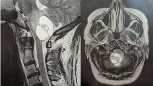
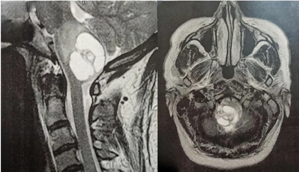
It's about a 47-year-old female patient who was presenting progressive weakness, difficulty in swallowing and ataxic gait of six months duration. There were no focal motor or sensory deficits. Her blood investigations were normal. The brain MRI showed a cystic lesion occupying the caudal part of the fourth ventricle and the dorsal medulla oblangata (Fig.1). We performed a midline suboccipital craniectomy in prone position, with C1 laminectomy. Under an operating microscope, a Y-shaped durotomy was performed, arachnoidal membrane opened, exposing the tumor which was shrunk by coagulation under copious irrigation.
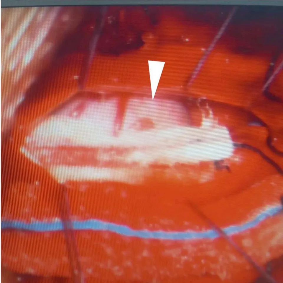
Subtotal excision of the tumor was performed limited by its adherence to the floor of the 4th ventricle and its high vascularity. The pathology exam concluded to a hemangioblastoma (Fig. 2).
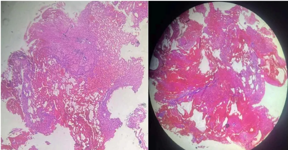
The patient presented a good evolution with no tumor progression in 6 months follow-up.
III. DISCUSSION
The cystic brainstem hemangioblastoma (HAB) is a rare seemingly harmless lesion but highly challenging to neurosurgeons. MRI is the investigation of choice, usually sufficient for preoperative evaluation, allowing to identify the location and the texture of the tumor and its relationship with the surrounding tissues. Tumors are hyperintense on T2-weighted images and isointense or hypointense on T1-weighted images with a marked homogeneous or heterogeneous enhancement after Gadolinium injection and serpentine areas consisting of the pathological vessels often seen at the periphery or within the tumor. However, MRI cannot clearly delineate the feeding arteries or draining veins of the tumor.
Hence the usefulness of Digital subtraction angiography (DSA) or Computed Tomography Angiography (CTA)(2).
In literature, cystic lesions were more frequent than solid ones, their survival was longer, recurrence less and postoperative state better. (3) In the past, surgical resection of HABs had often led to high morbidity and mortality rates (4). As a result, more and more patients with brainstem hemangioblastomas were referred for either conventionally fractionated irradiation or gamma knife radiosurgery. Some recent series of radiosurgery for hemangioblastomas report high tumor control rates (5). However, along with its potential complications, radiation rarely results in an integral cure. Some studies show it is not adequately reliable for the control of HAB cysts, but can be an effective treatment for solid tumors, especially those in eloquent regions (6). Therefore, microsurgical resection remains the first option, especially in cystic lesions.
During surgery, a superficial part of the lesion often appears once the dura is opened and facilitates progressing deeper. Any attempt of partial surgical removal must be avoided due to the rich vascularization of these tumors. Instead, it is crucial to find and follow the arachnoidal plane that, under high magnification, marks the boundary between the lesion and the normal neural tissue. Intraoperative bradycardia may occur, and it is always resolved either pharmacologically or by reducing distraction of the surrounding nerve pathways during dissection of the lesion. Intraoperative bleeding is not a major concern if the lesion is handled gently and not entered during its progressive devascularization conducted by properly low-set bipolar coagulation. Whenever hemorrhage occurs during tumor dissection, cottonoid compressive hemostasis is the means to control it until devascularization is completed. (7)
Major causes of the mortality and morbidity in surgical removal of HABs have been attributed to the location, size and solid type of the tumor, and profuse bleeding during the operation (8). With the aid of improved neuroimaging, preoperative tumor embolization (9), and microsurgical techniques, total surgical resection of hemangioblastomas can be performed nowadays with a mortality less than 5% (10).
Regarding the functional outcome, we found no difference in literature between VHL and non-VHL patients with surgically treated medulla oblongata hemangioblastomas. (11)
Postoperative respiratory control as well as constant monitoring of cardiopulmonary function and cerebrospinal fluid pressure are necessary and have contributed much to make surgery of this type feasible.
IV. CONCLUSION
Cystic brainstem hemangioblastoma is a scarce histologically benign but challenging lesion with surgical difficulties. Through our case and literature review, we concluded that surgery is the mainstay treatment for brainstem hemangioblastomas mainly cystic ones and that a thorough study of its vascularization via angiography or CTA with a preoperative tumor embolization can help achieve a total tumor resection with good functional outcome and low mortality.
Disclosures
Conflicts of Interest
We attest that there are no known conflicts of interest associated with this manuscript nor with its publication.
Sources of funding
This work has no sources of support/funding
Ethical approval
This work raises no ethical issue and was carried out after the consent of the patient and her parents.
The study is exempt from ethical approval in our institution.
Consent
Written informed consent was obtained from the patient for publication of this case report. A copy of the written consent is available for review by the Editor-in-Chief of this journal on request.
Research Registration (for case reports detailing a new surgical technique or new equipment/technology):
Our case-report is not a 'First in Man' study, so is not subject for registration in clinical trial websites.
Conflict of Interest
The authors declare no conflict of interest.
Ethical Approval
Not applicable
Data Availability
The datasets used in this study are openly available at [repository link] and the source code is available on GitHub at [GitHub link].
Funding
This work did not receive any external funding.
References
Cite this article
Related Research


Special Issue
Launch a focused special issue to highlight research, emerging trends, and expert insights in your academic field.