



IntelliPaper
Abstract
Invasive listeriosis is relatively rare, but is one of the deadliest food-borne infections, affecting pregnant women, their fetuses and newborn infants, the elderly and
mmunocompromised people. The aim of this study was to research the impact of the COVID-19 pandemic on invasive listeriosis in the metropolis. Loci and whole-genome sequencing with subsequent bioinformatic analysis were used for the study of clinical and food Listeria monocytogenes isolates revealed in 2018–2022. The results indicate the crucial change in the spectrum of the L. monocytogenessequence types (ST) causing invasive listeriosis during the COVID-19 pandemic, with slight changes in the ST spectrum of the food isolates. An increase in sensitivity to previously non-human L. monocytogenes genotypes, namely ST8, 21, 37, 391, and 425, was observed. L. monocytogenes of ST20 and 425 carried plasmids with virulence factors (VF), in addition to the 42 VF identified in the genomes with the vip gene exclusion in the genomes of ST7, 8, 21, and 37. Perinatal listeriosis cases were associated with the new hypervirulent L. monocytogenes of ST1, 4, and 219 compiled with old ST6. These data indicate the need for the more stringent control of food products for high-risk groups.
Explore Digital Article Text
I. BACKGROUND
Listeria monocytogenes is a saprophyte that is ubiquitously distributed in the environment. With the modern development of the food industry, L. monocytogenes has become an actual food-borne pathogen, being psychrophilic and salt-tolerant . Entering the human body through foodstuffs and ready-to eat (RTE) foods, L. monocytogenes causes fecal carriage, as was detected in 10% of humans from an independent cohort of 900 healthy asymptomatic donors , or listeriosis at high levels of contamination (> 1000 cfu/g) . A more severe form of the disease is invasive listeriosis, which has a high mortality rate (20%–30%) . This form of the disease affects the elderly, the immunocompromised (patients undergoing treatment for cancer, AIDS and organ transplants), pregnant women and their unborn or newborn infants . Defining the age limits of the elderly group, Mook et al. concluded that age >60 years was associated with an increased risk for listeriosis in England and Wales . According to Goulet et al., the age of the high-risk group with no underlying conditions was >65 years in France . Immunosenescence in the elderly is associated with declines in adaptive and innate immunity . Immunocompromised persons have suppressed T-cell-mediated immunity as a consequence of the treatment . The decrease in the number of helper T cells in the second and third trimesters of normal pregnancy is evidence of immunodeficiency in pregnant women . In addition, in pregnancy, in the second and third trimesters, the gastrointestinal (GI) tract undergoes a series of changes, the root cause of which is a decrease in intestinal motility . GI motility plays a key role in mucus renewal. The mucus is the first line of defense against the infiltration of microorganisms . It coats the interior surface of the GI tract, lubricates luminal contents and physically entraps bacteria, sweeping ingested bacteria out of the small bowel into the colon . Decreased GI motility results in the enrichment of the mucus with Proteobacteria and Actinobacteria (in October 2021, the valid names became Pseudomonadota and Actinomycetota ), and in the reduction in the health-related bacterial abundances . Alterations in the mucus microbial community result in a decrease in the production of protective factors including short chain fatty acids (SCFAs), in particular, acetate and formate, which have antimicrobial properties against enteric pathogens , and butyrate, which has anti-inflammatory effects .
Neurological disorders, stress, and antibiotic treatment are also accompanied by a reduction in the mucus-associated gut microbiota layer and mucus secretion . The damage to the GI mucosa could be caused by viral pathogens often having seasonal patterns . Schwartz et al. were the first to analyze the association of invasive listeriosis with the consequences of a viral infection in Philadelphia between 1 December and 1 April 1987, i.e., during the period of the epidemic rise in the incidence of influenza virus in the Northern Hemisphere . The subsequent years of intensive study into the microbial communities in different environmental niches within human bodies demonstrated the effects of viral infections on both the gut and respiratory microbiome. Influenza infection, for instance, has been found to result in significant changes in the gut microbiome, despite the lack of detectable virions in the GI tract . Furthermore, according to Bortel et al. pulmonary infection triggers the production of IL-10 by Ncr1+ cells, which could be a factor that increases the survival of invasive L. monocytogenes .
At the end of 2019, humanity faced a new virus, SARS-CoV-2, which caused severe respiratory coronavirus disease 2019 (COVID-19), reaching pandemic levels on March 11 2020 [20]. In 2020, Xiao et al. and other researchers provided evidence for the GI infection of SARS-CoV-2 [21,22,23]. A cell receptor for SARS-CoV-2, angiotensin-converting enzyme 2 (ACE2) protein, was demonstrated to be abundantly expressed in the glandular cells of gastric, duodenal, and rectal epithelia, supporting the entry of SARS-CoV-2 into the host cells [21, 23]. Among the hospitalized patients infected with SARS-CoV-2, tested positive for SARS-CoV-2 RNA in their stool, and more than of patients with SARS-CoV-2 remained positive for viral RNA in feces even after the test results for viral RNA in the respiratory tract became negative [21]. Furthermore, SARS-CoV-2 infection causes an alteration of intestinal permeability, resulting in enterocyte dysfunction [22]. These data suggest a direct effect of SARS-CoV-2 on the gut microbiota, in addition to in-direct effects via the gut-lung axis [24, 25].
Previously, we showed the impact of respiratory viral infections on the incidence of invasive listeriosis in a metropolis in 2018/2019. L. monocytogenes of phylogenetic lineage (PL) II prevailed in the groups of cases that occurred when the epidemic threshold for influenza was crossed during the 2018/2019 season. Listeria pneumonia identified in the senior age group occurred during the season of autumn acute respiratory viral infections and was primarily caused by L. monocytogenes of PLI [26]. It is strains of PL I and II that cause the majority of human illness, despite the fact that the population of L. monocytogenes consists of four divergent lineages (I–IV) [27].
The aim of this study was to research the impact of the COVID-19 pandemic on the incidence of invasive listeriosis in the same metropolis, comparing the spectrum of the Listeria genotypes that previously caused the disease with the spectrum of the Listeria pathogens identified during the COVID-19 pandemic.
II. RESULTS
The monitoring of the clinical and food-borne L. monocytogenes isolates started in November 2018 in a large metropolis with a population of 20.4 million (https://mosstat.gks.ru/folder/64634 / (accessed on 24 July 2022)). The COVID-19 pandemic divided our study into two periods: before the pandemic (November 2018–October 2019) and during the pandemic (March 2020–January 2022). Since SARS-CoV-2 was constantly mutating during the pandemic, we characterized the stages of the pandemic by the number of genotypes (pangolineages) circulating at the same time.
1. The analysis of the change in SARS-CoV-2 pangolineages in the Moscow metropolis.
The large amount of data on SARS-CoV-2 sequences related to the Moscow region, presented in the GISAID EpiFluTM Database (http://gisaid.org / (accessed on 24 July 2022)) including our own data (EPI_ISL_421275, EPI_ISL_1708505, EPI_ISL_1708506, EPI_ISL_1708510 - EPI_ISL_1708519, EPI_ISL_1731043 - EPI_ISL_1731045, EPI_ISL_2322757 - EPI_ISL_2322762, EPI_ISL_2356912, EPI_ISL_2356913, EPI_ISL_3102159, EPI_ISL_9058873, EPI_ISL_9058988 - EPI_ISL_9058990, EPI_ISL_9058992), indicates that the pandemic period included in our observation can be divided into two stages (Figure 1) in accordance with the number of simultaneously circulating pangolineages of SARS-CoV-2. At the first stage, from the beginning of the pandemic until the beginning of June 2021, various pangolineages were circulating: B.1.1 B.1.1.523, B.1.1.397, B.1.1.294, B.1.1.336, the Moscow endemic variants B.1.1.317, B.1.1.141, and subsequently Alpha B.1.1.7 (VOC, the variants of concern) [28].
{"image_source":{"path":"images/f6c89592ef8631ffb91c145dec061f2e024a7f8a7a5d795b90d7f37c8ea6a19f.jpg"},"content":"","chart_caption":[{"type":"text","content":"Figure 1: The stages of the COVID-19 pandemic. VOC—the variants of concern"}],"chart_footnote":[]}
At the second stage, from the beginning of June 2021 to the end of January 2022, VOC dominated in the Moscow region, successively replacing each other. The frequency of Delta (B.1.617.2) strain among the Moscow region samples had been growing since March 2021, and reached 80.0% in June, and 98.0% in early July 2021 [29, 30]. The Omicron strain (B.1.1.529), which appeared on December 3 [29], reached a frequency of 49.2% on January 4 and 90.1% on January 14 (https://www.youtube.com/watch?v=u-_NDK4DCLU / (accessed on 24 July 2022)). 2. Comparison of L. monocytogenes of two periods of time: before the pandemic and during the pandemic.
{"image_source":{"path":"images/bfed8122cc773fc52f49e43286a0f8abb12c6c158c06873aa21e50cd57528ff2.jpg"},"content":"","chart_caption":[],"chart_footnote":[]} {"image_source":{"path":"images/94cc4623444d557647d19ef113d825d7be1bf58fc6f73b13c3efa9b861db48c5.jpg"},"content":"","chart_caption":[],"chart_footnote":[]}
{"image_source":{"path":"images/fe8aa05583b5198fbdd351497697fdcea02ba2c7b4d39556ca5b79f21567cecc.jpg"},"content":"","chart_caption":[],"chart_footnote":[]} {"image_source":{"path":"images/a864cd770164dfd29827920c42ba7c6f82b83b6eb4a3ddaf617c1a96f6454026.jpg"},"content":"","chart_caption":[],"chart_footnote":[]}
{"image_source":{"path":"images/be55e1157df589b3ba59642d3a3898129fcb26292df9f2893995d64749fbe322.jpg"},"content":"","chart_caption":[{"type":"text","content":"b L. monocytogenes genotype spectrum in three observation periods"}],"chart_footnote":[]} a1) before the COVID-19 pandemic, 2018-2019; a2) during the COVID-19 pandemic, March 2020–May 2021; a3) during the COVID-19 pandemic, June 2021–January 2022; a4) during the COVID-19 pandemic as a whole.
b) L. monocytogenes genotype spectrum in three observation periods: I—before the COVID-19 pandemic, 2018-2019; II—during the COVID-19 pandemic, March 2020 May 2021; III—during the COVID-19 pandemic, June 2021 - January 2022. The genotypes of the first phylogenetic lineage are highlighted with hatching and shadow. H—the Shannon diversity index; PLI—phylogenetic lineage I; ST—sequence type.
Figure 2: L. monocytogenes genotypes identified during the observation periods.
In the period before the pandemic, the autochthonous ST7 of PLII prevailed, while ST6 (PLI) was the second most prevalent (Fig. 2a1, 2b), (Table S1). If ST7 is typical in Russia (with the exception of the Far East Federal District (FD)) for isolates from food and the environment [31,32], then the sources of ST6 have not yet been found. L. monocytogenes of this genotype is absent in food from domestic production in the Moscow region [31]. In perinatal listeriosis, only listeria of these two STs were found (Fig. 3a), while in invasive listeriosis in the older age group (meningitis and sepsis), three more STs of PLII and two STs of PLI added diversity (Fig. 4a). In general, during the period before the pandemic, L. monocytogenes of STs of PLII prevailed, which is typical for the population of listeria in most of the territories of the Russian Federation. Only in the Far East FD did listeria of PLI predominate both in the environment and among the clinical isolates [33, 34].
{"image_source":{"path":"images/62c3ca10db560f8aa4d63719276bcd9555fdbc80b0cf670c7b1c83dbe0aa14de.jpg"},"content":"","chart_caption":[],"chart_footnote":[]} {"image_source":{"path":"images/afc874661871b6332507b32678c0360e5a8c45286501f07673283baf6e5934d6.jpg"},"content":"","chart_caption":[],"chart_footnote":[]}
H—the Shannon diversity index; PLI—phylogenetic lineage I; ST—sequence type
Figure 3: L. monocytogenes genotypes causing invasive perinatal listeriosis.
a Invasive listeriosis (meningitis, sepsis) 2018-2019 {"image_source":{"path":"images/24a9fa55a557dfc013b7a6642321b59c5d6f1d984aad941335063e756e6440f7.jpg"},"content":"","chart_caption":[],"chart_footnote":[{"type":"text","content":"b Invasive listeriosis (meningitis, sepsis) 2020-2022"}]}
{"image_source":{"path":"images/c62f68b4f9475cbf1ef48f50d9e4eb06cf4bcb853ed75f877e869cfcf14a1924.jpg"},"content":"","chart_caption":[],"chart_footnote":[{"type":"text","content":"a)—before the COVID-19 pandemic, 2018-2019; b)—during the COVID-19 pandemic. H—the Shannon diversity index; PLI—phylogenetic lineage I; ST—sequence type"}]}
Figure 4: L. monocytogenes genotypes causing meningitidis and sepsis.
From the eight genotypes of PLII, only two STs (7 and 14) coincided with those identified in the period before the pandemic (Figure 2b). From the STs that appeared during the pandemic (Figure 2a4), ST451 was attributed only to perinatal listeriosis. ST451 was new in the Central FD in January 2021. In August 2021, the strain with the same ST and internalin profile (IP), including , , , and , was isolated from a lightly salted salmon fillet imported from the Faroe Islands. However, in 2016/2017, ST451 was also isolated in the Northwestern FD in the case of perinatal listeriosis (ID 37701) [35], and from chilled beef of unknown origin (ID 42266) [35]. It should be noted that comparative analysis of the sequences of internalin A demonstrated that isolates of ST451 had the same N187S substitution in the leucine-rich repeat (LRR) 5 domain as the strains of PLI. This is important for internalin A–E–cadherin interaction, which is exploited by for the invasion of enterocytes and to cross the placental barrier [36].
The two more STs, 21 and 8, were revealed not only in the perinatal listeriosis group (Figure 3b), but in the meningitis and sepsis group too (Figure 4b). Previously, ST21 was not observed in human listeriosis. In 1975, an isolate of this genotype was isolated from a goat in the North Caucasian FD of Russia [32], but it differed from the human isolate by the locus of internalin C in the IP. The L. monocytogenes isolates of ST8 were previously found in food (meat, fish, dairy) in Russia, but were absent in clinical cases of listeriosis. The isolates of Clonal Complex (CC) 8 were detected in meat (ST16, ID 42269) [35] and in the environment (ST758) [34], and in cases of meningitis (ST2096) before the COVID-19 pandemic [26]. All isolates of CC8 but one (ST758, IP34), had the same IP53. During the COVID-19 pandemic, L. monocytogenes ST8 was revealed in four clinical cases (Figures 3b and 4b), and one of them had a fatal outcome.
The greatest diversity of the PLII genotypes was noted in the older age group (meningitis and sepsis) during the pandemic (Figure 4b), and each of the genotypes was associated with a case of listeriosis with a fatal outcome (Table S1). ST20 was first detected in human listeriosis in the Central FD, but in 2007 an isolate of this genotype was isolated from the crucian carp (Carassius carassius) [34]. The IP of clinical and environmental isolates were identical. However, in 2016 L. monocytogenes ST20 was isolated in the North-western FD in the case of perinatal listeriosis (ID 42272), later, in 2017, a strain of this ST was isolated from chilled beef of unknown origin (ID 42270), and then, in 2018, it was isolated from wastewater (ID 42274) [35].
L. monocytogenes of ST425 is also new in human listeriosis in Russia but was isolated in the environment (from red deer (Cervus elaphus) feces) in 2009 [34]. However, in this case, clinical and environmental isolates differed in the internalin B locus of the IP. A single-nucleotide variant (SNV) in the same position of the alleles relative to the most common for the PLII allele 14 (A/T, A/C) leads to the same amino acid substitution (Ile/Leu).
ST37, found in food and food processing environments (FPEs) in 2017 (ID 37695) [35], and in 2019-2021 (ID 49371, 49372, 76388) [31], was isolated for the first time in 2021 in a case of human listeriosis. Note that food and clinical isolates had the same IP.
Finally, ST391 (CC89) was discovered for the first time, but it is a single locus variant (SLV) of ST1547 (CC89) identified in human listeriosis in 1997 [32]. The internalin B locus distinguishes the IP of these genotypes.
Thus, we note a significant role of the PLII STs in the group of diseases "meningitis and septicemia". Evaluating the L. monocytogenes genotypes in the older age group for the entire observation period (Fig. 4b), it should be noted that the proportion of the PLI is low, but the diversity index is high, which is provided by the PLII STs.
3. Comparison of L. monocytogenes of two stages of the COVID-19 pandemic
Comparing the causative agents of invasive listeriosis in the two periods of time, before the pandemic and during the pandemic, we saw a significant difference in the diversity of genotypes, in the representation of phylogenetic lineages, and in the number of deaths (43.8% before the pandemic and 81.2% during the pandemic in the group of the meningitis and septicemia [37]). Noteworthy is the sharp decrease in age in the group with meningitis and septicemia: from 59 to 33 years (Figure 5).
{"image_source":{"path":"images/6222b7de4e20ef72895a04f1c905709079f4a366494c5ae90d6094315683ff18.jpg"},"content":"","chart_caption":[{"type":"text","content":"Figure 5: The age of patients with invasive listeriosis manifesting as septicemia and meningitis."}],"chart_footnote":[]}
Since the pandemic period can be divided into two stages according to the number of circulating pangolineages, we compared the diversity of the L. monocytogenes genotypes between the pandemic stages (Figure 2a2, 2a3). As can be seen from the data in Figure 2a2, the increase in the diversity of the L. monocytogenes STs and in the proportion of the PLI genotypes occurred precisely at the first stage of the pandemic. At the second stage of the COVID-19 pandemic (Figure 2a3), the diversity of the L. monocytogenes STs was equal to the data for the period before the pandemic, and the proportion of the PLI genotypes was even lower than before the pandemic. However, the list of STs changed dramatically: all genotypes revealed at the second stage of the pandemic are new for clinical cases in the Central FD (Figure 2b).
4. Internalins' diversity in the different phylogenetic lineages
The internalins, as factors of the pathogenicity of listeria, are important for the bacteria–human cells interaction, and listeria adaptation . As a result, we note a large number of IP in the batch of Russian isolates, with the new ones among them in recent times presented in Table 1.
Table 1: New internalin profiles (IP) from this research
| CC | ST | inlA | inlB | inlC | inlE | IP | Phylogenetic Lineage |
| CC21 | 21 | 12 | 14 | 7 | 8 | 56 | II |
| CC11 | 451 | 22 (MW538938) | 23 (MW538939) | 6 | 6 | 58 | II |
| CC26 | 26 | 7 | 14 | 6 | 6 | 59 | II |
| CC4 | 219 | 23 (MW965279) | 8 | 4 | 3 | 60 | I |
| CC89 | 391 | 15 | 14 | 17 | 6 | 61 | II |
| CC90 | 425 | 13 | 24 (MZ486423) | 13 | 8 | 62 | II |
| CC403 | 403 | 9 | 23 | 21(OM240823) | 17(OM240824) | 66 | II |
Different internalins contribute to the diversity of IP of the clinical isolates in two phylogenetic lineages (Table 2). In the group of the clinical isolates of PLI, the locus was the most diverse, according to the H value, and was the least diverse locus. At the same time, in PLII, both and were the most diverse loci. It was the locus that distinguished isolates of ST21 when comparing the bacteria that caused human and goat listeriosis.
Table 2: Shannon's diversity index for internalin alleles of clinical L. monocytogenes isolates
| Phylogenetic lineage | Shannon's diversity index (H) | |||
| inlA | inlB | inlC | inlE | |
| I | 2,5 | 1,8 | 1,1 | 1,4 |
| II | 3,1 | 1,4 | 2,3 | 1,4 |
5. Virulence factors
The virulence of L. monocytogenes depends on its ability to colonize the gut and disseminate within the host . The key genes and gene products involved in microbial virulence were named as virulence factors (VFs) . A number of well-studied VFs are also known as pathogenicity factors, such as internalins, because virulence and pathogenesis represent two sides of one coin. The former is viewed from the point of view of the microorganism and the latter from its effect on the host . The virulence factor database (VFDB) (http://www.mgc.ac.cn/VFs/ (accessed on 24 July 2022)), which is an extensive collection of VFs from the important bacterial pathogens , was used for the VF analysis of L. monocytogenes, revealed during the monitoring.
Whole-genome sequencing (WGS) was performed for the isolates of the repeated genotypes and new genotypes for the clinical cases or for the Listeria population in Russia. Thus, the cohort of the sequenced isolates included L. monocytogenes of 12 STs, four of which belonged to PLI, and eight to PLII (Table S2). As a result, 45 virulence genes related to 12 VF classes were identified (Table 3). The VF classes "Adherence", "Invasion" and "Regulators" were the most numerous: seven, seven and nine genes, respectively, were revealed in all analyzed genomes. Only one VF of the "Invasion" class which was encoded by the vip gene (bold font in Table 3) was absent in the genomes of ST7, 8, 21, and 37. The vip protein (lmo0320) is an LPXTG surface protein, but does not contain LRRs, and thus does not belong to the internalin family [42]. According to Cabanes et al., vip is required for entry into some eukaryotic cells via the endoplasmic reticulum resident chaperone Gp96 [42]. Doumith et al. demonstrated the presence of lmo0320 in seven L. monocytogenes strains of the serovar 1/2a isolated in cases of human sporadic or epidemic listeriosis [43].
Table 3: Virulence factors revealed in the sequenced genomes of L. monocytogenes isolates. Bold font—virulence factor gene, which is absent in the genomes of ST7, 8, 21, 37
| VFclass | Virulence factors | Related genes | VFclass | Virulence factors | Related genes | VFclass | Virulence factors | Related genes |
| 1. Adherence | D-alanine-polyphosphoribitol ligase | dltA | 5. Intracellular survival | Lipoate protein ligase A1 | lplA1 | 10. Regulation | AgrA/AgrC | agrA |
| Fibronectin-binding protein | fbpA | Oligopeptide-binding protein | oppA | agrC | ||||
| GW autolysin | ami | Post-translocation chaperone | prsA2 | CheA/CheY | cheA | |||
| Internalin F | inlf | Sugar-uptake system | hpt | cheY | ||||
| Internalin J (LPXTG protein) | inlj | 6. Invasion | Autolysin (GW protein) | aut | LisR/LisK | lisK | ||
| Listeria adhesion protein | lap | Cell wall hydrolase | iap/cwhA | lisR | ||||
| lapB | Cell wall teichoic acid glycosylation protein | gtcA | Positive regulatory factor | prfA | ||||
| 2. Bile resistance | Bile-salt hydrolase | bsh | Internalin A (LPXTG protein) | inlA | VirR/VirS | virR | ||
| 3. Enzyme | Metalloproteinase | mpl | Internalin B (GW protein) | inlB | virS | |||
| PC-PLC | plcB | Internalin P | inlP | 11. Surface protein anchoring | Lipoprotein diacylglyceryl transferase | lgt | ||
| PI-PLC | plcA | Lipoprotein promoting entry protein | lpeA | Lipoprotein -specific signal peptidase II | lspA | |||
| Serine-threonine phosphatase | stp | Virulence protein (LPXTG protein) | vip | Sortase A | srtA | |||
| 4. Immune modulator | Inlc | inlc | 7. Iron uptake | Hemoglobin binding protein | hbp2 | Sortase B | srtB | |
| Inlk | inlk | 8. Nucleation-promoting factor | ActA | actA | 12. Toxin | Listeriolysin O | hly | |
| LntA | lntA | 9. Peptidoglycan modification | OatA | oatA | ||||
| PdgA | pdgA |
In our cohort, the vip gene was absent in the genomes of the isolates (ST7, 8, 21, 37) that caused serious consequences or even the death of patients (Table S1). In the reference genome of L. monocytogenes EGD-e (serotype 1/2a), the vip gene (lmoo320) is flanked by lmoo319 (phospho-beta-glucosidase) and lmoo321 (a protein of unknown function). The size of the vip gene is 1200 bp. In the genomes of ST7, 8, 21, and 37, the intergenic region between the genes encoding phospho-beta-glucosidase and uncharacterized protein is 275 bp. These data suggest the absence of the ORF (open reading frame) for vip in the mentioned genomes.
6. Plasmids as source of genes essential for cellular survival
The plasmids can help L. monocytogenes in its adaptation to survive both in the environment as well as within the body of humans and other animals, mediating heavy metals, heat resistance and virulence . However, as demonstrated by Lebrun et al., only 13% of strains from humans and animals with listeriosis harbored plasmids .
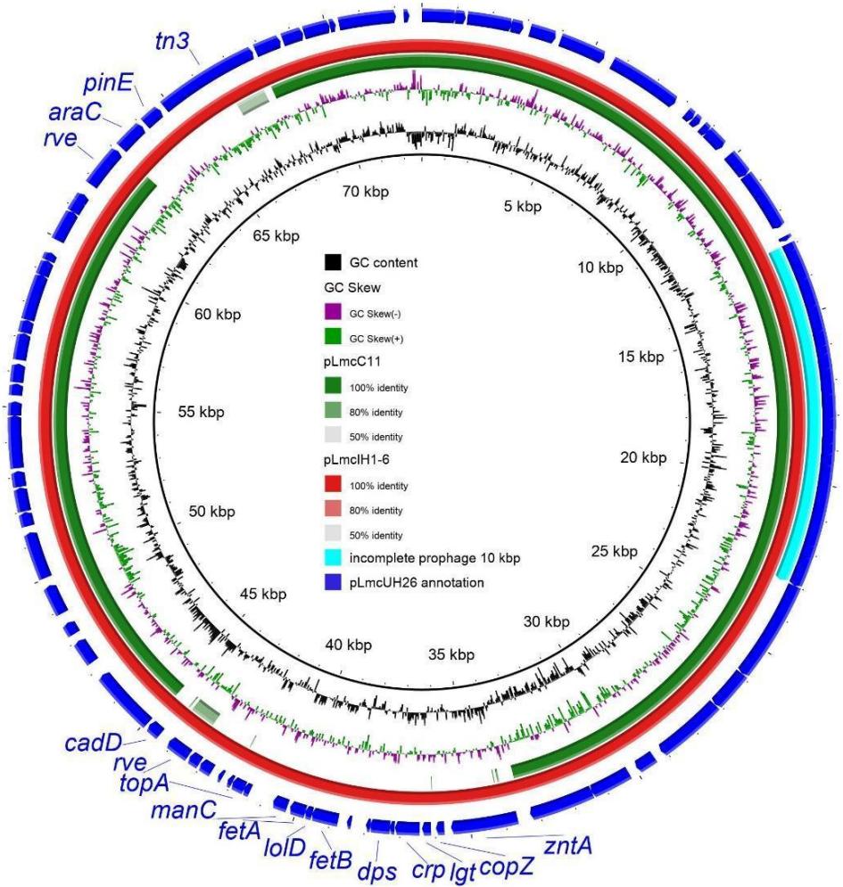 Figure 6: The comparison of L. monositogenes and L. welshimeri plasmids by using BLAST Ring Image Generator (BRIG) with blast-2.7.1.
The blue ring—pLmcUH26 (L. monocytogenes GIMC2079: LmcUH26, ST425); the gene names highlighted in blue refer to the two regions of difference annotated by the RAST; the red ring—pLmcIH1-6 (L. monocytogenes GIMC2088:LmcIH1-6, ST20); and the green ring—pLmcC11 (L. welshimeri GIMC2049:LmcC11, ST2331). The position of the incomplete prophage is shown by an aqua-colored segment.
In our WGS research, 5 isolates from 35 of the PLI and the PLII bore plasmids: four isolates were ST425, CC90, PLII and one isolate was ST20, CC20, PLII. These plasmids (accession numbers MZ869810, MZ869811, OM867528 - OM867530) were 72,763 bp in length (Table 4). We designate them here as pLm. The plasmids contained the incomplete prophage 10 Kb, a part of the Listeria phage A006 (Figure 6). Sixteen ORFs of prophage included the post-segregational killing system, encoded by a co-transcriptional gene pair of Phd antitoxin (prevent host death protein) and Doc (death on curing protein) toxin, which is involved in maintaining the plasmid in host cells [47, 48]. This set of Listeria adaptive genes is known also as the mazEF-like toxin-antitoxin system, which modulates the growth in the response to specific stress factors [49]. Another pair of genes encoding CadA and CadC proteins constituted the cadmium efflux system. One more interesting gene encoded the CRISPR-associated protein Cas5.
The same incomplete prophage we revealed earlier in a plasmid 57,530 bp in length (Accession Number MZ869809) was from the L. welshimeri strain of ST2331 isolated in the meat processing plant (Table 4). We will designate it here as pLw.
The alignment percentage (AP) of the six plasmids was more than 89% and the average nucleotide identity (ANI) was more than 99%. The GC content of identified plasmids was 36%. The number of coding sequences (CDS) varied from 67 in pLw to 86/88 in pLm.
The main region of differences between pLm and pLw was a pathogenicity island including 17 ORFs (Figure 6). First of all, these ORFs encoded proteins for the copper homeostasis, which is particularly acute during infections (copper-translocating ATPase and copper chaperone). The levels of copper fluctuate in the host from high in the gallbladder to low in the liver and spleen [50]. Thus, the plasmid coding the copper homeostasis proteins helps in adapting to these different niches, which is important for a successful infection.
The second group of genes encode the proteins contributing to the regulation of the iron concentration: the complex of the ABC transporters and the permease responsible for the iron export, and the Dps protein (DNA protection during starvation). The Dps, a ferritin-like protein, forms dodecamers that can bind approximately 500 iron molecules . Moreover, Dps has a dual role in protecting the cells against oxidative stress, either by binding directly to DNA, or by sequestering iron, thus avoiding oxidative damage , because in the presence of iron, superoxide and hydrogen peroxide can be transformed into the highly reactive hydroxyl radical .
One more gene of this region encoded deoxythymidine diphosphates (dTDP)-4-dehydro rhamnose 3,5-epimerase, or cupin RmlC. This protein's synthesis is the precursor of L-rhamnose, which is used by the . monocytogenes serogroup for the modification of the peptidoglycan-attached teichoic acids (wall teichoic acids). Such modification is significant for . monocytogenes virulence [53].
The smaller region deleted in pLmcC11 is represented by three genes encoding transposase, site-specific DNA recombinase and AraC family transcriptional regulator.
The comparison of pLm with the Listeria plasmids deposited in GenBank showed high similarity to the pLIS19 (MZ089999), pLIS38 (MZ127847), and pLIS43 (MZ147615) plasmids of L. innocua strains, isolated from food and city environments in 2003-2017 in Poland (Table 4). The ANI and AP among these plasmids are more than 99%.
The pLw showed more than 99% similarity to the plasmids of L. innocua strains isolated from food and to the plasmids of L. monocytogenes strains isolated from food and from clinical sources in different years and different geographical locations (Table 4). The L. monocytogenes strains belonged to ST3 of PLI and to different STs of PLII. The oldest clinical strain was isolated in 1967 in Germany . The 13 plasmid replicons mentioned above (Table 4) were included in the analysis. The neighbor-joining tree (Figure 7) was built on the basis of the ANI of whole-genome alignment and revealed two closely related groups of plasmid replicons. Group I, which is located at the top of the figure, includes smaller plasmids, which are distributed according to the source of isolation. In group II, in the lower part of the figure, there are larger plasmids, and the most different among them is the plasmid of the strain isolated from the environment.
Table 4: GenBank plasmids with high similarity to those found in the study.
| Size | Plasmid name | GenBank ID | Strain | Species | ST | CC | Phylogenetic lineage | Isolation date | Source | Country | Reference |
| 72763 bp | pLmcUH26 | MZ869810 | GIMC2079:LmcUH26 | L.monocytogenes | 425 | 90 | II | 2021 | Homosapiens (blood) | Russia | this research |
| pLmcUH27 | MZ869811 | GIMC2080:LmcUH27 | L.monocytogenes | 425 | 90 | II | 2021 | Homosapiens (blood) | Russia | this research | |
| pLmcH67_6 | OM867530 | GIMC2090:LmcH67_6 | L.monocytogenes | 425 | 90 | II | 2021 | Homosapiens (cerebrospinal fluid) | Russia | this research | |
| pLmcH67_8 | OM867529 | GIMC2095:LmcH67_8 | L.monocytogenes | 425 | 90 | II | 2021 | Homosapiens (blood) | Russia | this research | |
| pLmcIH1-6 | OM867528 | GIMC2088:LmcIH1-6 | L.monocytogenes | 20 | 20 | II | 2021 | Homosapiens (cerebrospinal fluid) | Russia | this research | |
| pLIS19 | MZo89999 | 297/05 | L.innocua | nd | nd | L.innocua | 2005 | food (ice cream) | Poland | [54] | |
| pLIS38 | MZ127847 | 10/03 | L.innocua | nd | nd | L.innocua | 2003 | food (food contact surface swab) | Poland | [54] | |
| pLIS43 | MZ147615 | Sr108 | L.innocua | nd | nd | L.innocua | 2017 | environment (city) | Poland | [54] | |
| 57530 bp | pLmcC11 | MZ869809 | GIMC2049:LmcC11 | L.welshimeri | 2331 | 2331 | OtherListeriaspecies | 2020 | meat processing plant | Russia | this research |
| pCFSAN044836 | CP045744 | CFSAN044836 | L.innocua | 448 | 448 | L.innocua | 2005 | food (minced meat) | Italy | [55] | |
| pPIR00545 | CP025561 | PIR00545 | L.monocytogenes | 199 | 199 | II | - | food | Switzerland | [56] | |
| pR2-502 | CP006595.1 | R2-502 | L.monocytogenes | 3 | 3 | I | 1994 | food | USA | [57] | |
| pLM1-2bUG1 | FR667692.1 | SLCC2755 | L.monocytogenes | 66 | 3 | I | 1967 | Homosapiens | Germany | [58] | |
| pl2015TE24968 | CP015985.1 | 2015TE24968 | L.monocytogenes | 7 | 7 | II | 2015 | Homosapiens (blood) | Italy | [59] | |
| pAUSMDU00000224_01 | CP045973.1 | AUSMDU00000224 | L.monocytogenes | 122 | 9 | II | 2009 | Homosapiens | Australia | Unpublished |
Listeria plasmids have been classified into two groups based on their replication protein RepA sequences . According to this classification, all of the analyzed plasmids belong to the repA_G1-Replication system group, which is the most common group . All of the analyzed plasmids also carried two loci involved in the active partitioning of plasmid molecules (PAR_1 and additional parA), and the loci coding Y-family polymerase components (UvrX and YolD) representing the POL_1 group. A whole conjugative transfer module is absent in the studied plasmids, except for the relaxase component of the mobL_1a type.
Of the functional genes that promote adaptation and are present in all plasmids, mention should be made of a region encoding a glycine betaine ABC transport system substrate-binding protein and an oxidoreductase (NADH peroxidase), and the gene for the ATP-dependent protease ClpL. The first group of the genes may play a role in responses to high osmolarity and oxidative stress, and was shown to be upregulated under high-salt conditions . The ATP-dependent protease ClpL was shown to be involved in the increased heat resistance of L. monocytogenes strains , and was also found to be upregulated in response to low pH .
No other additional genes, such as arsenic resistance loci, fluoride efflux transporters, or antibiotics resistance loci, or virulence factors, have been found in plasmids.
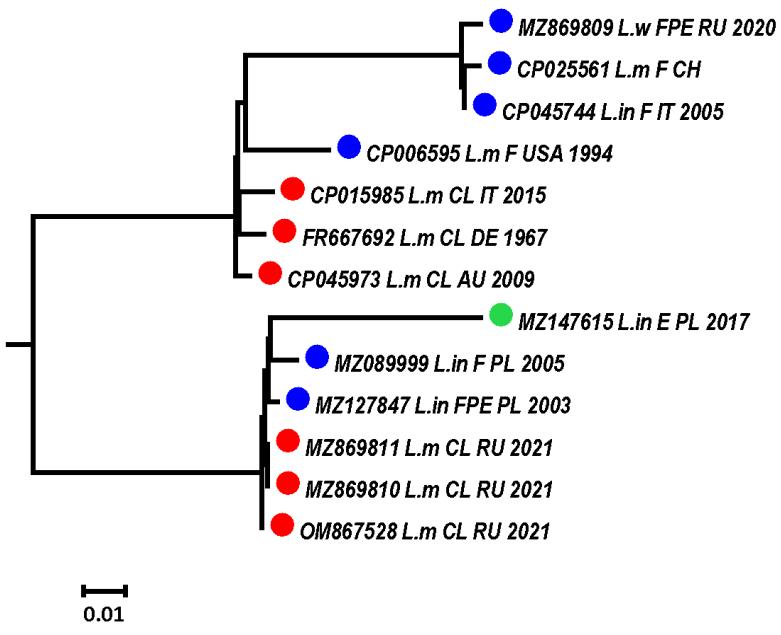 Figure 7: The neighbor-joining tree of the Listeria plasmid similarity based on the average nucleotide identity (ANI).
The Listeria species are labeled: L.m—L. monocytogenes, L.w—L. welshimeri, L.in—L. innocua. The countries of the isolates' origin are labeled: AU—Australia, CH—Switzerland, DE—Germany, IT—Italy, PL—Poland, RU—Russia, USA—USA. The sources of isolates are indicated as F—food, FPE—food production environment, CL—clinical. The color of the circles means: red—clinical isolate, blue—food isolate, green—environment isolate.
7. Core genome comparison
During the monitoring, especially in the pandemic period, we observed repeated genotypes among clinical isolates. To determine the epidemic relationship between isolates of the same genotype, we used the approach of cgMLST genotyping, developed by Moura et al. and having the discriminatory power required for epidemiological surveillance . The criteria for assignment to an epidemic outbreak was seven or fewer allelic mismatches between cgMLST profiles, including 1748 loci.
{"image_source":{"path":"images/52558b4fefa69a279417f11e123d81f9e424b3a37dbf9567a42792b0e3268353.jpg"},"content":"","chart_caption":[],"chart_footnote":[]} {"image_source":{"path":"images/b7931527b6189a990e972bf207f8a840f89b893e7311eb02e53ecb19e5b50119.jpg"},"content":"","chart_caption":[{"type":"text","content":"Differensis in cgMLST loci, isolates of PL II"},{"type":"text","content":"Figure 8: A number of allelic mismatches between cgMLST profiles for isolates of the same ST (sequence type). a)—phylogenetic lineage I (PLI), b)—phylogenetic lineage II (PLII)."}],"chart_footnote":[]}
The results of the comparison are shown in Figure 8. Among the isolates of PLI (Figure 8a), complete coincidence of the profiles was observed for isolates of ST219 attributed to perinatal listeriosis. Both strains were isolated in March 2021. One strain was isolated from a newborn, and a second strain was from a woman in the seventh week of pregnancy. The second group of isolates of PLI with minimal differences in cgMLST profiles belongs to ST4. The isolate from July 2020 (fatal) differs from the isolate from October 2020 (newborn) and the isolate from March 2021 (fatal) in three loci. The last two isolates have the same cgMLST profiles. The two isolates of ST6 (August 2019 and September 2020, both from newborns) have only two allelic mismatches, but the earlier isolate (March 2019, newborn) differs in 13-15 loci.
In PLII (Figure 8b), the isolates of ST8 (December 2021 (newborn) and January 2022 (meningitis, alive)), and ST21 (September 2021 (pregnant women) and December 2021 (septicemia, fatal)) had the same cgMLST profiles. Attention is drawn to strains of ST425. The four isolates shown in the figure are divided into two pairs according to the places of hospitalization. The isolates from April 2021 (septicemia, fatal) and June 2021 (meningitis, alive) were isolated from COVID-19 patients in one hospital with an interval of one and a half months, and had identical cgMLST profiles. The same profile is seen in the isolate from September 2021 (meningitis, fatal) from the second pair of the patients, which were from another hospital. The second isolate from this pair, from August 2021 (meningitis, alive), has one difference in the cgMLST profile from the three previous isolates of ST425. Thus, the isolates of ST219, 4, and two isolates of ST6 from the PLI, and the isolates of ST21, 8 and 425 from PLII can be attributed to epidemic outbreaks.
However, the isolates of ST37, often revealed during the COVID-19 pandemic in the group with meningitis and sepsis, had more than seven mismatches in the cgMLST profile (Figure 8b). The presented isolates, from March 2020 (meningitis, fatal), July 2021 (meningitis), and July 2021 (meningitis, fatal), were from three different hospitals. The isolates from July 2021 had fewer distinct loci (23) compared to the isolate at the start of the pandemic, March 2020, (34 and 37). As such, the three patients had different sources of the infection.
Previously, we obtained similar results for the isolates of the autochthonous ST7, which also belongs to PLII and was the most common in the pre-COVID-19 period (Figure 2a1) [26]. We compared the following isolates of ST7: the clinical isolates from June 2020 and February 2019 and a food isolate from May 2018. L. monocytogenes "February 2019" caused premature birth and pneumonia in a newborn, and listeria "June 2020" caused the death of an elderly patient recovering from COVID-19. The clinical isolates differed in 20 loci of cgMLST profiles, and both had 23 allelic mismatches with the food isolate. As such, three isolates of ST7 had no epidemiological link, but were closely related. The most considerable common mutation of the three isolates was the nine-nucleotide deletion in the MFS transporter gene (lmo2172) sequence, which distinguished this group of isolates from other food isolates and from the clinical L. monocytogenes ST7 previously isolated in Russia [26].
8. Possible functional role of mutated genes
The comparison of ST7 clinical isolates revealed 20 distinct loci coding for the proteins required for the metabolism of bacteria parasitizing in the human body. First of all, there were two virulence factors: listeriolysin O (lmoo202) and hexose phosphate transporter (lmoo838). According to the classification of pathways and functional systems using categories of the Clusters of Orthologous Genes (COG) [61], 20 loci can be classified into 12 COG categories (Figure 9); note that a quarter were related to the category "Carbohydrate transport and metabolism".
COG categories {"image_source":{"path":"images/3b7b8dbb5da2fa4f9cb3a661ae9391a626b7c6becb29e13b90cbf9a77a45d351.jpg"},"content":"","chart_caption":[{"type":"text","content":"Figure 9: Distribution of genes differing in mismatches between the core genomes of L. monocytogenes ST7 according to the Clusters of Orthologous Genes (COG) category."}],"chart_footnote":[]}
Four genes belonging to this category were transporters of two types: secondary and ATP-dependent transporters, as follows from the analysis using the TransportDB 2.0 [62]. Secondary active transporters, lmoo831 and lmoo838, have malonate/malate and hexose phosphate as a substrate, respectively, and couple the transport of substrates against their concentration gradients with the transport of other solutes down their concentration gradients [63]. Two other transporters, lmo0980 and lmo2123 (ATP-dependent), belonged to the ABC family most represented (89.7%) among the transporters of L. monocytogenes (http://www.membranetransport.org/ (accessed on 24 July 2022)). If lmo2130 was responsible for the transport of maltose/maltodextrin, then lmo0980 was classified as a multidrug resistance transporter.
The fifth gene of this COG category was glucosamine-fructose-6-phosphate aminotransferase (Imo0727), an important point in the biosynthesis of amino sugar-containing macromolecules. It has been marked by Zhou et al. as the gene encoding the bile-induced protein of L. monocytogenes wild-type strain EGD .
It is interesting that in the COG category "Translation, ribosomal structure and biogenesis", there were two tRNA synthetases (RS), PheRS (lmo1222) and AlaRS (lmo1504), included in the Class IIc, showing distinctive quaternary features. These enzymes are hetero- and homo-tetramers . Moreover, Phe-RS is indispensable for growth in all organisms .
The third gene in this category is lmo2620, encoding 50S ribosomal protein L5. Prokaryotic 5S rRNA interacts with the ribosomal protein L5, forming 5S ribonucleoprotein particles, and enhances protein synthesis by the stabilization of the ribosome structure [66].
In the category "Replication, recombination and repair", the delta subunit of DNA polymerase III (lmo1481) is remarkable. It is present in 5–7-fold excess over the gamma complex, and probably performs the clamp unloading function, sparing the limited number of gamma complex and polymerase III molecules in the cell for other important functions such as processive DNA replication. Additionally, the delta subunit does not require ATP for its activity, so the delta-catalyzed unloading reaction cannot be quenched with hexokinase and glucose as described for the gamma complex and polymerase III [67].
The next gene, lmo1646 (exonuclease SbcD), is part of the heterotetrameric SbcCD complex, which cleaves DNA hairpin structures. SbcC might constantly check the replication fork for misfolded DNA and associate with SbcD only when DNA repair is necessary, so a small amount of protein SbcD is required for the complex activity .
In the category "Signal transduction mechanisms", two loci distinguish clinical strains of ST7. Response regulator LiaR (lmo1022) is a component of the LiaFSR system, including membrane-bound inhibitor protein LiaF, sensor histidine kinase and a cognate response regulator. Additionally, LiaR is the less abundant component of this system. The LiaFSR system strongly responds to the presence of a number of cell wall antibiotics, such as bacitracin, daptomycin, nisin, ramoplanin and vancomycin, and is weakly induced by other more unspecific stresses that interfere with envelope integrity, such as alkaline shock and secretion stress .
The arginine phosphatase (lmo2540) is the next important protein for bacteria stress response. It modulates the arginine phosphorylation states of regulators such as the stress response regulator CtsR and the global regulator MgrA, particularly in response to oxidative stress .
Thus, using the example of the most represented categories of COG, we have shown the importance of genes affected by mutations for the vital activity of L. monocytogenes ST7.
Two loci distinguishing isolates of ST6 (August 2019 and September 2020, both from newborns) also belonged to the category "Carbohydrate transport and metabolism": lmo1728—cellobiose phosphorylase, and lmo2761—beta-glucosidase. These enzymes catalyze anaerobic (phosphorolytic) and aerobic (hydrolytic) pathways of the cellobiose catabolism, respectively. The phosphorolytic pathway requires one less ATP for each molecule of cellobiose to be metabolized by glycolysis, so it performs better than the hydrolytic pathway in terms of product yield in stressful conditions .
Three loci distinguishing the isolates of ST4 "October 2020 (newborn)" and "March 2021 (fatal)" from the isolate "July 2020 (fatal)" belonged to the three COG categories: "Transcription" (Imo0520—NagC transcriptional regulator), "General function prediction only" (Imo2049—nucleotidyltransferase), and "Translation, ribosomal structure and biogenesis" (Imo2596—30S ribosomal protein S9). Consider the most impressive locus Imo0520. In E. coli, as demonstrated Bréchemier-Baey et al., NagC is the transcriptional repressor of the catabolism of the mucin-derived sugars N-acetylglucosamine, and the activator of the locus of enterocyte effacement, which is critical regulator for the colonization of the intestine by bacteria [72].
9. The genotypes of L. monocytogenes isolated from food
The search for the source of infection that caused the cases of invasive listeriosis prompted us to analyze L. monocytogenes isolated from food in the metropolis during the monitoring period (Table S3).
{"image_source":{"path":"images/9f738ea429bc19b8ace5b4f3344a6cbf3981077cdda8441881160f7587436e9f.jpg"},"content":"","chart_caption":[],"chart_footnote":[]} {"image_source":{"path":"images/f5f7f67a7e007272bf91ec9ba21ba6a6ec8d6afb159d6dd69d9009f02ee32f5c.jpg"},"content":"","chart_caption":[],"chart_footnote":[{"type":"text","content":"Food sources of L. monocytogenes"}]}
{"image_source":{"path":"images/937aedb65831c40a25f365f18b6e9351c72ba998fe4544f22febbf5ca300d1dd.jpg"},"content":"","chart_caption":[],"chart_footnote":[]} a)—before the COVID-19 pandemic, 2018-2019; b)—during the COVID-19 pandemic; and c—the food sources of the L. monocytogenes isolates. H—the Shannon diversity index; PLI—phylogenetic lineage I; ST—sequence type.
Figure 10: L. monocytogenes from food sources.
L. monocytogenes was most commonly isolated from fish, followed by chicken and meat (Figure 10c). All food isolates were subdivided into two groups according to the isolation data: before the pandemic and during the pandemic (Figure 10).
The proportion of genotypes that coincided in the two periods of observation, namely ST7, 9, 37, 121, and 155, was 69% and 68%, respectively. The differing genotypes were more diverse during the COVID-19 period (5ST vs. 3ST), which led to a slight increase in the Shannon diversity index from 2.8 to 3.1 (Figures 10a, 10b). The proportion of PLI increased insignificantly during the pandemic, and ST288 of PLI was replaced by ST3, which had not previously been found in Russia. As such, the genotypes of L. monocytogenes infecting foods did not change significantly during the COVID-19 pandemic. In the final list of the genotypes of the clinical isolates of the second stage of the COVID-19 pandemic, only ST8 and ST37 coincide with the list of the food isolates' genotypes.
III. DISCUSSION
Listeriosis is relatively rare, but is one of the deadliest food-borne infections. In Russia in 2020, clinical diagnosis of listeriosis was laboratory-confirmed in 42 patients, and six cases were fatal (https://rospotrebnadzor.ru/ (accessed on 24 July 2022)). In Moscow, 18 cases of listeriosis were registered in 2020, and 21 cases in 2021 (https://77.rospotrebnadzor.ru/ (accessed on 24 July 2022)). The analysis of food included 6883 samples subjected to laboratory control in the departments of Rospotrebnadzor in Moscow in 2020, and 0.6% of samples were positive for L. monocytogenes (https://77.rospotrebnadzor.ru/ (accessed on 24 July 2022)). However, persons with recognized underlying diseases such as liver disease, cancer, diabetes and COVID-19 in the case of listeriosis can be counted as having an underlying disease in the clinical report. In perinatal listeriosis, neonatal complications (respiratory failure and heart failure in newborns, cerebral ischemia, etc.) can also be registered as the main diagnosis. Therefore, cases of listeriosis may be underreported in statistics.
Our study included 50 clinical and 37 food L. monocytogenes isolates revealed from November 2018 to January 2022 in the metropolis. The obtained results clearly indicate a change in the spectrum of the Listeria genotypes causing invasive listeriosis during the COVID-19 pandemic.
L. monocytogenes ST7 (PLII) and ST6 (PLI), which previously typically prevailed among the clinical isolates in the metropolis (Figure 2a1), were changed by the variety of the PLI genotypes (ST1, ST4, ST194, ST219) and the PLII genotypes (ST8, ST20, ST21, ST37, ST391, ST425, ST451) (Figure 2a4). L. monocytogenes of ST8 and ST425 were previously found in food and in the environment, but during the pandemic, L. monocytogenes of these genotypes were isolated for the first time in cases of human listeriosis in Russia along with ST21, ST37, and ST391. In addition, we observed an increase in the number of fatal cases of invasive listeriosis in the group with meningitis and sepsis (43.8% vs. 81.2%), as well as a sharp decrease in age in this listeriosis group: from 59 to 33 years.
Almost all new genotypes of the clinical isolates revealed during the COVID-19 pandemic in the metropolis were previously described in the works of European researchers. The L. monocytogenes diversity which related to 1185 cattle abortion cases in Latvia during the period of 2013–2018 included 27 different ST overall [74]. Four of them, ST29, 37, 451 and 7, covered more than half of the L. monocytogenes isolates; ST8, 20, 21, 391, and 425 were also detected [74]. Kubicová et al. (Slovakia, 2010 – 2020) described ST451 as the most prevalent in the cohort (15.3%) as a whole, and especially in food (14.8%) and animal isolates (17.5%) [75]. In food, CC11-ST451, followed by CC7, CC14, and CC37, were the most prevalent CCs in the milk sector, and CC9 and CC8 were most prevalent in the meat sector [75]. In the Czech Republic, ST29, 37, and 451 were the most frequent genotypes of L. monocytogenes, isolated from vacuum-packed steak tartare in 2013–2016 [76]. In 2019, as non-human isolates, L. monocytogenes ST8, ST21, and ST451 were mentioned in the work of researchers from Austria [77]. They found ST21 to be more associated with vegetables, and ST451 with dairy products [77]. Finally, during the COVID-19 pandemic in Italy (September and October 2020), a nosocomial outbreak of listeriosis caused by an ST451 strain of L. monocytogenes was recorded [78]. This outbreak involved one immunocompromised patient and three cancer patients hospitalized in different units of the same hospital. The source of contamination was identified in the hospital kitchen, where a meat slicer used to prepare meals tested positive for the same ST of L. monocytogenes. This was the first report of an outbreak of listeriosis caused by ST451 in Italy [78].
In our study, the epidemic connections were shown for the PLI isolates (ST219, ST4, ST6), and also for new clinical isolates of ST21, 8 and 425 (PLII), which can be attributed to epidemic outbreaks because of similar cgMLST profiles (0–3 allelic mismatches of cgMLST). The strains of ST425 were isolated from four COVID-19 patients in two hospitals with an interval of 5.0 months. The sources of outbreaks have not been identified.
L. monocytogenes of all genotypes revealed in the clinical cases were analyzed for the presence of 42 VFs. All VFs but one (vip) were observed in all of the analyzed isolates. The vip involved in cell invasion [42] was absent in the isolates of ST7, 8, 21, and 37, which were associated with clinical cases (perinatal listeriosis, septicemia, meningitis), including fatal ones. We found confirmation of our results in the works of a number of researchers. The lack of the vip gene in FPE L. monocytogenes isolates of ST8 and ST37 was noted by Alvarez-Molina et al. from Spain [79]. In the research into the isolates obtained from cattle abortion cases by Šteingolde et al. (Latvia), the list of vip-missing genotypes was expanded and additionally included ST29, 120, 226, 391, 403, 573 (PL II) and even ST 2, 4, and 6 (PL I) [74]. Thus, the thesis about the obligatory presence of the vip gene in the genomes of isolates of the PLI and PLII [42, 43] needs to be revised.
In addition to the main 42 virulence factors, Maury et al. noted the following features of the PLI isolates with high clinical frequency (CC1, CC2, CC4, and CC6), which were named as hypervirulent: 1) the over-representation of genes involved in replication, recombination, and repair, which may indicate a higher exposure to genotoxic conditions; and 2) the over-representation of genes involved in cell wall and membrane biogenesis, which may reflect the selection of genes involved in interactions with the host [80]. In our investigation, the cases of perinatal listeriosis were associated with hypervirulent L. monocytogenes of ST6 during the pre-COVID-19 period. Listeria belonging to the additional genotypes of CC1 (ST1) and CC4 (ST4 and ST219) caused the perinatal listeriosis during the COVID-19 pandemic. According to Charlier et al., L. monocytogenes of CC1, CC2, CC4, are overrepresented in maternal–neonatal (MN) infections in France, with more than two-thirds of cases being due to one of these CCs. [81]. Among them, CC4 is the most associated with MN infections, with 20% of CC4 isolates being of MN origin [81].
The plasmids can become an additional source of virulence factors. The presence of plasmid replicons in the clinical L. monocytogenes isolates is not a frequent phenomenon . In our study, the plasmids were found in five isolates: four isolates of ST425 and one of ST20. All of them were new in clinical cases. More often, the discussion of Listeria plasmids takes place in the context of the ability of Listeria spp. to colonize various environments, especially the FPE, with a focus on the genes that provide resistance to sanitizers , heavy metals or elevated temperatures . However, according to Chmielowska et al., there is evidence that the presence of some cadAC resistance cassettes in Listeria can influence other phenotypic traits, such as virulence and biofilm formation, which suggests that these heavy metal resistance determinants may have additional functions . Other genes encoding the proteins contributing to copper homeostasis, the regulation of iron concentration, the modification of wall teichoic acids and even in-creased heat resistance may facilitate the increase in virulence and the spread and survival of Listeria in the human body. Thus, on the one hand, if, during the COVID-19 period, invasive listeriosis is caused by listeria of genotypes previously typical of food isolates, in addition to containing plasmids, then we can confirm that SARS-CoV-2 has a serious damaging effect on the defense system of the gastrointestinal tract. On the other hand, L. monocytogenes of ST425 (of one epidemic cluster, according to cgMLST analysis) caused disease not only in people in the older age category, but also in younger people (36 and 47 years old); therefore, isolates of this genotype can hardly be classified as hypovirulent, and their virulence factors, including those introduced by plasmids, are subject to further study.
IV. CONCLUSIONS
The COVID-19 pandemic has had a significant impact on the health of the human population, promoting bacterial infections in weakened organisms. Listeriosis, as one of the deadliest foodborne infections, completely changed the portrait of those L. monocytogenes genotypes that caused invasive listeriosis both in the perinatal group and in the group with meningitis and sepsis, according to monitoring in a metropolis with a population of 20.4 million. The rejuvenation of listeriosis and increase in mortality in the group with meningitis and sepsis, the emergence of new genotypes of PLII, previously classified as food and hypovirulent, and new genotypes of L. monocytogenes of PLI in the perinatal group indicate the need for the even more stringent control of food products for the high-risk groups regarding invasive listeriosis.
V. MATERIALS AND METHODS
5.1 Bacterial isolates
Fifty clinical L. monocytogenes isolates (19 isolates from the first time period: November 2018–October 2019; 31 isolates from the second time period: March 2020–January 2022) were obtained from microbiological laboratories of the Moscow laboratory diagnostic centers. All L. monocytogenes were isolated from the samples of the patients with invasive listeriosis: perinatal listeriosis–44%, meningitis and listeria septicemia–56%. Isolates were submitted in the Bacterial Isolate Genome Sequence Database for L. monocytogenes (BIGSdb-Lm) (https://bigsdb.pasteur.fr/listeria/ (accessed on 24 July 2022)), ID: 42973-42983, 45724-45730, 49373-49375, 75929, 75931, 75932, 76308-76311, 76389, 77384-77386, 78377, 78378, 78657-78660, 78714, 82478-82480, 82484-82486, 82489-82492, 82494 (Table S1).
The Listeria isolates from food (ID: 42984-42998, 45731, 49370-49372, 75930, 75933, 76312, 76385-76388, 78379, 78713, 78715, 78717, 82479, 82481-82483, 82487, 82488, 82493) and food processing environments (ID: 49188-49202, 49206) were used in the comparison groups (Table S2).
5.2 Methods
5.2.1 DNA isolation
For genotyping, listeria isolates were grown on BHI agar overnight at . One loopful of cells was suspended in of the lysis buffer (0.25% SDS and 0.05 M NaOH), warmed up for 15 min at , and of the bidistilled water was added. The resulting lysate was stored at temperature . For the whole-genome sequencing, listeria isolates grown on BHI agar overnight at were suspended in the BHI broth and grown overnight at the same temperature. The cells were centrifuged, and the washed pellet was used for DNA isolation according to the Monarch Kit instructions (New England Biolabs, Ipswich, Massachusetts, USA).
5.2.2 MultiLocus Sequence Typing (MLST) and Multi-Virulent-Locus Sequence Typing (MvLST)
For MLST, we used a combination of primers and , laboratory-designed ldh primers, and our modification of PCR and sequencing protocols . New alleles and new sequence types (ST) for MLST genes were controlled by the team of curators of the Institut Pasteur MLST system (Paris, France).
The MvLST scheme including four internalin genes (inlABCE) chosen earlier by was used with our modification . The alleles of the internalin genes were numbered in the order of the detection, submitted in GenBank and published in . For each L. monocytogenes isolate, the combination of four detected alleles (in order inlABCE) received an internalin profile (IP) number. New alleles and IP from this research are presented in Table 1.
5.2.3 Whole genome sequencing (WGS)
The genomes of the strains with ST6, ST7, ST451, and new ST for the Russian listeria population (ST4, 194, 219), and new ST for clinical listeria isolates (8, 21, 425), were sequenced on the Illumina platform. The Nextera DNA Flex Library Prep (Illumina, San Diego, CA, USA) protocol was used for the library's preparation. Sequencing was performed on MiSeq and NextSeq 500/550 (Illumina, San Diego, CA, USA).
5.2.4 Data analysis
CLC Genomic Workbench v.20.0.4 (QIAGEN, Germantown, MD, USA) and SPAdes v.3.13.0 (St. Petersburg genome assembler, Russia, URL: http://cab.spbu.ru/software/spades/ (accessed on 24 July 2022)) were used for genome assembling. CGView Server (http://stothard.afns.ualberta.ca/cgview_server/ (accessed on 24 July 2022)) was applied for the visualization of assembling results and for the genome comparison [87]. The software Rapid Annotations Subsystems Technology (RAST) and SEED were used for genome annotation [88, 89]. Prophage sequences were revealed with the help of PHASTER (PHAge Search Tool Enhanced Release, https://phaster.ca/ (accessed on 24 July 2022)) [90].
WGS data are available in GenBank: Bio Project PRJNA605697. Previously published ST7 genomes (GenBank Accession Numbers CP060433-CP060435) were used for the comparison. The plasmid sequences of the strains GIMC 2079:Lmc UH26, GIMC2080:Lmc UH27, GIMC2090:LmcH67_6,
GIMC2095:LmcH67_8 (ST425) and GIMC2088:LmcIH1-6 (ST20) were submitted to GenBank (Accession Numbers MZ869810, MZ869811, OM867528 - OM867530).
The core genome MLST scheme of 1748 loci (cgMLST) was used for L. monocytogenes genome characterization [91] on the basis of an open bioinformatics platform (https://bigsdb.pasteur.fr/listeria/(accessed on 24 July 2022)). Alleles which were not characterized by the computational tool due to their difference from the registered sequences were searched by comparing genomic data with alleles of the reference EGD-e strain using BLAST NCBI. The identified sequences of the corresponding alleles of the isolates of the same ST were compared by the number of differences with the closest match. New alleles were considered identical if there were the same substitutions.
The assignment of genes to different Clusters of Orthologous Genes (COG) categories was carried out with NCBI Blastp and the COG database/Conserved Protein Domain Family database .
The comparison of the plasmids pLmcIH1-6 and pLmcC11 with the reference pLm-cUH26 was performed by using BLAST Ring Image Generator (BRIG) with blast-2.7.1+ [92].
CLC Genomics Workbench v. 21.0.1 with the Whole Genome Alignment 21.0 plugin was used for plasmid alignment and tree building. The Whole Genome Alignment 21.0 plugin works by identifying seeds (short stretches of nucleotide sequence that are shared between multiple genomes but not present multiple times on the same genome) in the plasmid. This option determines the number of nucleotides required for a seed to be included in the later stages of the alignment. The minimum initial seed length was 15 bp. From the initial extended seed matches, a distance matrix between the input plasmid was calculated. This distance matrix was used for the subsequent pairwise processing, where the most similar plasmid sequences were processed first. Proceeding iteratively on the most similar pair of plasmids, the tool extended and merged seed matches to create longer alignment blocks. The minimum alignment block length was 100 bp.
The Create Average Nucleotide Identity Comparison tool was useful for obtaining a quantitative measure of the similarity between plasmids. This tool took whole-genome alignment as input, and for each pair of plasmids, the regions aligned between the two plasmids were identified. The toll was launched using the following parameters: the minimum similarity fraction—0.8 and the minimum length fraction—0.8. On the basis of Create Average Nucleotide Identity Comparison tool data, the neighbor-joining tree was constructed.
The analysis of virulence factors was performed using the Virulence Factors Database (VFDB, http://www.mgc.ac.cn/VFs/ (accessed on 24 July 2022)) and VF analyzer (http://www.mgc.ac.cn/cgi-bin/ VFs/v5/main.cgi?func=VFanalyzer/ (accessed on 24 July 2022)) [41].
The Shannon diversity index (H) was used to characterize L. monocytogenes diversity and was calculated by the formula:
where pi is the proportion of the number of isolates of genotype i (ni) relative to the total number of isolates (N): pi = ni/N.
List of abbreviations
ACE2 - angiotensin-converting enzyme 2
AIDS - acquired immunodeficiency syndrome
ANI - average nucleotide identity
AP - alignment percentage
BIGSdb-Lm - Bacterial Isolate Genome Sequence Database for L. monocytogenes
CC - Clonal Complex
CDS - coding sequences cgMLST - core genome MLST
COG - Clusters of Orthologous Genes
Dps protein - DNA protection during starvation protein
FD - Federal District
GI - gastrointestinal tract
IP - internalin profile
LRR - leucine-rich repeat
MLST -. MultiLocus Sequence Typing
MvLST - Multi-Virulent-Locus Sequence Typing
ORF - open reading frame
PL - phylogenetic lineage
RAST - Rapid Annotations Subsystems Technology
RTE - ready-to eat
SCFAs - short chain fatty acids
SLV - single locus variant
SNV - single-nucleotide variant
ST - sequence types
VF - virulence factors
VOC - variants of concern
WGS - whole genome sequencing
Supplementary Materials: Table S1: The clinical L. monocytogenes isolates; Table S2: Virulence factors revealed in L. monocytogenes genomes by the VF analyzer; Table S3: The food L. monocytogenes isolates.
Declarations
-
Ethics approval and consent to participate. The authors of the study conducted only with microorganisms provided by the clinical laboratories. Consent to the study in the clinic was issued by patients during hospitalization.
-
Consent for publication. The publication of the research data was approved by the Biomedical Ethics Committee at the Gamaleya National Research Center for Epidemiology and Microbiology, Ministry of Health of the Russian Federation.
-
Availability of data and materials. Data of the MLST are available in the Bacterial Isolate Genome Sequence Database for L. monocytogenes (BIGSdb-Lm) (https://bigsdb.pasteur.fr/listeria/) under the IDs indicated in the methods section. WGS data are available in GenBank: Bio Project PRJNA605697, GenBank Accession Numbers: CP060433- CP060435, MZ869810, MZ869811, OM867528 - OM867530, MZ869809
-
Competing interests. There are no competing interests to declare.
-
Funding. This research received no external funding.
-
Authors' contributions. Conceptualization, O.L.V., E.A.K., O.A.G., A.R.M. and I.S.T.; methodology, O.L.V., N.N.R., M.S.K. and E.I.A.; software, N.N.R., M.S.K. and E.I.A; validation, O.L.V., N.N.R., M.S.K. and E.I.A.; formal analysis, A.V.K., A.N.T.; investigation, O.L.V., N.N.R., M.S.K., E.I.A., A.V.K., A.N.T. and T.I.K.; resources, O.L.V., E.A.K., O.A.G., A.R.M. and I.S.T.; data curation, M.S.K., N.N.R., and O.L.V.; writing—original draft preparation, O.L.V., N.N.R., M.S.K.
and E.I.A.; writing—review and editing, O.L.V., N.N.R., M.S.K. and E.I.A.; visualization, M.S.K., N.N.R., and O.L.V.; supervision, O.L.V.; project administration, I.S.T.; funding acquisition, O.L.V., E.A.K., O.A.G., A.R.M. All authors have read and agreed to the published version of the manuscript.
- Acknowledgements. The authors are grateful to the laboratory assistants of the microbiological laboratories of the Moscow laboratory diagnostic centers for providing L. monocytogenes isolates for the study: Burmistrova E.N., Dudnikova S.V., Gambaryan K.U., Mammadova A.M., Nechaeva S.A., Orlova O.E., Pokidysheva A.Y., Prokopaeva M.A., Pronina T.V.
Conflict of Interest
The authors declare no conflict of interest.
Ethical Approval
Not applicable
Data Availability
The datasets used in this study are openly available at [repository link] and the source code is available on GitHub at [GitHub link].
Funding
This work did not receive any external funding.
References
Cite this article
Related Research



Special Issue
Launch a focused special issue to highlight research, emerging trends, and expert insights in your academic field.
