



IntelliPaper
Abstract
Introduction: The kidney is one of the most frequently injured organs during blunt abdominal trauma, and the probability increases significantly if there is any renal pathology, such as hydronephrosis. The ureter Injury, which is extremely uncommon in blunt trauma, also increases its incidence in the presence of previous renal pathologies. Case report: Patient J.R.A, male, 30 years old, brought to the HMMSJP hemodynamically stable by SIATE after suffering a automobile x truck accident as a driver and the vehicle overturning. A contrast computed tomography (CT) scan of the abdomen, chest and skull CT scan showed a large retroperitoneal collection adjacent to the left kidney, with pelvis distension and a positive “claw sign”. It was concluded that the patient had grade V renal injury, and exploratory laparotomy was indicated. During surgery, a total lesion of the ureter was visualized approximately 10 cm from the ureteropelvic junction (UPJ) with subsequent total left nephrectomy and distal ureter ligation. Discussion: in this case, the patient's kidney was previously hydronephrotic, due to UPJ stenosis, being more prone to serious injuries in the trauma. There was also ureter injury, which is extremely uncommon in blunt trauma and should be treated as early as possible. Kidney injury should always be suspected in patients with a history of direct impact to the abdomen, with consequent CT scan. Patient management aims at hemodynamic stabilization, preservation of renal tissue and prevention of complications, which can be non-operative, if the injury is low-grade and hemodynamically stable, or surgical, if the injury is high-grade or hemodynamically unstable. Conclusion: Blunt kidney injury is more common in patients with previous renal alterations, while ureteral injuries are mostly due to iatrogenic events during surgery or penetrating trauma, being uncommon in blunt trauma. The correlation between anamnesis, physical examination and imaging tests is essential for the appropriate and individualized management of the patient.
Explore Digital Article Text
I. INTRODUCTION
Injuries caused by traffic accidents are responsible for approximately 1.35 million of deaths worldwide and 50 million of non-fatal injuries, most of which the people involved ends up beeing incapacitated. In Brazil, trauma is the third leading cause of overall mortality, with injuries occurring in traffic accounting for 40,000 deaths and more than 150,000 victims being injured with a high degree of severity annually. The incidence is more prevalent in males and young adults, mainly between 15 and 44 years of age.
The kidney is the organ of the genitourinary tract most frequently injured during trauma, which affects up to 10% of patients hospitalized for abdominal trauma. Renal trauma mainly affects men, in 72-93% of cases, it is more frequent in the younger population, between 31 and 38 years old, and pathological polycystic kidneys, with tumors or with hydronephrosis secondary to stenosis of the ureteropelvic junction are more prone to trauma, even in lower energy impacts.
Kidney trauma can occur through three mechanisms: blunt trauma, penetrating trauma and high-speed deceleration. Blunt trauma is responsible for 71-95% of kidney injuries, the cause of which comes mainly from car accidents and falls. Most kidney injuries are mild, while severe trauma is more prevalent in patients with penetrating trauma than in those who have suffered blunt trauma (27-68% vs. 4-25%).
The most used classification of renal trauma is the one proposed by the American Association for the Surgery of Trauma (AAST), revised in 2011, which grades renal injuries from I to V with progressive severity (Table 1).
Table 1: Classification of the American Association for the Surgery of Trauma, 2011 Source: Table adapted from the 2011 American Association for the Surgery of Trauma Classification
| Graduation of Kidney Injury | Description of Injury |
| GRADE I | Subcapsular hematoma, non-expanding, without laceration in the parenchyma, and there may be microscopic or macroscopic hematuria, but without changes in urine tests. |
| GRADE II | Parenchymal laceration < 1 cm in depth, without urinary extravasation, and with non-expanding perirenal hematoma. |
| GRADE III | Laceration > 1 cm deep, without collecting system damage and without urinary extravasation. |
| GRADE IV | Laceration in the cortex, medulla and collecting system, possibly with renal artery or vein injury, with contained hemorrhage. |
| GRADE V | Kidney is completely fragmented (shattered) and there is avulsion of the renal hilum, which cuts off the organ's blood supply. |
Injury to the ureter occurs in less than 1% of traumatic genitourinary injuries due to external causes, and in most cases it is caused by a gunshot wound. Traumatic ureteral injury is usually secondary to iatrogenesis during surgery, with external causes accounting for only 20% of all injuries to these organs. It is common to have an association with lesions in other organs such as the small intestine, colon and kidney. Blunt ureteral injury is extremely uncommon and is more frequent in children with congenital anomalies of the genitourinary tract, such as UPJ stenosis and secondary hydronephrosis. Definitive treatment varies according to the topography and extension of the lesion; when the diagnosis is made intraoperatively, immediate surgical correction is the most appropriate approach. The most frequent complication is prolonged urinary fistula, which occurs in up to 25% of cases, and may present as a urinoma, abscess, or peritonitis.
Table 2: Classification of Ureteral Lesions, proposed by the Brazilian Society of Urology (SBU) Source: table adapted from the book "Fundamental Urology" by SBU, 2010
| Graduation of Ureteral Injury | Description of the Injury |
| GRADE I | Bruise; contusion or hematoma without devascularization. |
| GRADE II | Laceration; < 50% transection |
| GRADE III | Laceration; > 50% transection |
| GRADE IV | Laceration; complete transection with devascularization < 2cm |
| GRADE V | Laceration; avulsion with > 2cm of devascularization |
II. CASE REPORT
J. R. A., 30 years old, male, 1.85 m, 92 kg, is brought by SIATE after suffering an accident with a car x truck as a driver, with the car overturning. The patient was brought in on a rigid board, with a cervical collar, his airways were patent and there was no pain on palpation of the cervical spine. Upon examination of the chest, expansion was preserved, there was no crackling or pain on palpation, and breath sounds were present bilaterally. Flaccid abdomen, diffusely painful on palpation, without signs of peritoneal irritation, with present bowel sounds and stable pelvis. The pupils were isochoric and isophotoreactive and the Glasgow Coma Scale was 15. At the time of hospital care, the patient was conscious and oriented in time and space, without neurological deficits, hemodynamically stable, reporting left abdominal pain. He denied traumatic brain injury, headache, nausea, vomiting and amnesia.
The victim denied comorbidities and use of continuous medication and was unaware of previous kidney pathology. He also denied tobacco use but reported moderate alcohol consumption (6 cans per weekend). There was no family history of neoplasms or organ malformations.
Computed tomography (CT) scans of the skull, chest and abdomen were requested. According to the high-energy trauma protocol established by the service, tomography is available 24 hours a day and is readily accessible, in addition, the patient was hemodynamically stable and needed imaging tests to complement the evaluation, opting for CT instead of FAST (Focused Assessment with Sonography for Trauma).
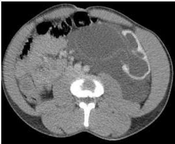
The computed tomography report of the abdomen (Figure 1) showed a large retroperitoneal collection adjacent to the left kidney, showing pelvis distention and the "claw sign". He also considered that the possibility of a large adjacent urinoma could be a differential diagnosis, suggesting further investigation. The spleen had normal dimensions, smooth contours and homogeneous attenuation. The exam also showed a small ascites, and there were no other significant alterations, including in the liver, bile ducts, pancreas, adrenals, retroperitoneal lymph nodes or in retroperitoneal vessels, with those cited being found in anatomical aspect and normal dimensions. Chest and skull tomography showed no alterations. From the data found in the anamnesis, physical examination and CT, it was concluded that it was a renal lesion grade V. It wasn't visualized ureteral lesion in the imaging exams due to massive leakage of urine and total distortion of the renal anatomy.
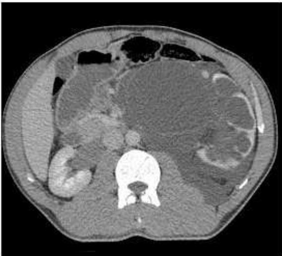
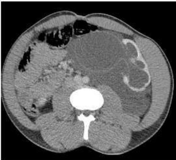
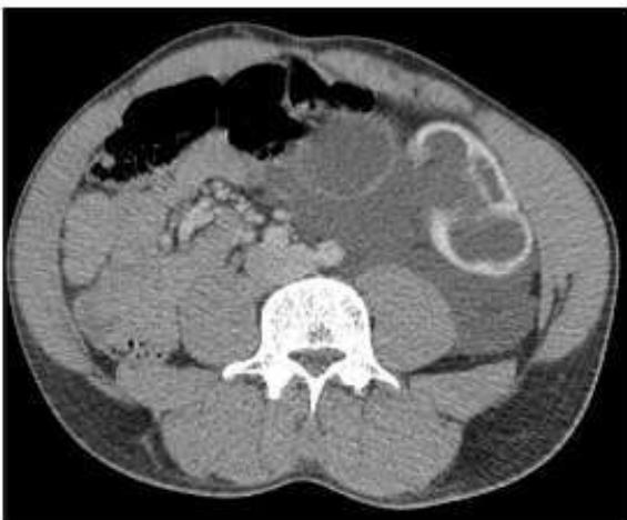
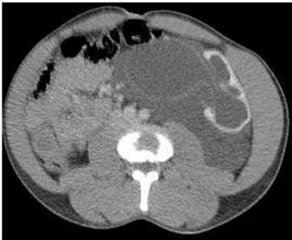 Figure 1: Computed Tomography of the Upper Abdomen
Copyright source, 2022
Therefore, the patient was referred and underwent exploratory laparotomy, left nephrectomy and distal ureteral ligation. During the surgical procedure, no intraperitoneal free fluid or changes in hollow viscera were found.
However, when exploring the left retroperitoneal region, a large amount of urine was drained and a hyperdistended renal capsule was found, in addition to the kidney with a grade V lesion, characterized by complete laceration with avulsion of the hilum and devascularization. In addition, the ureter was patent in the renal hilum, with a complete section 10 cm from the ureteropelvic junction, with a grade V urethral lesion, and a distal ligation was performed. No other injuries were found. The anatomical specimen removed from the patient during surgery is shown in Figure 2.
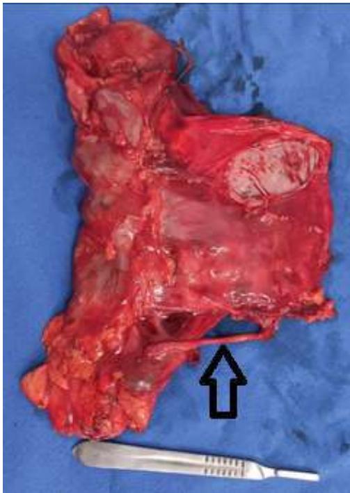 Copyright source, 2022. Arrow: left ureter
Figure 2: Anatomical Part
The product of the nephrectomy was sent for anatomopathological study, whose report showed signs of rupture of the renal tissue, with ischemic changes and hydronephrosis, in addition to the absence of malignancy, while in the ureter, focal stenosis and complete section of this were observed, suggesting stenosis of the UPJ (junction pyelic ureter).
III. DISCUSSION
In the case observed, the patient's kidney was previously hydronephrotic due to UPJ stenosis, despite not having a previous diagnosis, a fact that increases the risk of more serious injuries and the need for nephrectomy in blunt trauma. However, despite the spleen being the most affected organ in this type of trauma, the patient did not have splenic injury. However, there was ureter trauma, which is extremely uncommon in blunt trauma, whereas ureter injury is usually secondary to iatrogenesis in surgical procedures or due to penetrating trauma, being the most common blunt injury in children with congenital anomalies of the urinary tract, such as UPJ stenosis, which causes hydronephrosis. Ureteral trauma, when identified during surgery, should be treated as early as possible by surgical correction.
Kidney trauma should be suspected whenever there is a history of an event with fast deceleration or direct impact on the flank region, back, lower chest and upper abdomen. In these cases, the physical examination may reveal bruises on the flank or upper abdomen, signs of peritoneal irritation, a palpable mass, ecchymosis or abrasions, and rib fractures. In this context, CT with intravenous contrast is the imaging method of choice for hemodynamically stable patients who are victims of both penetrating trauma and blunt trauma, as it allows the definition of the location and severity of the injury, as well as allows the visualization of associated traumas.
FAST, on the other hand, is useful to demonstrate the presence of free fluid in the cavity, but it is inferior to CT in terms of resolution and ability to define renal trauma.
The management of the patient victim of renal trauma has as priority the control of blood loss, the preservation of the renal tissue and the prevention of complications. While in the past it was thought that the best way to control bleeding and prevent nephrectomies was through surgery, in recent decades the management of renal trauma has evolved towards prioritizing non-operative approaches. A recent meta-analysis has shown that the management of non-operative treatment (NOT) is the treatment of choice for low-grade renal trauma, and it should also be considered the first-line treatment of high-grade blunt trauma and penetrating trauma, as it is associated with lower mortality rates, shorter hospital stay hospitalization and similar morbidity rates. Absolute indications for surgery involve: hemodynamic instability and unresponsiveness to aggressive resuscitation due to renal hemorrhage, grade V vascular injury, and expanding or pulsating perirenal hematoma found during laparotomy performed due to associated injuries to other organs. Relative indications for surgery include, among others: large urine extravasation (spontaneous resolution in most cases), presence of non-viable renal tissue and arterial thrombosis installed for more than 4 hours.
IV. CONCLUSION
It was possible to conclude that patients with previous renal alterations are more susceptible to more severe injuries, even in blunt trauma, where the most affected organ would be the spleen.
Injury to the ureter is extremely uncommon in blunt trauma, the main cause of which is iatrogenic. The case described above is atypical because the patient had left kidney injury, which is anatomically close to the spleen, but did not present injury to this organ, but a complete section of the ureter. For the definitive diagnosis, the correlation between data from the anamnesis, physical examination, imaging tests, intraoperative findings and the result of the anatomopathological analysis was essential.
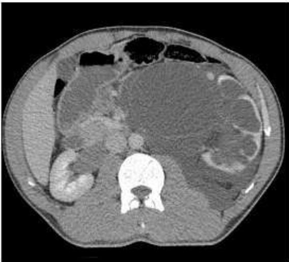
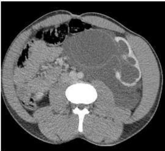
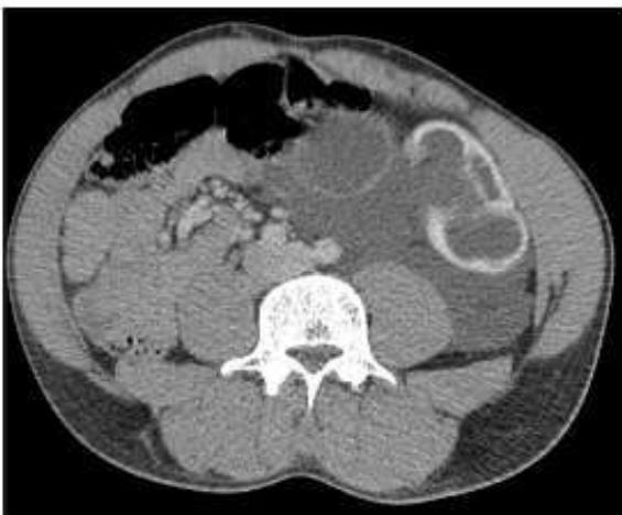 Figure 1: Computed Tomography of the Upper Abdomen
Conflict of Interest
The authors declare no conflict of interest.
Ethical Approval
Not applicable
Data Availability
The datasets used in this study are openly available at [repository link] and the source code is available on GitHub at [GitHub link].
Funding
This work did not receive any external funding.
References
Cite this article
Related Research




Special Issue
Launch a focused special issue to highlight research, emerging trends, and expert insights in your academic field.
