



IntelliPaper
Abstract
Peritoneal mesothelioma (PeM) represents around one fifth to one third of all mesothelioma. The incidence of PeM is approximately one per 1,000,000. Giving the fact of non-specificity of the symptoms, the diagnoses is usually delay. Multimodality treatment with adjuvant chemotherapy and cytoreductive surgery (CRS) and hyper thermic intraperitoneal chemotherapy (HIPEC) improve the long term survival. Herein, this is a presentation of patient diagnosed with peritoneal mesothelioma with a review of literature.
Explore Digital Article Text
I. BACKGROUND
Malignant mesothelioma is a rare but fatal tumor that primarily arises from the pleura but to a lesser extent from the peritoneum (1). Peritoneal mesothelioma (PeM) represents around one-fifth to one-third of all mesothelioma (2). The incidence of PeM is approximately one per 1,000,000 (2,3). Similar to all types of malignant mesothelioma, PeM has an attributable risk with exposure to asbestos (4). The majority of patients with PeM present with abdominal distention with or without pain, weight loss, and some presents with ascites (2,3). Given the fact of non-specificity of the symptoms, the diagnosis is usually delayed (3). The survival rate of PeM was around 5 to 12 months, but since the introduction of cytoreductive surgery (CRS) and hyperthermic intraperitoneal chemotherapy (HIPEC), long-term survival can be achieved (1,3).
II. CASE PRESENTATION
A 44 year old female with no significant past medical history presented with four weeks history of progressive abdominal distention and pain. This was not associated with nausea or vomiting, and no history of fever. She had never smoked and had no known previous exposure to asbestos.
The rest of the systemic review was unremarkable. On clinical examination, she was hemodynamically stable, had positive ascites with no palpable mass and no pedal edema but normal cardiovascular and respiratory system examinations. Laboratory investigation, including complete blood count (CBC), urea and electrolytes, liver function tests (LFTs), hepatitis, inflammatory markers, and viral and autoimmune screens, were all normal. She underwent a CT abdomen and pelvis, which showed massive ascites and omental nodular thickening with multiple scattered lymph nodes (Image 1A and 1B). Also, there were few hypodense liver lesions suggestive of hepatic cysts, and a large heterogenic enhancing mass in continues with the uterus, most likely representing subserosal leiomyoma. Diagnostic laparoscopy with an omental biopsy was performed. The histopathological examination showed epithelioid peritoneal mesothelioma. Ascetic fluids were taken for assessment; however, the sample was not enough. The case was discussed in the multidisciplinary meeting, and the conclusion was to treat the patient with systemic chemotherapy and repeat CT for assessment of operability. She received neoadjuvant chemotherapy with a total of four sessions of Pemetrexed with carboplatin.
She responded well and the ascites resolved with improved general well-being. Computed Tomography repeated, and showed resolution of the once noted ascites, small residual omental peritoneal soft tissue nodularity and stable previously seen liver cysts and uterine mass. After that, she underwent Exploratory laparotomy + cytoreductive surgery (CRS) and hyperthermic intraperitoneal chemotherapy (HIPEC). The intraoperative finding was no peritoneal deposits, peritoneal cancer index (PCI) of 12, multiple small deposits over the small bowel (Image 2), small liver cyst, and uterine fibroid. The histopathological examination of the omentum and soft tissue of the small bowel showed involvement by malignant mesothelioma, but a decrease in cellularity in comparison to the initial biopsy consisting of the chemotherapy response.
Then she had regular follow up with surgery and oncology departments, where she received adjuvant chemotherapy with the same neoadjuvant regimen where she had good response to the treatment in terms of clinical and radiological findings. A follow-up CT after surgery showed no evidence of intra-abdominal local recurrence or distant metastasis (Image 3)
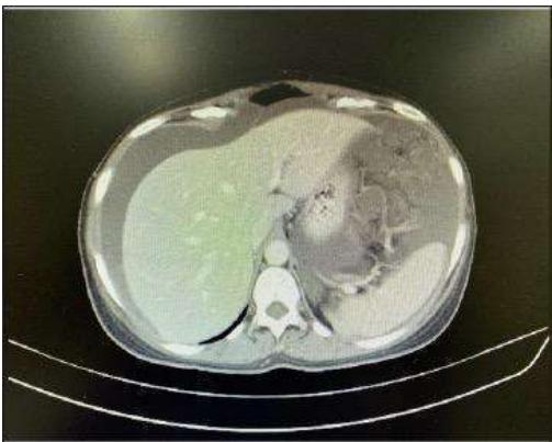
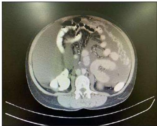 Image 1: Represents preoperative CT finding Image 1a: showing ascites with multiple liver cysts and Image 1b is showing the omental thinking with multiple lymphnodes
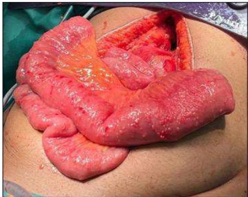 Image 2: Intra operative finding of multiple small deposits over over small bowel
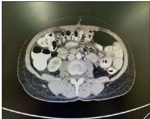 Image 3: Post operative CT finding of resolution of the ascites and no evidence of local recurrence
III. DISCUSSION
Mesothelioma is a tumor that originates from the mesothelial surface lining cell of the serous cavity, with the pleural being the most commonly involved serosal membrane (3). Peritoneal mesothelioma (PeM) represents less than 10% of malignant mesothelioma (4). In the old literature, cases of PeM were reported to have a poor prognosis with a median survival of less than one year whereas recent studies reported a median survival of 60-90 months (5,6).
The main risk factor for the development of malignant mesothelioma is a history of asbestos exposure. This risk is less prominent in the case of PeM compared to pleural mesothelioma (1). Other factors have also been implicated in the development of PeM, like radiation, peritonitis, and Simian virus 40 (SV40) (3,7). However, our patient had none of these risk factors.
The clinical presentation is usually not specific and poses a struggle and difficulty in the diagnoses. The most commonly reported symptoms and signs are; abdominal pain, abdominal distention, ascites, mass and tenderness (1,8). Imaging modalities like Ultrasonography and CT scan can provide essential information during the process of a mesothelioma diagnosis. However, to confirm the diagnosis of PeM, a histological examination is needed, which can be established through either open or laparoscopic surgery (2). The immunohistochemical analysis helps in increasing the diagnosis accuracy. Multiple Immunohistochemical markers were linked to PeM, including; calretinin, epithelial membrane antigen (EMA), Wilms' tumor-1 protein (WT-1), antimesothelial cell antibody-1, cytokeratin 5/6, thrombomodulin and mesothelin. Also, tumor markers like CA - 153 and CA - 125 have been helpful in the diagnosis and monitoring of this disease (9,10). However, no immunohistochemical examination was done for our patient where the histological examination was enough to confirm the diagnosis.
The histopathological examination of this patient revealed epithelioid peritoneal mesothelioma.
Different studies showed that epithelial PeM confers a more favorable prognosis and overall survival among different histological subtypes whereas the sarcomatoid and biphasic had the worst prognosis (13).
The staging peritoneal cancer index (PCI) is used as a measure of disease spread. Thirteen abdominal regions are used to score the PCI for tumor size and distribution. A lower PCI score is associated with a good prognosis (14). In the presented patient, the PCI was 12, which considered a low PCI score that might play a role in the excellent response to the treatment.
Over the last decades, the management of Peritoneal mesothelioma was evolving. With the multimodality treatment, including cytoreductive surgery (CRS) and hyperthermic intraperitoneal chemotherapy (HIPEC), the median survival rate increased up to 60-90 months (5,6). In the present case, neoadjuvant chemotherapy was used as systemic therapy with four sessions before the CRS+HIPEC surgery, which showed a dramatic reduction in the size of mesothelioma deposits that were seen in the initial laparoscopic surgery for a biopsy. The standard first-line systemic chemotherapy is Pemetrexed with cisplatin or carboplatin (12). The same standard chemotherapy was used on our patient. However, multiple studies showed that using systemic chemotherapy does not increase the long-term survival rate but its benefit in palliative treatment and consider an alternative therapy for inoperable cases (11).
IV. CONCLUSION
The case highlighted the importance of keeping peritoneal mesothelioma a differential diagnosis in a patient who presents with ascites, even if it is uncommon. A multidisciplinary approach with multimodality treatment including Cytoreductive surgery with HIPEC and chemotherapy is needed for better outcomes in patinet with epithelioid histology.
V. DISCLOSURE
The authors declare no conflicts of interest. The consent from the patient was taken before writing up the case.
ACKNOWLEDGEMENTS
Department of Surgery, Royal Hospital, Muscat, Sultanate of Oman.
Conflict of Interest
The authors declare no conflict of interest.
Ethical Approval
Not applicable
Data Availability
The datasets used in this study are openly available at [repository link] and the source code is available on GitHub at [GitHub link].
Funding
This work did not receive any external funding.
References
Cite this article
Related Research




Special Issue
Launch a focused special issue to highlight research, emerging trends, and expert insights in your academic field.
