




IntelliPaper
Abstract
Over time, neck lifting has undergone a notable evolution. Initially, techniques focused on treating the skin and subcutaneous cellular tissue (SCT), resulting in excessive tension on these structures, producing suboptimal results and unaesthetic scars.
Over time, techniques involving plication of the platysma muscle (PM) in its lateral and medial regions were introduced, aiming to reposition deep structures through compression and tension.
However, these approaches had limitations as the results tended to deteriorate quickly due to the stretching of the platysma fascicles.
Today, a more advanced approach has been adopted in neck treatment, shifting from focusing on tension on the platysma to reducing deep structures. The deep neck lifting technique addresses the reduction of superficial and deep fat compartments, as well as the digastric muscles and the submandibular gland, avoiding excessive tension on the PM sutures. This methodology allows for more stable and long-lasting results over time.
Explore Digital Article Text
I. INTRODUCTION
Over time, neck lifting has undergone a notable evolution. Initially, techniques focused on treating the skin and subcutaneous cellular tissue (SCT), resulting in excessive tension on these structures, producing suboptimal results and unaesthetic scars.
Over time, techniques involving plication of the platysma muscle (PM) in its lateral and medial regions were introduced, aiming to reposition deep structures through compression and tension. However, these approaches had limitations as the results tended to deteriorate quickly due to the stretching of the platysma fascicles.
Today, a more advanced approach has been adopted in neck treatment, shifting from focusing on tension on the platysma to reducing deep structures. The deep neck lifting technique addresses the reduction of superficial and deep fat compartments, as well as the digastric muscles and the submandibular gland, avoiding excessive tension on the PM sutures. This methodology allows for more stable and long-lasting results over time.
Applied Anatomy
We can describe the anatomical structures in planes:
Plane 1: Skin
Plane 2: The SCT (superficial fat) contains adherent septa extending from the superficial face of the PM (perimysium) to the deep face of the dermis, forming a unit or combined layer with the skin. This plane can be separated by sharp dissection accompanied by dilation; however, a thin layer of fat remains firmly attached to the PM.
PM: platisma muscle
SCT: subcutaneos cellular tissue.
DAO: depressor anguli oris
DLI: Depressor labii inferioris.
SMG: submandibular gland.
ECM: esternocleidomastoideo. ABDM: anterior belly digastric muscle.
FN: facial nerve
Plane 3: Represented by the PM, which, in its natural aging process, undergoes gradual atrophy and descends from the zygoma, giving rise to the surgical plane called SMAS. At age 30, the PM is located at the level of the dental alveolar line.
The PM has 5 portions: 1. Preparotid;
-
ECM;
-
Modilolar;
-
Labial;
-
Mandibular. The fibers of the platysma have a longitudinal direction in its cervical and facial portions. In contrast, in the facial portion near the dental alveolar line and the modiolus, a change in direction to horizontal, parallel to the maxilla, can be observed. We believe that some of these fibers correspond to the risorius muscle (Figure 1). In its anterior portion, after passing the mentonian ligament, it is located below the depressor anguli oris (DAO) muscle and gives rise to the depressor labii inferioris (DLI). We can assert that the DLI is a continuation of the PM, sharing the same anatomical plane, fiber direction, and innervation from the cervical branches of the facial nerve.
In the midline, there are anatomical variations of decussation, which De Castro classified as:
-
Type I (75%): PM separated in the suprahyoid region, with its junction 1-2 cm from the chin.
-
Type II (15%): Fibers meet completely at the level of the thyroid cartilage, up to the chin.
-
Type III (10%): Fibers fully separated, with subcutaneous insertion into the chin muscles.
In Type I and III cases, deep supra-hyoid fat herniation is often observed.
The PM in its lower part is attached to the clavicular and acromial adipose tissue, and superiorly it is fixed by the mentonian ligament and has firm attachments to the tail of the parotid gland posterolaterally.
Plane 4: An areolar space that can be separated, with the ceiling being the platysma and the floor being the superficial layer of the deep cervical aponeurosis.
Plane 5: Corresponds to the superficial layer of the deep cervical aponeurosis, which extends upward to the pre-masseteric-parotid fascia and posteriorly to the aponeurosis of the sternocleidomastoid muscle (ECM). All vascular and nerve elements run beneath this fascia, up to the anterior limit of the masseter, where the zygomatic, buccal, and marginal branches of the facial nerve (FN) emerge. In the neck region, at the posterior limit of the submandibular gland, the cervical branches of the FN perforate the aponeurosis and adhere vertically to the deep face of the PM.
In the anterior suprahyoid region, there is dense and fibrous deep fat attached to the cervical fascia, with significant lymphatic content. They can be differentiated into pre-digastric and inter-digastric. In the lateral portion, there is a triangle formed by the mandibular border and the digastric muscle with its two bellies. In this sector, the aponeurosis forms the capsule of the submandibular gland.
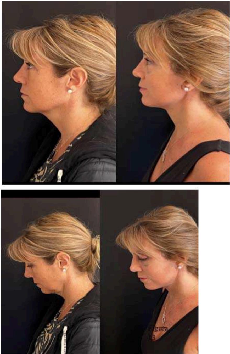
Plane 6: This plane involves the mylohyoid muscle and the anterior belly of the digastric muscle (ABDM) (Figure 2), which evolves with slight atrophy and a consequent descent of the buccal floor, and with it, a descent of the submandibular gland (SMG), which is sometimes accompanied by secondary hypertrophy due to fat deposits from aging or increased BMI. All these structures descend in the aging neck (Figure 3).
Function of the PM: It is a flat sheet that connects to the skin through perimysium and retinacula (lamination) and covers the deep structures. In facial expressions, it is responsible for the downward movement of the lower lip along with the DLI. During aging, this muscle tries to maintain its anatomy by supporting the lax deep structures, acquiring greater tonicity and shortening, leading to the formation of bands that are initially dynamic and later static, also contributing to the formation of a more obtuse, aged neck.
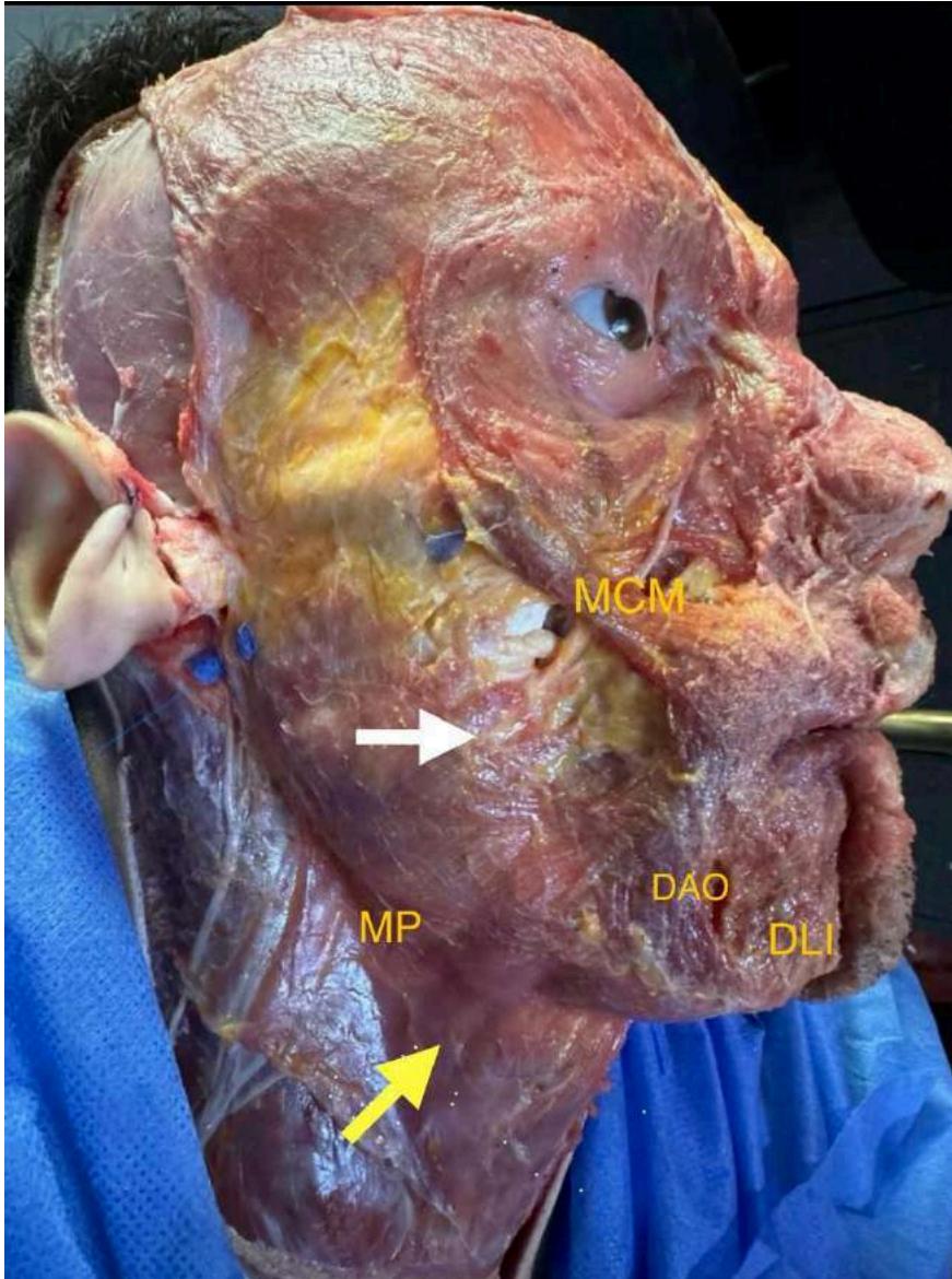
Figure 1: (young specimen): The platysma muscle can be seen with its cervical portion and longitudinal fibers (yellow arrow) and its superior facial portion near the zygoma, with horizontal fibers (white arrow), corresponding to the risorius muscle.
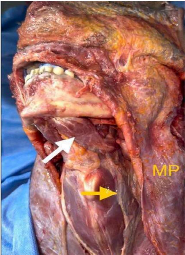
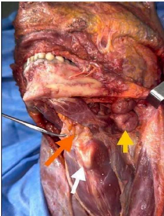 Figure 2: White arrow: ABDM (anterior belly of digastric muscle); yellow arrow: resected edge of the deep cervical aponeurosis, superficial layer
Figures 2 and 3: Dissection of a specimen with an obtuse neck where we can observe a ptotic ABDM (anterior belly of digastric muscle) protruding below the mandibular border, as well as a ptotic SMG (submandibular gland).
II. CERVICAL CLASSIFICATION
Aesthetic Neck: All authors describe it as having a cervicometal angle of 105 to 120 degrees, a defined mandibular border with some horizontal depth from the lower mandibular border to the neck, a slight visibility of the anterior edge of the sternocleidomastoid (ECM), a marked gonial angle, and a more visible thyroid cartilage in men.
Unaesthetic Neck: We further classify it as follows:
-
Congenitally Obtuse: This type of neck, with an angle greater than 120 degrees, is associated with retrognathia and poor positioning of the hyoid (more anterior and inferior). It is observed in young individuals (Figure 3.1).
-
Lipodystrophic Obtuse: Young patients with increased body mass index and/or ethnic variants with predominance of lipodystrophic necks, typically ranging in age from 20 to 35 years.
-
Aged Obtuse Neck: Begins with skin laxity and platysmal atrophy, generally starting from age 45, followed by ptosis of the buccal floor and observable fat redistribution starting from age 55, with the presence of jowls.
Grade 1: Skin laxity with or without lipodystrophy in Plane 2.
Grade II: Addition of platysmal bands.
Grade III: Additional deep fat component.
Grade IV: Additional ptosis of the SMG (submandibular gland). 4. Short Neck: Excluding syndromes such as Klippel-Feil, Turner Syndrome, and necks that appear short due to morbid obesity.
Congenitally short necks are those where the ratio between the clavicular-menton distance represents less than 24% of the clavicular-vertex distance. These patients frequently present with ptosis and hypertrophy of the SMG, submental bulging due to deep fat and ABDM (anterior belly of digastric muscle), as well as laxity in the infrahyoids: sternothyroid muscles.
5. Secondary Neck: We can differentiate two types of secondary necks:
a) Secondary to Energy Treatments: E.g., radiofrequency, ultrasound, subcutaneous laser. In these cases, we observe reabsorption of fat tissue in Plane 2 as well as varying degrees of platysmal atrophy, which we believe are related to the number of sessions the patient undergoes.
b) Secondary to Previous Surgery: Liposuction, lifting, other procedures.
IV. PHYSICAL EXAMINATION
Performed with the patient seated, observing and palpating cervical structures.
-
Static and dynamic platysmal bands.
-
Chin ptosis.
-
Skin laxity, thin skin with atrophy of the PM, visible deep structures.
-
Superficial fat gives the appearance of a uniformly bulging full neck that obscures the contraction of the PM and does not move with swallowing.
-
The deeper anterior fat, which is more compact and fibrous, moves with swallowing and is mostly distributed centrally and vertically. Laterally, the ABDM and then the SMG can be observed; while they can be examined, their final assessment is intraoperative.
V. TREATMENT
Once the diagnosis is made, we determine the type of neck we are dealing with. We can describe 5 types of procedures for neck treatment:
-
Cervical Liposuction: This technique, either alone or combined with energy devices, is suitable for lipodystrophic necks in patients under 35 years old who do not present with SMG ptosis, retrognathia, or jowls.
-
Isolated Neck via Submental Approach: Indicated for obtuse necks in young patients with some degree of retrognathia, deep fat lipodystrophy, and mild hypertrophy and ptosis of the submandibular gland, with a present gonial angle and without jowls.
-
Isolated Neck via Submental and Lateral Approach: The same as the previous case but with a lack of definition of the mandibular angle (Figure 3.1).
-
Cervicofacial Lift with Lateral Approach Without PM Delamination: For patients with a good cervicomental angle, presence of jowls, mid-facial ptosis, some cervical skin laxity without bands, and De Castro Type I platysma decussation.
-
Cervicofacial Lift with Lateral and Submental Approach, With or Without PM Delamination: For patients with facial and cervical ptosis, obtuse neck, or short neck.
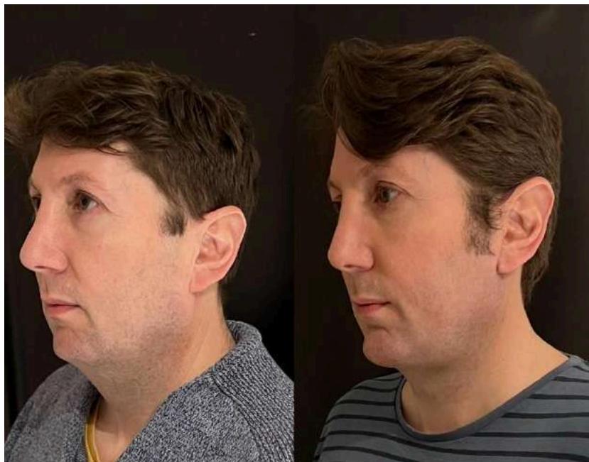
Figure 3: Yellow arrow: Ptosis of the SMG (submandibular gland), which is located below the mandibular border, reaching the level of the hyoid bone. White arrow: Thyroid cartilage. Orange arrow: Hyoid bone and deep interdigastric fat held with a Halsted clamp.
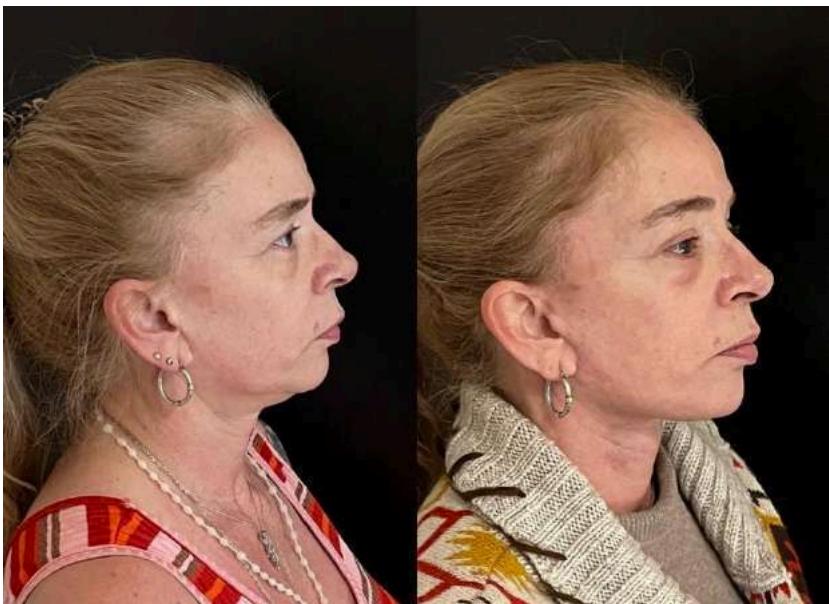
Figure 3.1: Pre and postoperative at 30 days. Congenital obtuse neck. Isolated submental and lateral treatment type III. Chin implant placement
VI. SURGICAL TECHNIQUE
We will describe the steps for the lateral and submental neck approaches:
Lateral Approach: Retroauricular incision: follows a line placed in the retroauricular groove, extending upward and horizontally across the mastoid when it becomes flat, then continuing towards the occipital area in a pre-pilose manner (Figure 7).
Dissection Limits:
-
Lateral Region: Lower limit 6 cm below the gonion (subcutaneous dissection).
-
Central Region: Lower limit at the upper border of the thyroid cartilage and upper limit at the mandibular margin (Figure 8).
Submental Approach: Make a slightly curvilinear incision at the posterior border of the mandibular bone at the submental level, 2.5 cm in length (Figure 4).
Separate the lower skin edge with a double hook, section Plane 1 and Plane 2 with a needle-tip electrocautery until the mentalis muscle fibers and PM (platysma muscle) are visible. Perform dilation and cutting with scissors laterally in Plane 2 above the PM, leaving a 5 mm layer of fatty tissue in the skin flap.
Finally, repeat this dissection in the central area. This allows for a uniform flap with even thickness of fat (Figure 5).
To achieve better visualization and working field, partially release the mandibular ligament subcutaneously laterally for about 2 cm. If the patient has significant submental skin laxity, a total release of the ligament is justified. This allows for better excursion and vertical traction from the SMAS (superficial musculoaponeurotic system) - facial platysma. Perform hemostasis of the area.
Incise along the midline with needle-tip electrocautery and dissect laterally below the PM, near its deep face, leaving the subplatysmal fat adhered to the deep plane. The limit is the external border of the ABDM (anterior belly of digastric muscle) and visualization of its tendon at the level of the hyoid bone.
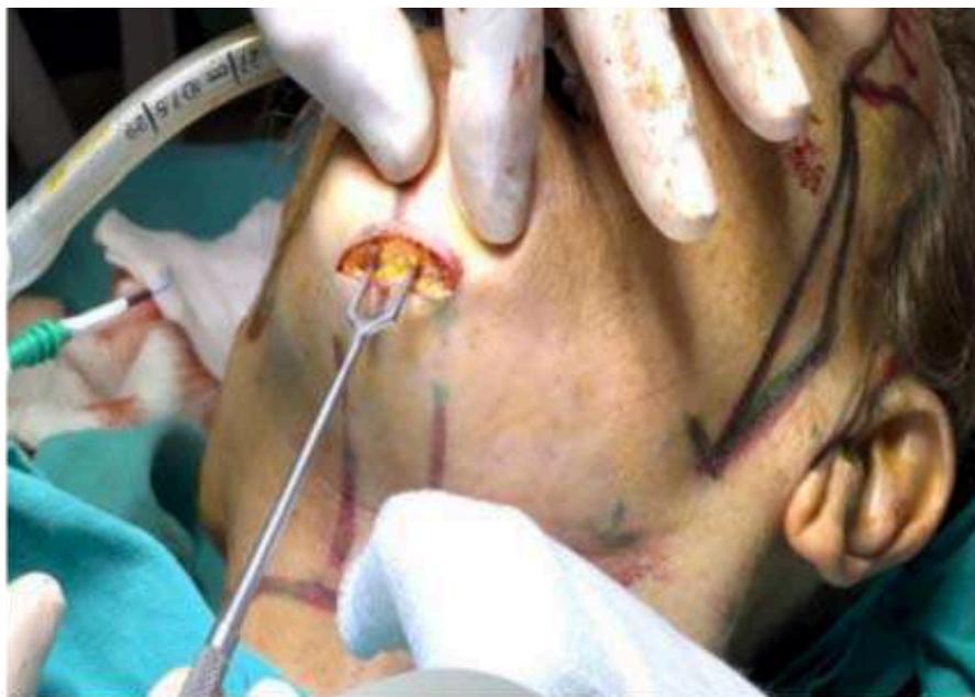
Figure 4: Slightly Curved Submental Incision along the Posterior Border of the Mandibular Bone
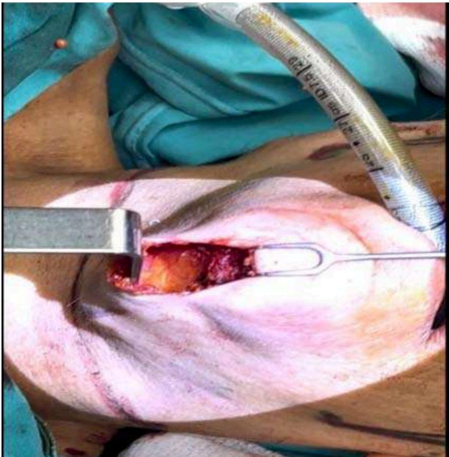 Figure 5: Anterior Cervical Dissection from the Submental Incision, Showing the Dermofat Flap
We now explain a series of steps that we believe provide better control in the uniform reduction of deep structures in obtuse necks:
-
Resection of the Deep Fat: Remove the deep fat located over the ABDM (anterior belly of digastric muscle).
-
Addressing SMG Hypertrophy and Ptosis: If there is hypertrophy and ptosis of the SMG (submandibular gland), locate these issues at the level of the digastric tendon by dissecting the deep face of the PM (platysma muscle) until reaching its capsule.
- Infiltrate with lidocaine and adrenaline to prevent bleeding.
- Incision and Dissection of SMG Capsule: Incise the SMG capsule on its inferointernal part with needle-tip electrocautery, then continue with intracapsular dissection using blunt maneuvers with a swab and hemostasis with needle-tip electrocautery.
Once the superficial pole is released, resect the portion below the mandibular border ("ptotic") with bipolar electrocautery (intensity 25), achieving progressive coagulation that gradually sections the gland.
Intermittently irrigate the area with saline to cool the tissues and prevent thermal injury to the cervical and marginal branches of the facial nerve (FN), which are close to the lateral surface of the glandular capsule.
-
Resection and Reduction of ABDM: Resect and reduce the ABDM tangentially. This provides two benefits: it reduces their volume and weakens them, aiding in the retraction and elevation of the hyoid bone.
-
Resection of Interdigastric Fat: Control the resection and reduction of interdigastric fat, leveling it with the remaining digastric bellies.
-
Bring PM Flaps to the Midline: Bring the PM flaps towards the midline and resect the excess that extends beyond it.
-
Suture Medial Edges of PM: Suture the medial edges of the PM in the midline, from the hyoid bone to the mental region.
Avoid infrahyoid suturing, as it can create resistance and affect the excursion and vectorization of the SMAS-platsima facial. 8. Horizontal Myotomy of PM: Perform a horizontal myotomy of the PM 1 cm below the hyoid bone, 2 cm in length, bilaterally to address platysmal bands and provide a hammock effect on the SMG during lateral traction.
Observations on the Technique:
-
Light Skin Laxity Only: When the neck presents only slight skin laxity without platysmal bands, lateral traction of the PM (platysma muscle) alone is sufficient.
-
Presence of Bands or Significant Submental Skin Laxity: When there are bands or noticeable submental skin laxity, a submental approach is necessary.
-
Neck Post-Surgery or Energy Therapy with PM Atrophy: For necks secondary to previous surgery or energy therapies with PM atrophy, precise dissection of Plane 2 in the submental triangle is required to ensure the skin adapts to the new deep structure. In the lateral region, modify the technique by advancing in plane 2 to the anterior border of the ECM (sternocleidomastoid muscle), then entering plane 4 to visualize the SMG, creating a lateral composite cervical flap.
The goal is to obtain a PM not delaminated from the skin (Figure 9), which can be retracted without tearing the atrophic platysma, allowing it to be fixed to the mastoid aponeurosis (Figure 10).
This technique is described as the Face and Neck Lift Preservation technique by Dr. Mike Nayak.
Special Considerations for Obtuse Neck:
-
Steps 2 and 3: These are only performed when palpation and observation reveal a ptotic and/or hypertrophic SMG.
-
Resection of SMG Capsule: Once the SMG capsule is opened, it must be resected;
otherwise, secondary ptosis will occur due to weakening.
-
Step 5: Avoid over-resection of the interdigastric fat to prevent a cosmetic complication known as "cobra neck."
-
Interdigastric Tendon Suturing: Suturing the interdigastric tendons can help mobilize the hyoid bone backward and upward, improving the cervicomental angle. Note that if the SMG was not previously addressed, this maneuver will make it visible.
-
Deep Cervical Fascia Release: Another approach is to release the deep cervical fascia at the superior border of the hyoid bone, allowing it to move posteriorly and superiorly, assisted by the posterior belly of the digastric muscle.
-
Step 8: Ensure that the myotomy is performed lower than the hyoid bone (1 cm); otherwise, the hammock effect from the lateral tension of the platysma on its fixation to the mastoid will be lost
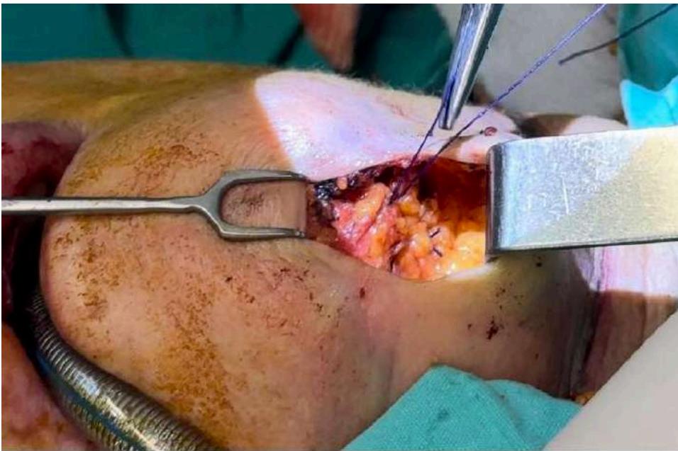
Figure 3.2: Pre and postoperative at 2 months. Treatment type V with reduction of SMG (submandibular gla
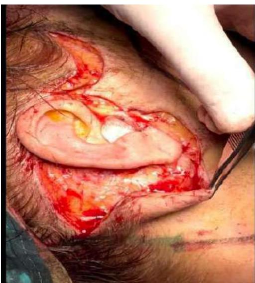
Figure 6: Medial Suturing of the PM, Suprahyoid Area, with Interrupted Stitches
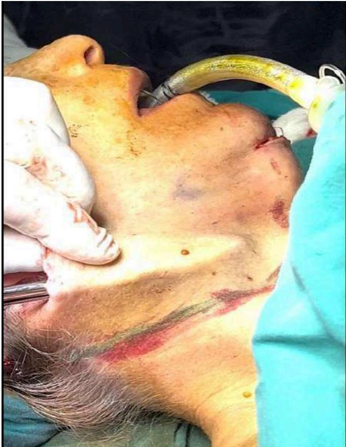
Figure 7: Dissection of the Flap in Plane 2, Retroauricular and Preauricular
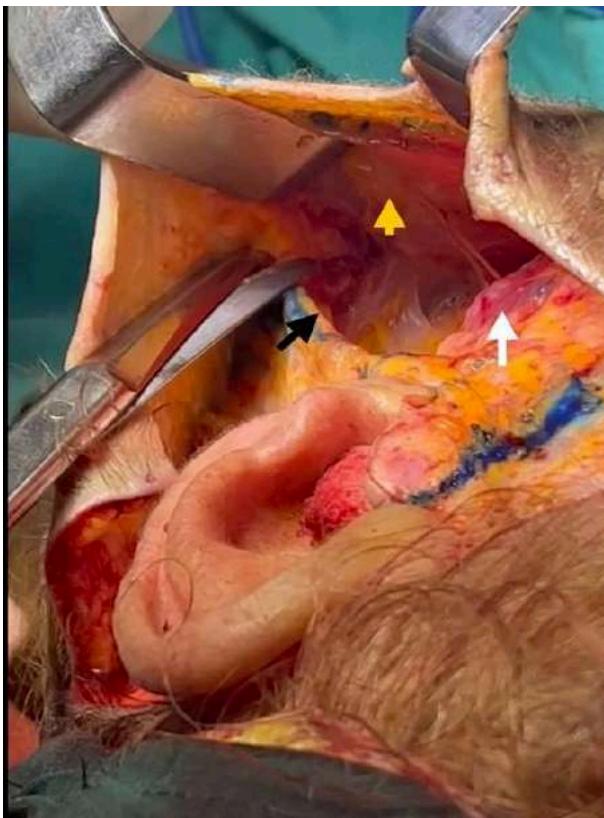 Figure 8: Lateral Dissection with Skin Delamination to the Platysma and Anterior Submental Dissection
Yellow Arrow: PM adhered to Planes 1 and 2.
White Arrow: Mandibular gonion with parotid-masseteric fascia.
Black Arrow: Anterior border of the ECM.
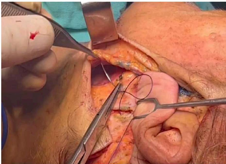 Figure 10: Suture Fixation of the Cervical Composite Flap to the Mastoid
VII. COMPLICATIONS
-
Hematoma: These can vary in size, with large blood collections being rare. Small, isolated collections are more common. The use of the Auersvald hemostatic net is very effective in preventing these.
-
Seroma: Generally occurs in the submental region. Treating deep fat alters lymphatic drainage. Immediate drainage is required through repeated punctures every 48 hours; if this is not feasible, a drain should be left in place.
-
Skin Retractions: Usually occur post-seromas or untreated small hematomas.
-
Asymmetries: Result from asymmetric reduction of deep structures.
-
Cobra Neck: Caused by over-resection of the interdigastric fat.
-
Facial Marginal Nerve Palsy: Can occur due to diathermy when resecting the superficial lobe of the SMG.
-
Sialomas: Rare but can be expected with reduction of the SMG.
-
Skin Necrosis and Suffering: Rare, typically observed with high concentrations of tranexamic acid in local anesthetic solutions. It can also occur in cases with prior energy treatments affecting the vascularization of Planes 1 and 2. In these cases, it is advised not to delaminate the PM from the skin.
-
Dry Mouth: Rare, occurring post-SMG reduction. The SMG, along with the sublingual gland and accessory glands, maintains mouth moisture, whereas the parotid gland only functions during mastication and stimulation of food bolus. Antidepressant medication can exacerbate this complication, which is usually transient and resolves within 3 to 4 months. Treatment includes administering pilocarpine.
VIII. POST-OPERATIVE CARE
-
Pre-Surgery Information: Inform the patient that a hard edema will develop in the submental region, lasting 4 to 6 weeks.
-
Post-Operative Education: Instruct the patient to avoid extreme neck rotation and to avoid opening the mouth wide.
-
Massage Restrictions: Avoid massages to reduce neck tension for 2 weeks to prevent elongation of the PM until it adheres to its new position.
-
Dietary Restrictions: For patients who have undergone SMG treatment, avoid alcohol and citrus fruits that stimulate salivary glands for 2 weeks.
-
Sleeping Position: For the first 48 to 76 hours, the patient should sleep semi-sitting at 25 to 30 degrees head elevation, with the nose pointing toward the ceiling to facilitate venous drainage.
Figure 11-12: Patient with an Aged Obtuse Neck and Their 20-Day Postoperative Result. Preservation Face and Neck Lift without Cervical Platysma Delamination
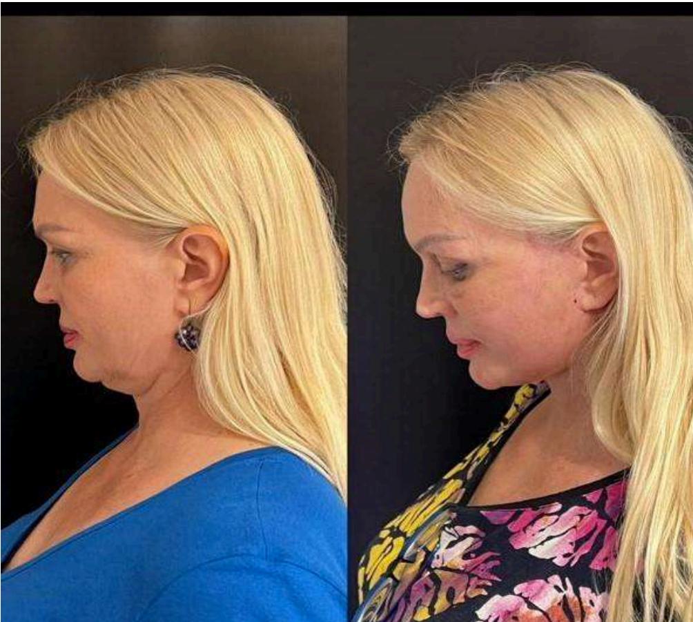
Figure 9: Posterior view of the Auricular Pavilion, Showing A Lateral Composite Cervical Flap without Delamination of the Pm
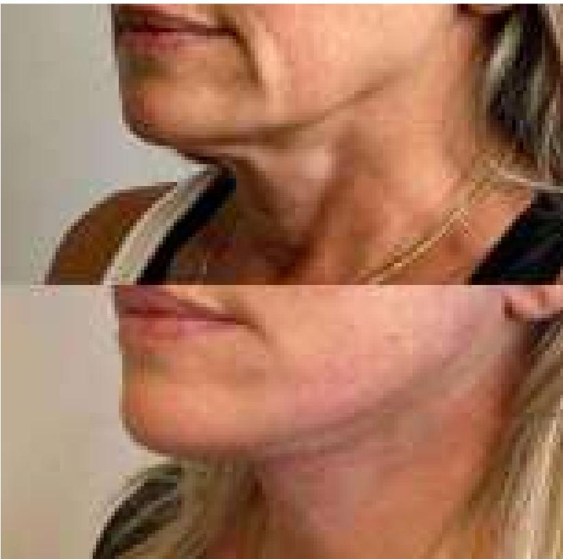
Figure 13: Pre- and Postoperative of a Patient with a Short Neck. Cervicofacial Lifting. Treatment type 5
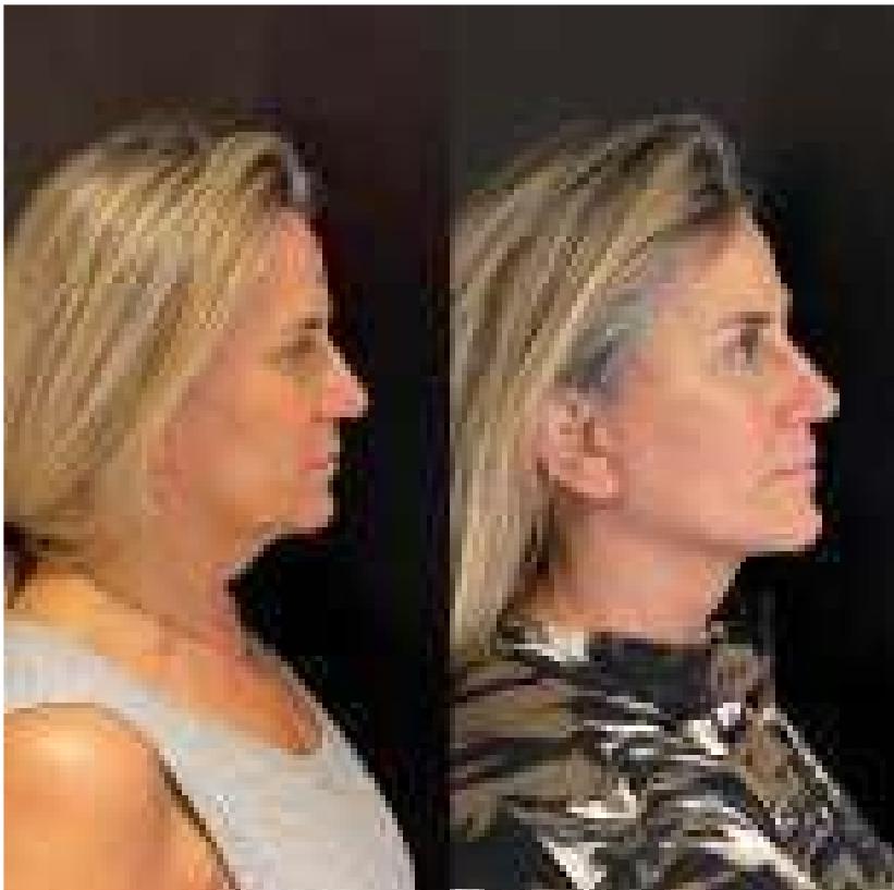
Figure 14: Pre- and 2-month postoperative. Treatment type 5 with platysma delamination
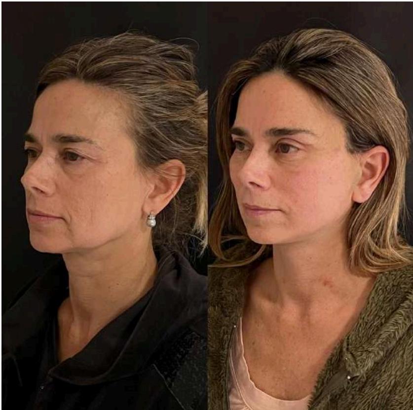
Figure 15: Pre- and 2-Month Postoperative, Type 5 with Cervical Platysma Delamination
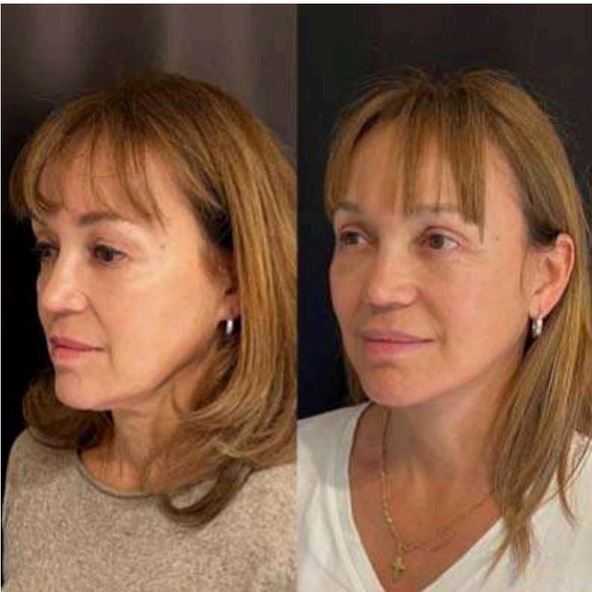
Figure 16: Pre- and 2-Month Postoperative. Cervicofacial Lifting with Lateral Approach, without Platysma Delamination. No Submental Incision. Procedure Type IV
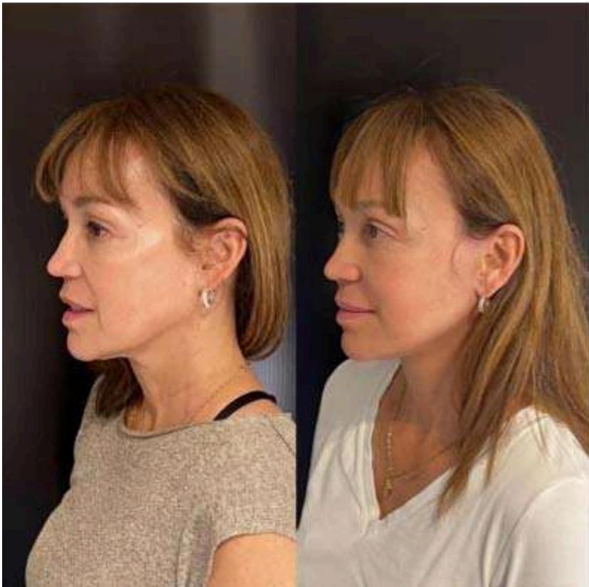
Figures 17-18: Procedure type IV. 6 Months Postoperative
VIII. CONCLUSION
Facial aging presents distinctive characteristics compared to neck aging. In the midface, we observe deflation or reabsorption of fat compartments, accompanied by progressive laxity. The lower third reflects this same aging, adding a descent of soft tissues that migrate to the neck, causing the appearance of jowls, loss of definition of the mandibular border, and the gonion.
Current aesthetic medicine tools have shown good results in these areas, especially in faces with deflation, through volumization of the midface and simulation of a new mandibular border and gonion in the lower third. However, neck aging is characterized by a fullness effect due to laxity and descent of deep structures, along with shortening of the platysma and redistribution of subplatysmal fat, creating an incompetence between the container and its content.
Aesthetic treatments based on energy that act on the skin, subcutaneous tissue, and PM do not fully meet the expectations of patients with obtuse necks, making this area a challenging unresolved issue in the aesthetic field.
Therefore, we believe that deep neck lifting techniques, being reductive, are the ideal option to address these issues. Neck rejuvenation requires a deep understanding of anatomical structures and their dynamics during aging. This approach allows for maximizing results and minimizing complications. Accurate diagnosis is crucial for proper planning and treatment. The external shape and contour of the neck reflect its internal anatomy, similar to what happens in aesthetic rhinoplasty. Thus, thorough treatment of the deep structures of the neck is essential for improving postoperative results and prolonging their durability.
In summary, the neck today represents a new and significant challenge for plastic surgeons.
To complement our study, we conducted two informational surveys directed at plastic surgeons specialized in deep cervicofacial lifting techniques, with the following relevant results:
Treatment of the Submandibular Gland (SMG):
-
60% of respondents regularly perform submandibular gland excision as part of neck treatment.
-
10% perform it preventively.
-
50% do it only in cases of glandular hypertrophy or ptosis.
-
The remaining 40% do not intervene directly in the gland, limiting treatment to ptosis with the platysma (PM) hammock technique.
Platysma Myotomy:
-
100% of respondents perform some form of platysma myotomy.
-
50% perform myotomy approximately 1 cm below the hyoid bone.
-
49% perform myotomy below the hyoid, combining it with other techniques:
-
18% combine it with an anterior myotomy of the sternocleidomastoid muscle (ECM).
-
31% combine it with a triangular resection below the hyoid.
-
1% of respondents perform a total transection of the platysma.
These results highlight the diversity of approaches and techniques used in neck treatment, reflecting different preferences and experiences of specialists in the quest for optimal aesthetic and functional results.
- L. Mike Nayak,6 and Ben Talei. (p.10) ↩
Conflict of Interest
The authors declare no conflict of interest.
Ethical Approval
Not applicable
Data Availability
The datasets used in this study are openly available at [repository link] and the source code is available on GitHub at [GitHub link].
Funding
This work did not receive any external funding.
References
Cite this article
Related Research



Special Issue
Launch a focused special issue to highlight research, emerging trends, and expert insights in your academic field.
