


IntelliPaper
Abstract
Phrenic nerve paralysis is a severe clinical condition with significant repercussions on respiratory mechanics and patient quality of life. It results from the interruption of diaphragmatic innervation, compromising the physiology of the muscle, reducing pulmonary expansion, and leading to alveolar hypoventilation. The phrenic nerve originates from the cervical roots C3 to C5, following a complex anatomical pathway that makes it vulnerable to iatrogenic injury during cervical or thoracic surgeries or anesthetic blocks. This study reviewed scientific evidence from the past decade, without language restrictions, using databases such as PubMed and SciELO and search terms including "phrenic nerve paralysis," "iatrogenesis," and "neurotization." The analysis integrated data from prospective studies and systematic reviews, highlighting diagnostic techniques such as dynamic ultrasonography and electroneuromyography. Therapeutic strategies ranged from conservative approaches to surgical interventions, including neurotization with autologous grafts and diaphragmatic plication. The findings conclude that preventing iatrogenic injuries requires standardized protocols, intraoperative monitoring, and equitable access to therapies. These measures are essential to reduce morbidity and mortality and improve clinical outcomes, particularly in vulnerable populations such as the elderly and patients with neuromuscular comorbidities.
Explore Digital Article Text
I. INTRODUCTION
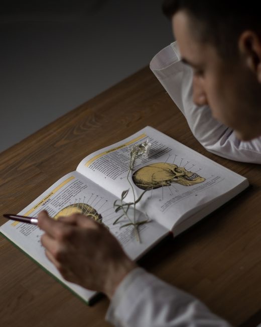
Phrenic nerve paralysis is a serious clinical condition with systemic repercussions on the patient's health1. It results from the interruption of diaphragmatic innervation, severely affecting respiratory mechanics and compromising the physiological contraction of the diaphragm, which prevents its ideal flattening and, consequently, decreases lung expansion2.
The phrenic nerve is a mixed nerve, usually originating from the cervical roots C3, C4, and C5 . However, in some cases, there are anatomical variations in its origin or path, which considerably increases the risk of surgical iatrogenesis in the absence of preoperative imaging studies .
Its path begins at the neck, descending anteriorly to the anterior scalene muscle and posteriorly to the internal jugular vein and subclavian artery . Upon crossing the mediastinum, it branches to innervate both hemidiaphragms, synchronizing their contractions during inspiration .
It is important to note that there is anatomical asymmetry between the right and left sides: on the right, the nerve accompanies the superior vena cava and the right atrium5. In some cases, the proximity of the nerve to the subclavian vein increases the risk of injury during venous procedures in the region4. On the left side, it crosses the aortic arch and continues over the left ventricle5. This relationship explains the incidence of postoperative phrenic paralysis in cardiac surgery9.
This superficial and complex path exposes the nerve to iatrogenic trauma, especially during cervical, thoracic, or cardiac surgery. Interscalene brachial plexus blocks are associated with transient diaphragmatic paralysis. Other causes include compression by mediastinal neoplasias and neuromuscular diseases, such as amyotrophic lateral sclerosis. Rare cases, such as granulomatosis with polyangiitis, may also manifest with phrenic paralysis as an initial symptom.
Anatomically, the diaphragm acts as a dynamic barrier between the thoracic and abdominal cavities, being the main motor of respiration5. The exclusive innervation of the diaphragm by the phrenic nerve highlights the interdependence between neural structure and muscle function5. When this connection is interrupted, the balance of intrathoracic and abdominal pressures is disturbed, resulting in alveolar hypoventilation and dyspnea8.
These changes can be evidenced by imaging tests, such as X-rays and ultrasounds, which reveal unilateral or bilateral elevation of the diaphragm and reduced respiratory excursion8. Clinically, a paradoxical movement is observed, in which the affected diaphragm is elevated during inspiration, exacerbating hypoxia1,8.
Therapeutic management varies according to etiology and severity. Conservative strategies include respiratory physiotherapy and non-invasive ventilation in most cases . In acute traumatic injuries, neurotization with autologous grafts is indicated, achieving functional recovery in half of patients over the course of one year . Techniques such as diaphragmatic stimulation are a promising therapeutic option in bilateral paralysis, although their high cost limits their accessibility .
This study aims to integrate current evidence on phrenic nerve injury, addressing two fundamental axes. First, the correlation between anatomical variations and risk of iatrogenesis. Second, we evaluate diagnostic methods with an emphasis on their clinical applicability and accuracy.
II. MATERIALS AND METHODS
The bibliographic method was used to collect scientific data from the last five years from medical sources in Portuguese, Spanish, and English, using the following platforms: SciELO, Google Scholar, and PubMed. Keywords such as 'Denervation,' 'Atrophy,' 'Fibrosis,' 'Hypoventilation,' and 'Neurostimulation' were searched. The study included a comparative table addressing the main causes of paralysis, anatomical alterations, histological changes and the associated functional impact.
Classical reference works were also consulted, such as Gray's Anatomy (Gray H. et al., 42nd ed., Elsevier, 2020), Ross MH, Pawlina W. Histology: Text and Atlas. 8th ed. Barcelona: Wolters Kluwer; 2023. and Clinical Embryology (Moore KL., 11th ed., Elsevier, 2021). In addition, guidelines from the American Thoracic Society (ATS) and studies from the National Institutes of Health (NIH) on nerve regeneration were included.
III. RESULTS
The review revealed findings consistent with the literature and medical epidemiology. It provides a systematic overview of cases of phrenic nerve injuries. Such injuries directly compromise diaphragmatic dynamics, resulting in physiological and systemic dysfunctions. Table 1 summarizes crucial data from the main studies, highlighting the type of study, sample, etiology of the injury, and clinical outcome.
Table 1: Phrenic Nerve Paralysis: Comparative Summary of Clinical Studies
| Author/Year | Type of Study | Sample | Etiology of the Injury | Clinical Outcome |
| Saba-Santiago et al. (2022)10 | Prospective | 78 patients | Interscalene block of the brachial plexus | Spontaneous recovery in most cases |
| Hu et al. (2024)9 | Integrative review | Data from 107 references | Surgical procedures (cardiac/thoracic) | Reduction of complications with intraoperative monitoring |
| Boussuges et al. (2020)8 | Systematic review | Data from 102 references | Diaphragmatic dysfunction | Accurate diagnosis via dynamic ultrasound |
| Supra & Agrawal (2023)11 | Narrative review | Data from 94 references | Traumatic injury to the phrenic nerve | Moderate success of nerve grafts |
| Dubé et al. (2016)13 | Clinical review | 65 surgical cases | Irreversible chronic paralysis | Symptomatic improvement after plicature |
Prospective data, such as those from Saba-Santiago et al, highlighted that of interscalene blocks resulted in transient diaphragmatic paralysis, with spontaneous resolution in most cases10. On the other hand, cardiothoracic surgical interventions, analysed by Hu et al, showed a reduction in complications when accompanied by intraoperative monitoring, reinforcing the importance of preventive strategies9.
In addition, advanced ultrasound techniques, such as speckle tracking, allow for more accurate assessment of diaphragmatic micro-movements compared to conventional ultrasound . This approach is particularly useful in the early detection of subclinical dysfunctions, such as in patients with chronic comorbidities that predispose them to diaphragmatic weakness .
As for therapeutic options, autologous nerve grafts, reviewed by Supra & Agrawal, achieved functional success in of cases, but with heterogeneity in the evaluation criteria between studies11.
At the same time, electroneuromyography of the phrenic nerve, performed with electrical stimuli near the C3-C5 roots and intramuscular recording in the diaphragm, allows parameters such as latency and amplitude of the muscle action potential to be quantified18.
Additionally, esophageal manometry assists in the assessment of transdiaphragmatic pressure, whose optimal difference reflects preserved function[17,19]. These findings corroborate the central hypothesis of the study, the complexity and anatomical variability of the phrenic nerve.
IV. DISCUSSION
Phrenic nerve paralysis is not just a technical complication, but a reflection of structural gaps in the safety of routine medical procedures1. The incidence of of transient paralysis after interscalene block is not merely a statistical fact, but rather an alarming indicator of how anesthetic protocols underestimate the anatomical vulnerability of the nerve2.
The emphasis on 'spontaneous recovery' in cases of transient paralysis masks an underlying problem, the normalization of iatrogenic risk. Patients may recover diaphragmatic function, but sequelae such as chronic muscle fatigue and exercise intolerance are often overlooked. Studies such as that by Hu et al, which celebrate a reduction in complications with intraoperative monitoring, fail to question why of cases are still exposed to predictable risks 1,9.
The sensitivity of dynamic ultrasound contrasts sharply with the reality of healthcare systems that still rely on static X-rays to assess respiratory dynamics[4,20]. Meanwhile, electroneuromyography, the gold standard for neuropathies, remains inaccessible in regions without specialists, deepening inequalities in the quality of care[18].
The success rate of nerve grafts is a warning sign: half of patients undergoing invasive procedures are left with functional sequelae, often without access to pulmonary rehabilitation or psychological support[19]. Diaphragmatic plication, despite improving symptoms in of cases, is a palliative solution that does not restore respiratory physiology, perpetuating dependence on medical interventions[20].
The ability of bulboprotuberant centers to mask symptoms through accessory muscles is not a physiological detail[22]. While young patients adapt to unilateral dysfunction, elderly patients or those with neuromuscular comorbidities face abrupt decompensation, which is often fatal[23].
V. FINAL CONSIDERATIONS
Phrenic nerve paralysis not only paralyses a muscle, but also chains the body to a silent struggle, where breathing becomes an act of resistance. Those who suffer from it face days marked by constant fatigue, the anguish of not being able to fill their lungs, and the fear that a simple movement will aggravate their weakness. It is a condition that robs them of their most basic autonomy, turning everyday life into a challenge.
However, amid this reality, there is a clear path forward: accurate diagnosis and timely treatment. An ultrasound revealing diaphragmatic elevation, adapted physiotherapy, or a properly placed pacemaker are not just medical procedures, but bridges to the recovery of a full life. Each successful step in managing this disease not only repairs a nerve, but also restores the freedom to breathe without fear.
ACKNOWLEDGMENTS
I would like to thank our advisor Silvio Stafi Filho for his constant dedication, availability, academic competence and scientific rigor, which were fundamental to the conception and writing of this article.
Conflict of Interest
The authors declare no conflict of interest.
Ethical Approval
Not applicable
Data Availability
The datasets used in this study are openly available at [repository link] and the source code is available on GitHub at [GitHub link].
Funding
This work did not receive any external funding.
Cite this article
Related Research


Special Issue
Launch a focused special issue to highlight research, emerging trends, and expert insights in your academic field.
