






IntelliPaper
Abstract
Objective: This manuscript aims is to present the difficulties involved in optimizing dosimetric distribution, based on six cases of patients who underwent radiotherapy before or after breast implant surgery. Materials and methods: Clinical characteristics, imaging, anatomical pathological and immunohistochemical data, treatment (neoadjuvant or adjuvant chemo- therapy, subcutaneous mastectomy surgery with axillary curage, radiotherapy before or after breast implant placement and difficulties in dosimetric coverage). Results: Patients aged 37, 30, 48, 32, 44, and 36 years were followed for invasive breast carcinoma of luminal B, triple-negative, Luminal B Her2-positive, triple-negative breast diagnosed by anatomical pathology and immuno-histochemistry. They received either neoadjuvant or adjuvant chemotherapy, or subcutaneous mastectomy with breast implant or breast prosthesis before or after radiotherapy. Radiotherapy with breast implants in the pre-pectoral, retro pectoral, and axilla clavicular CTVs was delivered at a dose of 50 Gy (2Gy per session) or 40.05 Gy (2.67Gy per session). Better optimization of dosimetric distribution was difficult to achieve in the pre-pectoral, retro pectoral, and axilla supra clavicular CTV, as the breast implant was considered a high-risk organ. Conclusion: Direct, permanent breast reconstruction using a breast implant or prosthesis is an increasingly popular treatment option. It minimizes the psychological, social, and aesthetic repercussions of mastectomy. However, there are still significant challenges to be overcome when adequately planning dosimetric distribution for radiotherapy
Explore Digital Article Text
I. INTRODUCTION
Over the past decade, breast cancer has become the most frequent cancer in women and the leading cause of death . However, data in the literature have confirmed a significant improvement in local control and overall survival in patients with locally advanced disease and, or lymph node metastases . [Approximately 40% of women with breast cancer will undergo mastectomy due to more advanced local and regional disease or individual preference , and others will receive post-mastectomy radiotherapy . The social and psychological pressures on women caused by mastectomies have diminished considerably with the advent of immediate or deferred breast reconstruction. This mastectomy resulted in the loss of female sexual characteristics. Today, around 50% of women who have had a mastectomy opt for breast reconstruction to improve their psychological, social, and sexual well-being . Breast reconstruction can be performed at the time of mastectomy (during the same anesthetic) or after several months or even years . With the frequent use of screening tests for various genetic mutations (BRCA1/BRCA2), mastectomy followed by immediate breast reconstruction (IBR) is increasingly indicated . Also, more and more patients, even in the context of post-mastectomy radiotherapy, are opting for immediate breast reconstruction to cope with the aesthetic and psychological consequences of breast loss . The difficulties of irradiating a reconstructed breast may lead to sub-optimal coverage of target volumes by the optimal radiotherapy dose, particularly when locoregional lymph nodes have to be treated along with the chest wall. And this can lead to increased amounts of organs at risk, such as the heart and lungs .
However, the current techniques for administering radiotherapy to breast cancer patients who have undergone reconstruction have improved, reducing the radiotherapy planning challenges that once seemed difficult to overcome . Despite improvements in reconstruction and radiotherapy techniques, complications remain without compromising oncological and cosmetic results . This work aims to show the constraints or difficulties associated with immediate breast reconstruction with radiotherapy. We report the cases of six patients, three of whom underwent subcutaneous mastectomy with immediate breast reconstruction and three of whom underwent deferred breast reconstruction.
Observation 1
We report the clinical observation of a 37-year-old female patient with a family history of breast cancer (sister treated for breast cancer at age 38 and aunt treated for breast cancer at age 60), smoker at 15 packs/year, and weaned alcoholic. She presented with a left breast nodule that had appeared three months previously, without mastodynia or nipple discharge. On clinical examination, she was in good general condition, had a type C cup breast, and palpated a nodule measuring approximately 2 cm at the union of the upper quadrants of the left breast, with no axillary or supraclavicular adenopathy. Mammography and breast ultrasound revealed a 21.8 mm nodular lesion at the junction of the upper quadrants, associated with a small focal lesion with no axillary adenopathy. The examination was graded ACR 5. Magnetic resonance imaging of the breasts revealed a tumoral process in the left breast with a 43.7 x 33 x 28 mm mass, diffuse micronodular enhancement of the superior- internal quadrant, and several nodules at the union of the upper quadrants. A microbiopsy of two nodules was performed. Histological examination and immunohistochemistry revealed, for the first mass, an invasive breast carcinoma, non-specific type SBR II, without in situ component or vascular emboli, with estrogen receptors at 100%, progesterone receptors at 0%, Her2 score 1+ negative and Ki67 at 30%; for the second nodule, invasive breast carcinoma, non-specific type SBR II, with no in situ component or vascular emboli, estrogen receptors 100%, progesterone receptors 0%, Her2 score 1+ negative and Ki67 20%. Thoracic- abdominal- pelvic CT scan and bone scan did not reveal any distant secondary lesions.
The clinical case was presented at a multidisciplinary consultation meeting. Subcutaneous mastectomy with sentinel lymph node and immediate breast reconstruction with breast prosthesis was proposed and performed. Histological examination and immunohistochemistry of the mastectomy specimen and axillary curage revealed a non-specific invasive breast carcinoma, grade SBR 1, measuring 1.5 cm long, with an associated intraductal component of intermediate nuclear grade and comedonecrosis estimated at 10% of tumor volume. There were no perivascular emboli. The deep margin was tumoral, and the other margins were healthy. There were no lymph node metastases at the oN+/3N sentinel node. In the immunohistochemical study, estrogen receptors were 80%, progesterone receptors 30%, Her2 negative, and Ki67 30%. The disease was classified as pT1NoMo Luminal B. The DX Oncotype was requested, and the RS score was 46. The patient then underwent three courses of Epirubicin 60mg per m2 - Cyclophosphamide 600mg per m2, and a course of paclitaxel 175mg per m2 (paclitaxel was discontinued due to anaphylactic shock resulting in two cardiac arrests). Adjuvant radiotherapy at a dose of 40.05 Gy (2.67 Gy per session in 15 sessions) on the pre-pectoral, retro pectoral, and axilla supra clavicular VTC with a boost of 13.35 Gy (2.67 Gy per session in 5 sessions) on the pre pectoral VTC was performed. Due to the breast implant, the dosimetric coverage of the target volumes (pre-pectoral CTV, retro pectoral CTV, and axilla sus clavicular CTV) was challenging to achieve. Optimization was necessary to achieve good dosimetric distribution while delivering less dose to the breast implant. Tamoxifen-type hormone therapy for five years was proposed, followed by clinical and radiological monitoring (mammography-ultrasound, magnetic resonance imaging of the breasts, and abdominopelvic ultrasound).
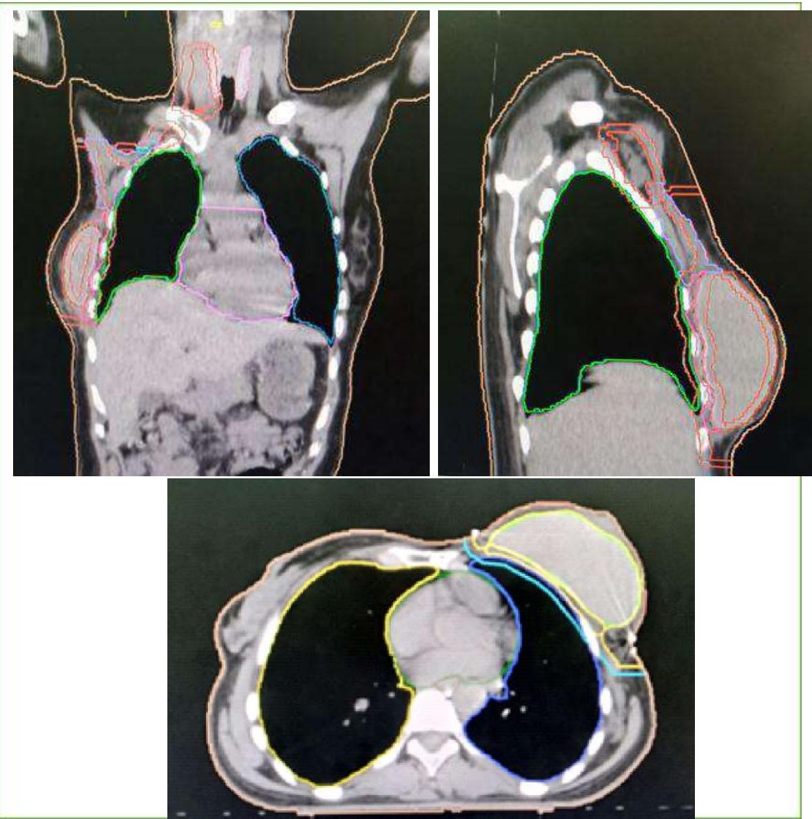 Figure 1: Delineation Images of Pre-Pectoral, Retro Pectoral, Axilla-Supra Clavicular CTV and Organs at Risk
Observation 2
A 30-year-old female patient with a medical history of lymph node tuberculosis treated in 2018 and family history (Uncles treated for (stomach cancer) was consulted in February 2019 for a right breast nodule evolving for about two years, without mastodynia or nipple discharge.
The patient's physical examination revealed a 1.5 cm nodule in the lower inner quadrant of the left breast and a 4 cm nodule in the lower inner quadrant of the right breast. Mammography-ultrasound revealed two nodules in the left breast, 12 mm in the lower internal quadrant and 9 mm in the upper external quadrant, and two nodules in the right breast, 7 mm in the lower outer quadrant and 45 mm in the lower internal quadrant. The examination was classified as ACR 3 on the left and ACR 4 on the right. The patient underwent conservative surgery on the right breast, with histological examination and immunohistochemistry showing invasive breast carcinoma of non-specific SBR grade 3 without vascular emboli, with a triple-negative molecular profile.
Magnetic resonance imaging of both breasts six weeks later revealed a right breast nodule classified as ACR BIRADS 6 and a left breast nodule classified as ACR BIRADS 3. The patient received neoadjuvant chemotherapy consisting of four courses of Epirubicin 90mg/m2 - Cyclophosphamide 600mg/m2 and 4 courses of Docetaxel 100mg/m2 with good tolerance. Surgery involving right mastectomy with axillary lymph node dissection (sentinel node technique) combined with immediate breast reconstruction with a prosthesis was performed. Anatomical pathology revealed no tumor proliferation. Adjuvant radiotherapy at 50 Gy in 25 sessions was performed on the pre-pectoral CTV. From the point of view of dosimetric distribution, coverage of the pre-pectoral CTV and retro pectoral CTV was challenging, as the breast implant was considered an organ at risk and should receive less dose. Monitoring was marked by a small nodule measuring 8 mm in the lower internal quadrant of the right wall prosthetic implant. The patient underwent revision surgery involving extraction of the nodule and replacement of the prosthetic implant.
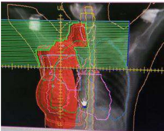 Figure 2: Digital Radiological Reconstruction Image of the Pre-Pectoral, Retro Pectoral, and Axilla-Supra-Clavicular CTV With Breast Implant
Observation 3
A 48-year-old female patient, nulligravida, non-menopausal, with a surgical history of thyroidectomy in 2010 and myomectomy in 2016, consulted for a nodule in the upper external quadrant of the left breast one year ago. On clinical examination, the patient was in good general condition. The breast examination revealed a 70 x 70 mm supra areolar mass of the left breast, rugged and mobile about the two planes, associated with a 30 x 20 mm left axillary adenopathy with no supraclavicular adenopathy and a 40 x 30 mm nodule straddling the right lower quadrants, mobile about the two superficial and deep planes, with a 10 mm axillary adenopathy and no supraclavicular adenopathy. A radiological consisting of mammography, breast ultrasound, and magnetic resonance imaging of the breasts was performed. Mammography revealed breasts of type C density. In the left breast, there was a well-limited, oval, inferior medial quadrant opacity measuring 28 x 36mm classified as ACR BIRADS 5. There were no suspicious lesions in the right breast. On breast ultrasound, a hypoechoic, lobulated, calcified tissue formation with peri-lesional infiltration measuring 37 x 21mm was found in the upper outer quadrant of the left breast, along with left axillary adenopathies, the largest measuring 32 x 17mm, and multiple bilateral breast cystic formations, the largest on the right, in the periareolar region, measuring 37 x 14. 5mm straddling the outer quadrants and lower outer quadrants; the largest on the left was located in the lower inner quadrant, measuring 31 x 14mm.
The examination was classified as ACR BIRADS 5 on the left and ACR BIRADS 3 on the right. Magnetic resonance imaging of the breasts revealed, in the left breast, a roughly rounded lesion straddling the upper quadrants, with irregular contours, measuring 40 x 40mm; an oval lesion, with irregular outlines, measuring 24.9 x 19mm in the deep retro-areolar region, associated with multiple bilateral cystic formations, the largest of which was located in the lower external quadrant, measuring 32.3 x 20mm, axillary adenopathies, the largest of which measured 40 x 23.6mm, and integrity of the pectoralis major muscle. In the right breast, there was a roughly rounded lesion with irregular contours measuring 16 x 13 mm straddling the lower quadrants, multiple cystic formations, the most voluminous straddling the outer quadrants measuring 43 x 22 mm, and a right axillary adenopathy measuring 11 mm in minor axis, suggesting multicentricity and bilaterality. The examination was classified as ACR BIRADS 4 on the right and ACR BIRADS 5 on the left.
A biopsy of the nodule straddling the upper quadrants and the retroareolar nodule of the left breast, with puncture of the left axillary adenopathy, and a biopsy of the lesion in the lower external quadrant of the right breast were performed. Histological examination revealed a non-specific invasive breast carcinoma with SBR grade II and the presence of a few clusters of carcinomatous cells in the lesion at the union of the upper quadrants of the left breast, with estrogen receptors at 5%, progesterone receptors at 1%, Ki67 at 80% and Her2 upbeat with a +++ score, and in the left retro-areolar mass with negative hormone receptors at 0%, Ki67 at 70% and Her2 upbeat with a +++ score. The nodule straddling the lower quadrants of the right breast revealed an invasive breast carcinoma, SBR grade II with estrogen receptor-positive at 30%, progesterone receptor-negative at 0%, Her2 upbeat score 3+++ and Ki67: 40%. Extension report with thoracic-abdominal-pelvic CT scan and bone scans revealed no distant metastatic lesions. The patient was a 48-year-old nulligravida patient with bilateral invasive nonspecific breast carcinoma. The patient was receiving neoadjuvant chemotherapy consisting of Epirubicin 90mg per m2 - Cyclophosphamide 600mg per m2 combined with Paclitaxel 175mg per m2 with double block (Trastuzumab 8mg per kilogram, then 6mg per kilogram from cycle 2 + Pertuzumab 840mg in cycle one then 420mg from cycle 2). Surgery was performed as a subcutaneous mastectomy with right and left axillary curage. Histological examination revealed mastoid breast parenchyma in the left breast, with fibrous and calcific changes and a histiocytic reaction, no residual tumor proliferation, and no lymph node metastases (oN+/7N). Histological examination of the right breast showed no residual tumor proliferation. Absence of lymph node metastasis (oN+/6N). The patient underwent radiotherapy at 40.05 Gy (2.67 Gy per session in 15 sessions) on the left and right walls and the left supra-clavicular axilla. The dosimetric distribution of CTV left wall, left axillary supraclavicular, and right wall did not encounter any difficulties with the VMAT technique. Six months after the end of radiotherapy, she benefited from a bilateral breast prosthesis.
Observation 4
Patient aged 32, menarche at age 12, genitally active with a regular menstrual cycle, no particular pathological history. She had been taking oral contraceptives for eight years. The patient was consulted in June 2020 for mastodynia with no palpable mass or mamelon discharge. Clinically, the patient's general condition was preserved, but there were no palpable masses in either breast or the right and left axillary and supra-clavicular areas.
Mammography revealed a predominance of fibro-glandular tissue in both breasts, with type c density. A periareolar opacity in the right upper-external quadrant with irregular contours containing amorphous microcalcifications and thickening and retraction of the periareolar plate. Right axillary adenopathy. There is no suspicious-looking nodular or stellate opacity on the left side. Mammary ultrasound revealed highly hypoechoic tissue formations with irregular outlines containing microscopic calcifications measuring 28 x 17 mm in diameter in the right breast. Hypoechoic right axillary adenopathies, the largest measuring 18.7 mm in long axis. Edematous infiltration of suitable conjunctiva-glandular tissue. No tissue, fluid, or attenuating image is visible in the left breast. Examination classified as ACR BIRADS 5. A true-cut breast biopsy was performed, and histological examination and immunohistochemistry noted a non-specific, poorly differentiated, SBR grade III invasive breast carcinoma infiltrating the lower external quadrant of the right breast without neoplastic emboli with negative hormone receptors, HER2 negative score of 0+, Ki67% not evaluated. Thoracoabdominal and pelvic CT scans and bone scans were unremarkable.
Neoadjuvant chemotherapy consisting of four courses of Adriamycin 60mg per m2 - Cyclophosphamide 600mg per m2, and four classes of Paclitaxel 175mg per m2 was administered. The patient underwent subcutaneous mastectomy, removing the aeromamellar plate, with immediate reconstruction and fitting of a prosthesis with a satisfactory aesthetic result. Histological examination revealed residual invasive breast carcinoma of the non-specific type classified as ypT2N2a, chevalier grade 3 stage TB, and staff NC classified as RCB III. Adjuvant chemotherapy with eight courses of capecitabine 1000 - 1250mg per m2 twice daily D1 to D14 was performed. She subsequently underwent radiotherapy at a dose of 50 Gy (2 Gy per session in 25 sessions) to the right pre-pectoral CTV, the proper axilla supra clavicular area. Difficulties related to dosimetric coverage were noted, as it was necessary to have a good distribution in the right pre-pectoral CTV, retro pectoral CTV, and axilla supra clavicular CTV while sparing the breast implant as much as possible.
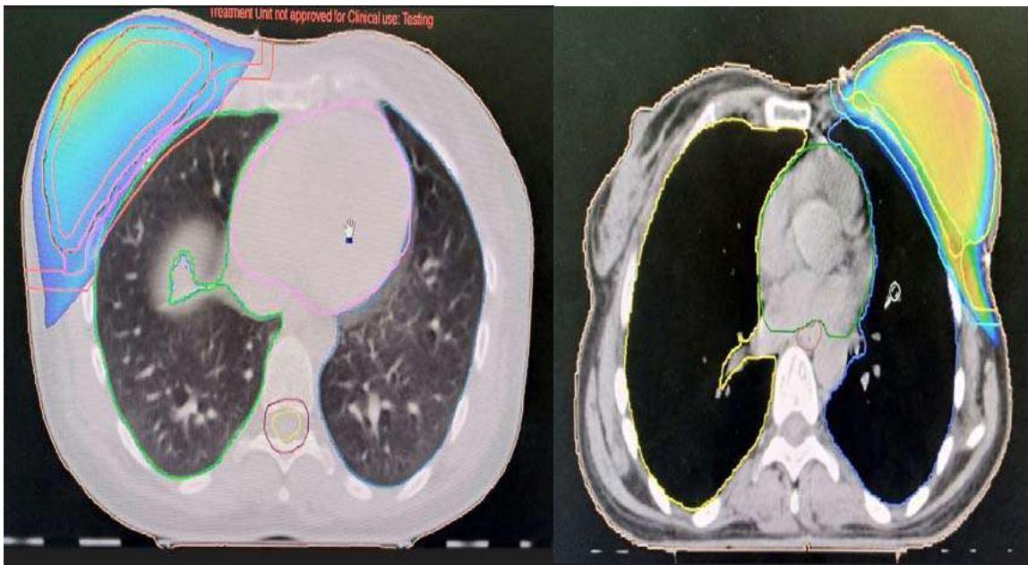
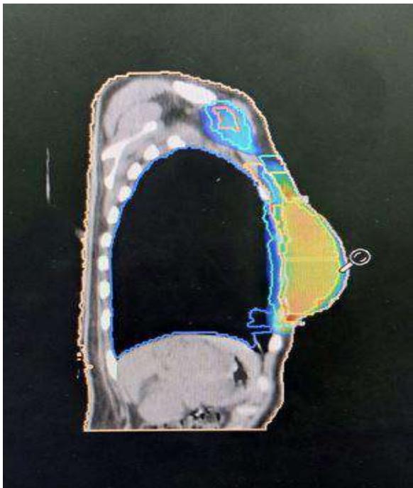 Figure 3: Dosimetric Distribution Images with the Breast Implant
Observation 5
We report the clinical observation of a 44-year-old patient, gender two and parity two, with no particular pathological history. She presented in July 2020 with right shoulder pain that had been evolving for about two months. Clinical examination revealed a patient in good general condition, with a left mastodynia associated with a nodule measuring approximately 3 cm on the left breast, rounded, mobile concerning the two superficial and deep planes, and without axillary adenopathy. Mammography and ultrasound revealed a right breast mass measuring 45 x 28 mm in large diameter, classified as ACR BIRADS 5. Histological examination and immunohistochemistry of the tru-cut biopsy fragments showed the presence of an invasive breast carcinoma of the left breast, SBR grade III without vascular emboli, with negative hormone receptors, negative Her2 score 1+, and Ki67 at 60%. A thoracic-abdominal-pelvic CT scan and bone scintigraphy were used to assess the extent of the disease, showing no distant secondary lesions. Neoadjuvant chemotherapy consisting of four courses of Adriamycin 60mg per m2 - Cyclophosphamide 600mg per m2, and four classes of Paclitaxel 175mg per m2 was administered, followed by subcutaneous mastectomy with left axillary lymph node dissection. On pathological examination, non-specific infiltrating breast carcinoma, 3.5cm large diameter, SBRIII, vascular embolus present, healthy margins, oN+/22N. Adjuvant chemotherapy (8 sessions of capecitabine 1000 - 1250mg per m2 twice daily D1 to D14) and adjuvant radiotherapy at a dose of 50 Gy to the left wall and supraclavicular area were performed, followed by placement of a left breast prosthesis.
Dosimetric distribution of the left wall and supra-clavicular size was obtained without great difficulty during radiotherapy sessions, as the patient had no breast implant at the time of radiotherapy.
Observation 6
A 36-year-old female, G4P3, non-menopausal, with no particular pathological history, was consulted for the appearance in August 2020 of a mass in the right breast associated with mastodynia. On clinical examination, the patient's general condition was unchanged. In the right breast, there was an approximately 3cm mass, which was rugged and mobile about the deep and superficial planes, and large homolateral axillary adenopathies, approximately 3cm in size and fixed about the deep plane. The rest of the clinical examination was unremarkable. Bilateral mammography showed a patch of overdensity in the lower internal quadrant of the right breast, spiculated in places and extending over 66mm on the front view, classified as BI-RADS 6, and left breast classified as BI-RADS 1. A biopsy of the right breast mass was performed, and the pathological anatomy examination noted a non-specific SBR grade 2 invasive breast carcinoma without vascular emboli. Immunohistochemistry showed estrogen receptors at 1%, progesterone receptors negative, Her2 negative, and Ki67 at 80%. A thoracic-abdominal-pelvic CT scan showed no secondary or suspicious bone lesions, and a 46 x 42 mm necrotic mass in the right breast, with multiple large homolateral axillary adenopathies measuring approximately 3.5 cm. The patient received chemotherapy, four sessions of Adriamycin-Cyclophosphamide, and four sessions of Paclitaxel, with three episodes of febrile neutropenia. The patient subsequently underwent subcutaneous mastectomy with right axillary curage. Histological examination revealed residual invasive breast carcinoma of non-specific type, SBR grade 3, with vascular emboli. The retro-mamelonal zone was non-tumoral, the surgical section boundaries were healthy, TILs were estimated at 10%, and an absence of lymph node metastases oN+ /9N, ypT1cNo classified RCB III. Immunohistochemistry showed negative hormone receptors, negative Her2, and 80% Ki67. She subsequently underwent adjuvant chemotherapy with capecitabine and 40.05 Gy of radiotherapy to the right wall and proper axilla supra clavicular area. One year after radiotherapy, the patient underwent deferred breast reconstruction.
II. DISCUSSION
Immediate breast reconstruction using implants or prostheses has become a widely used technique for breast reconstruction after mastectomy in recent years [9]. According to the Korean Breast Cancer Society registry, breast reconstruction surgeries almost tripled between 2002 and 2013 [8, 10]. In this Korean Breast Cancer Society 2017 report, the percentage of breast implants after mastectomy was for women aged under 40, for women aged 40-59, and for those aged 60 and over [8,11]. Breast reconstruction is an essential component of treatment. Studies have consistently demonstrated improved in quality-of-life indices and aesthetic results [12, 13]. Most reconstructions performed after mastectomy for breast cancer are stent- or implant-based [12, 14]. One commonly used technique has been the initial placement of a subpectoral tissue stent for patients who may require radiotherapy after mastectomy. This stent is then replaced by a permanent implant months after the end of radiotherapy [12]. However, in recent years, immediate direct implant reconstruction has been increasingly used for breast cancer patients undergoing mastectomy [12, 15]. Although immediate breast reconstruction by implant or prosthesis is a safe and effective technique, limited data are available on outcomes for patients undergoing post-mastectomy radiotherapy following immediate and permanent implant reconstruction [12, 16, 17]. Immediate and permanent implant reconstruction offers patients several opportunities, the possibility of a single surgery (mastectomy with reconstruction and breast implant placement) [12,18]. Secondly, it provides a more enhanced aesthetic result due to the preservation of the submammary fold, enabling a more natural appearance and also the possibility of adjusting the position of the scar . More importantly, for women, it offers enormous psychosocial benefits by restoring femininity and improving vitality, sexuality, and quality of life . However, the benefits of such an approach must be weighed against the oncological outcome and complications . For patients who prefer immediate implant reconstruction, it is essential to counsel patients on the potential difficulties associated with radiation planning and toxicities . It has an acceptable rate of post-operative complications and, from an oncological point of view, is considered safe, with a local recurrence rate ranging from 2% to 10% . Complications following irradiation of a breast implant for immediate breast reconstruction include the risk of capsular retraction, unsatisfactory aesthetic results, and replacement of the prosthetic breast implant. As for breast implant placement after radiotherapy, the various complications are more complex and painful expansion, risk of thoracic deformation and prosthesis exposure, and unsatisfactory aesthetic results.
III. CONCLUSION
Direct, permanent implant reconstruction is increasingly used for patients undergoing mastectomy. It has become an essential part of the multidisciplinary management of breast cancer patients because of the importance of the psychosocial functions of the symmetrical breast mound. Post-mastectomy radiotherapy reduces recurrence and improves survival. The presence of reconstructed tissue or implants can compromise radiotherapy planning and adversely affect its final results. However, reconstruction should not impact radiotherapy dose, fractionation, or irradiated areas. Multidisciplinary collaboration between radiation oncologists, surgical oncologists, plastic surgeons, and medical oncologists is imperative to provide the best care for our patients.
Conflict of Interest
The authors declare no conflict of interest.
Ethical Approval
Not applicable
Data Availability
The datasets used in this study are openly available at [repository link] and the source code is available on GitHub at [GitHub link].
Funding
This work did not receive any external funding.
References
Cite this article
Related Research



Special Issue
Launch a focused special issue to highlight research, emerging trends, and expert insights in your academic field.
