




IntelliPaper
Abstract
Practical training is essential for learning and mastering various medical approaches, being a differential for quality professional training. As a result, over the years, cadavers and live animals have been used for such execution, and these options are ethically questionable in several areas. Currently, there are emerging techniques that seek to offer an alternative to the use of animals in education in an economically viable and skillful way. Using computed tomography images, free software for tissue segmentation, polygon mesh editing and a 3D FDM printer with PLA filament to create biomodels that are biosafety, rigid to support orthopedic training, recyclable and biodegradable. With these tools it was possible to set up a virtual and physical database of canine and feline skeletal anatomy with direct application in academic teaching in veterinary medicine.
Explore Digital Article Text
I. INTRODUCTION
Corpses have always been a common choice in veterinary education over the years for training various clinical surgical techniques, however this option is always dependent on fresh specimens, free of infectious contagious pathogens, zoonotic and/or pathological dependent. Another option is the use of live animals, exposing the patient to an error rate on the part of unexpected candidates, which can generate irreversible injuries and even lead to death, conflicting with ethical issues. However, in recent years, there has been a growing trend in universal medical education to place more emphasis on the practical teaching of clinical and surgical procedures more commonly performed in private practice, thus requiring new means to achieve this goal of finding both physical and digital alternatives that are not harmful to animals for application in teaching (GRIFFON, 2000; KNIGHT, 2007; WARAN et al., 2013; REIS et al., 2017; MASHARI et al., 2018).
The use of Computed Tomography (CT) combined with the use of CAD software and the Rapid Prototyping (RP) technology has been a bold and innovative method to generate synthetic biomodels analogous to biological, and can be used easily with students and veterinary medical teams for several trainings. These techniques have been providing clinicians and surgeons with the planning and best approach for each situation encountered in the routine and which instruments to adopt to treat the patient in the most efficient and safe way (PLOCH et al., 2016; BORDELO et al., 2018).
Virtual biomodeling is the necessary step to obtain a digital model, which reproduces the morphological characteristics identical to that of a determined anatomical structure found in vivo. 3D modeling software uses images from CT, magnetic resonance exams or photogrammetry to build a replica of a structure in the polygon mesh language (FREDIEU et al., 2015; BORDELO et al., 2018). Prototyping, or physical biomodeling, is obtaining physical biomodels through 3D printing, building a model from scratch through the extrusion of melted material with overlapping layers on the Z axis, being the technique of Fused Deposition Modeling (FDM) the most economically accessible (SILVA & GAMARRA ROSADO, 2014). 3D printing is a unique tool when used with medical images, having a considerable positive impact in the fields of modern medicine. Pieces can be made to assist complex surgeries, training professionals in delicate surgeries, enabling the creation of personalized pieces, with high morphological realism of the anatomical structures, providing clarification for preoperative planning and their training in several areas (GRAUVOGEL et al., 2012; WARAN et al., 2013; PLOCH et al., 2016; SHI CHEN, 2017). Physical and digital anatomical models can be created in a short period of time according to the skill of the team involved or a professional facilitator, in addition to being able to use free digital editing software, reducing the cost of producing the models (HESPEL et al., 2014; FREDIEU et al., 2015; BORDELO et al., 2018).
The availability of these materials reduces the need for biological or commercial anatomical models, being applied to an alternative form of study, promoting good assimilation and allowing students to move between the physical and virtual world in a dynamic and interactive way (SILVA & GAMARROROSADO, 2014; FREDIEU et al., 2015; REIS et al., 2017). It can also be applied to clarify tutors or guardians, exploring treatment options and approaches, performing simulations with the structures to be addressed. Alternative methodologies to animal studies is a necessity within the scientific community, with the 3Rs program (reduce, recycle and reuse), a goal to be achieved in research, and met through the use of biomodels created by RP (FREDIEU et al., 2015; PLOCH et al., 2016; SINGHAL et al., 2016; BORDELO et al., 2018).
The accuracy of biomodels is suitable for orthopedic applications, enabling training and planning for both simple and complex surgeries such as total hip arthroplasty, for example, in addition to generating the opportunity to tangentially handle the model and make immediate comparisons (FREDIEU et al., 2015; GRAUVOGEL et al., 2012; MASHARI et al., 2018). The use of 3D FDM printers promotes low production costs and allows the generation of clinically identical replicas, with no distortions, providing dimensional accuracy compared to other PR technologies (HESPEL et al., 2014). This represents an advance in the accessibility of new technologies for use by surgeons in training prior to surgery, reducing the use of animals, valuable time and costs in the operating room (HARRYSSON, 2003; ASMA and EDDINE, 2014; MASHARI et al., 2018).
The objective of this study was to create a canine and feline bone bank, both physical and digital, for theoretical and practical educational purposes in the veterinary surgical clinical area in a fast, economically viable manner, with realistic quality and portability, helping to reduce the need for use of animals and corpses in veterinary orthopedic training.
II. ANIMAL MODEL
For this experiment, a cadaver of an adult dog and an adult cat without defined breed were used as experimental models, both obtained from ethical sources at the Veterinary Pathology sector of the Veterinary Hospital of the Universidade Federal de Mato Grosso-Cuiabá. The canine specimen had no bone fractures or deformity, the feline specimen had multiple complete fractures in its right pelvic limb due to the cause of death, being run over a car.
III. METHODS
The specimens were properly dissected by hand with a scalpel, removing all muscles and soft tissues, preserving only bone structures. The canine specimen was completely disarticulated, preserving the skull, pelvis and limbs. The feline specimen was dissected in two stages, dislocating the limbs and discarding the fractured one. In the first moment, the complete spine separated from the long bones was used, in the second moment the long bones were disjointed and isolated for further data capture.
The bones were scanned using the SOMATON Spirit Siemens® device in the Diagnostic Imaging sector at HOVET-UFMT Cuiabá, using the helical computed tomography technique, with the Kv and mAs values generated automatically by the equipment in each protocol adopted. In the first moment, the pelvic and thoracic limbs of the dog were scanned, using the Long Bones protocol, bone window with 1.5 mm spacing between the slices with 0.5 mm image overlap. In a second moment the dog's skull, jaw and pelvis was scanned under the Skull Bone protocol, bone window with 1.5 mm spacing and 0.5 mm overlap between slices. To capture the data of the felid the same protocols as that of the dog had been used, only alternating the arrangement of the parts for capture, making possible the capture in addition to the limbs, pelvis, skull and the fully articulated spine (Figure 1).
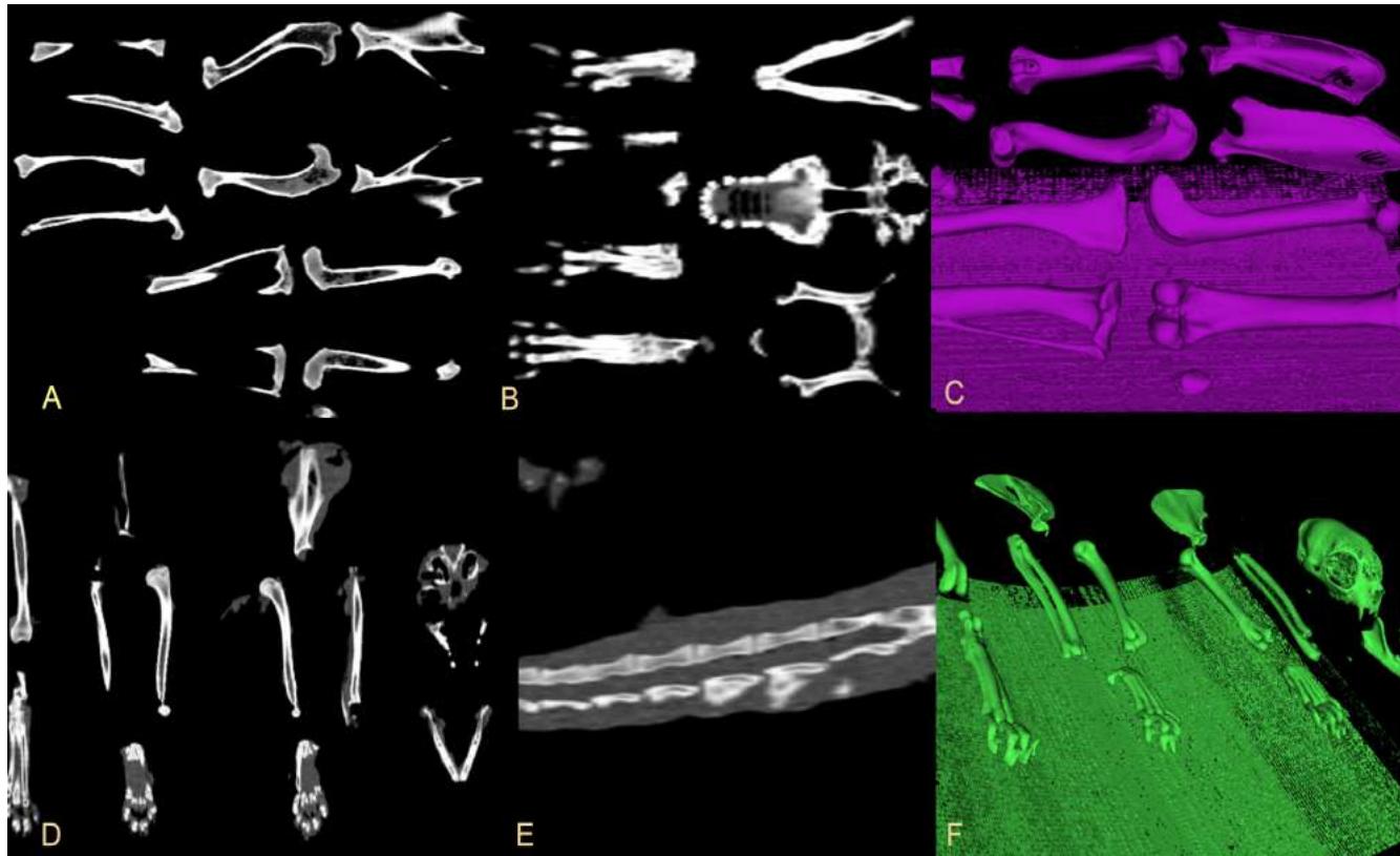 Figure 1: Tomographic projections analyzed in the InVesalius software. (A) Canine long bones in bone window with dorsoventral projection. (B) Dorsoventral projection with bony window of the skull, pelvis and canine paws. (C) With the Housted threshold applied, it is possible to create a 3D polygon mesh of the bone structures with the desired density. (D) Dorsoventral projection of uptake of long bones, skull, pelvis and feline paws. (E) Laterolateral projection of the spine showing the medullary canal in the healthy feline lumbar area. (F) 3D projection of feline feline polygon mesh exportable in.stl format.
All images were saved on CD-R in DICOM (Digital Imaging and Communications in Medicine) format and subsequently analyzed by the free software InVesalius®, allowing to view the tomographic images, apply image filters and also regulate the threshold value of the Hounsfield scale generating the segmentation of different types of tissues present in the exam. With the region of interest segmented, a polygon mask is generated creating an interactive 3D surface that allows it to be saved in an STL (Stereolithography) file.
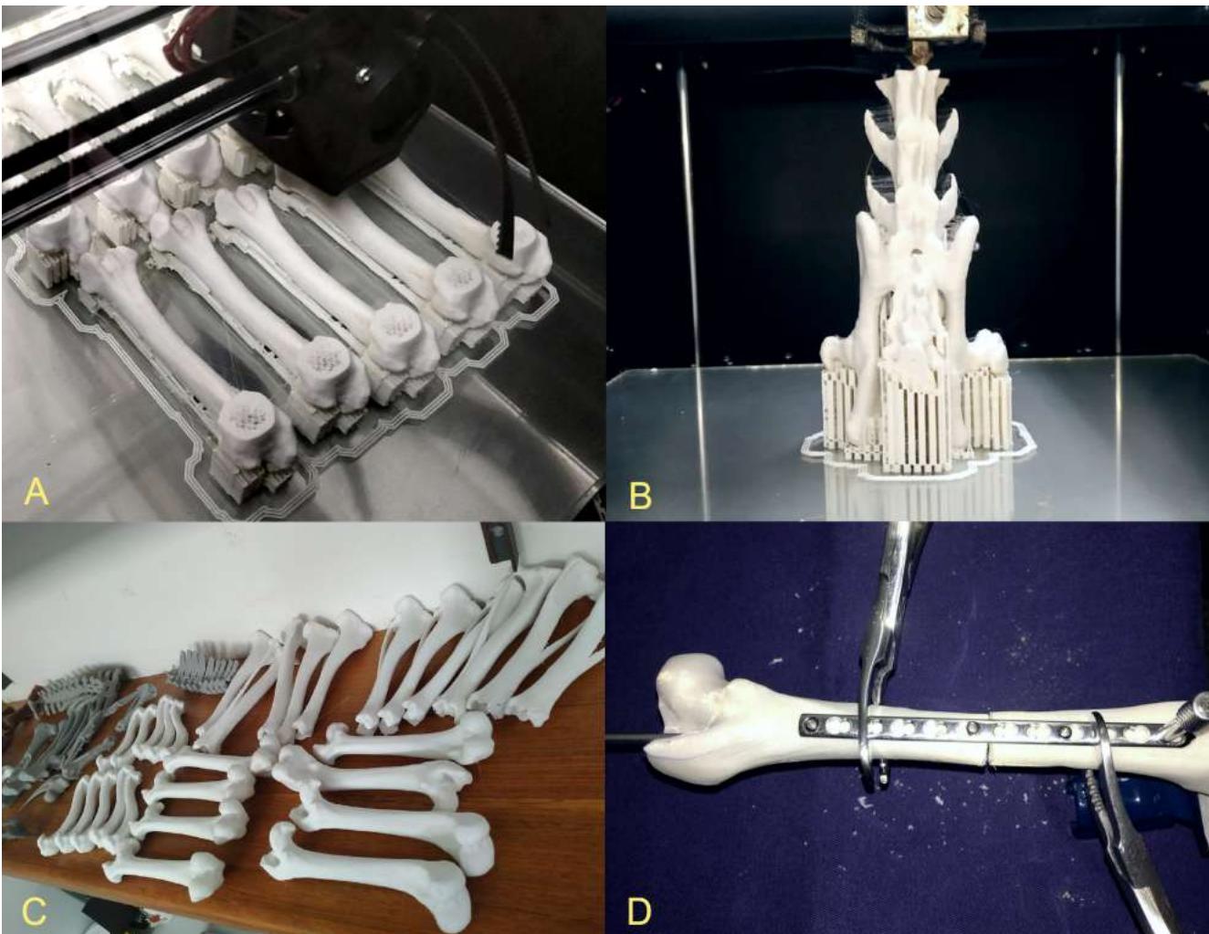
Using the Meshmixer® educational version software,.stl files were imported allowing them to be isolated, smoothed, mirrored, transformed into solid objects and to fill in gaps when necessary. After completing the virtual biomodeling, a digital library was created where all the bones were saved separately in folders on the central computer of the operating room, with copies in external HD and cloud (Figure 2). With the stored virtual biomodels, it is possible to sequence the FDM RP using a 3D printer of the GTMax3D® Core A3v2 model. Each file was opened separately in the Simplify3D® software and previously stipulated the total size of the piece, its positioning, supports and infill. For all models, the 3DX® filament composed of Polylactic Acid (PLA) with 1.75 mm in diameter was used under the following protocol described by Lima (2019); automatic support, 40% filling, 3 layers of detailing, thickness between layers of 200 microns, heating the extruder nozzle to 195°C with extrusion speed of 30 mm/s and nozzle displacement speed of 40 mm/s. After the print definition, the files were saved in.x3g format on an SD card and attached to the 3D printer to perform the PR. At the end of the 3D printing, each piece received a manual finish, with the support brackets removed with metal clamps followed by the finish with sandpaper for wood in the protruding areas, in order to generate greater smoothness and realism (Figure 3).
With a digital and physical bank of canine and feline bones assembled, undergraduate and graduate students at the UFMT School of Veterinary Medicine were able to use the biomodels in theoretical classes to understand the anatomy of the limbs and their classifications for fractures, followed by a practical class of orthopedics fractures using all the necessary equipment (drill, surgical forceps, saw, plates, drills, pins) to perform osteotomies in techniques with simple and blocked plates, external and cerclage fixation. The skulls, jaws, vertebral column with delimitation in the pelvic area were used to exemplify theoretical and practical classes of veterinary anesthesia about loco-regional block in the maxillary, mentonian, infraorbital, palatal and incisor pterygopalatine fossa. In the pelvis, an epidural block simulation was performed between the seventh lumbar and the first sacral, where students were able to train on the biomodel first, then on cadavers and later on routine patients at the hospital.
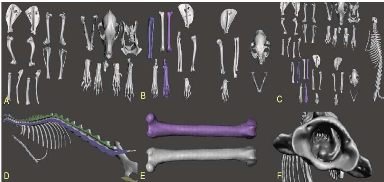 Figure 2: Interactive 3D data edited in the MeshMixer software forming the virtual database. (A) Canine bone bank. (B) Feline bone bank, including the digitally created right pelvic limb highlighted in cold tones. (C) Complete bone bank, including long bones, skull, pelvis, canine and feline limbs in addition to the feline spine. (D) Feline spine after digital dissection, showing the healthy spinal canal and nerve foramens, ideal to understand the points of anesthetic blocks loco regional. (E) Using the healthy part of the feline pelvic member, could mirror it creating the absent member analogous to the scanned, evidenced bone created in purple hue. (F) The 3D pieces allow virtual explorations of the anatomical details, in the image the medullary canal is highlighted, being seen craniodorsal through the base of the atlas and the exit of the nerve branches through the lateral foramina.
 Figure 3: Physical biomodels bank created from 3D FDM printing with PLA filament. (A) Multiple biomodels being produced simultaneously to meet the demand of undergraduate classes. (B) Vertebral column in the pelvic portion being printed, the positioning of the piece is essential for a quality impression. (C) Bank of physical bones assembled for use in the medical school routine. (D) Orthopedic training of variable techniques using surgical tools required in the physical biomodels.
IV. RESULTS
The bank of digital and physical bones has refined the quality of theoretical-practical classes in anatomy, anesthesia, general veterinary orthopedics and orthopedic surgical training for undergraduate and graduate students. Because its content is digital and physical, students were able to access digital models on their smartphones before and during classes. Spatially understanding the patient's anatomy and the area of interest, in addition to being able to carry out practical training for various situations several times. Participants reported at the end of the school semester that they were much more confident to perform the surgery in vivo after having had multiple planning and training classes on synthetic biomodels.
The capture of the feline spine allowed those involved to understand the clinical anatomy and cases of spinal pathologies using sectional CT and 3D images. This prototype also ensured that students understand the minutiae of vertebral anatomy, visualizing the shape and diameter of the foramina and intervertebral spaces, understanding how to orient themselves between the spinous processes to designate the correct area and depth of anesthetic application as suggested by Mashari (2018). Guiding a practical theoretical epidural anesthetic blocking class without pressure before the class had contact with routine patients. The capture of the skulls and mandibles proved to be very satisfactory to reproduce via 3D printing the details of the bone structures and facial foramina, incorporating them also in anesthetic and surgical classes.
The various prototypes also provided simulations of specific fractures in regions of interest, as well as their correction using fixing plates, drills, pins and the technique of fracture reduction cerclage. The physical biomodels are resistant to support special orthopedic materials serving for training and surgical planning, allowing to elaborate the desired alteration and perform its practical surgical correction. Ensuring adequate training in orthopedics and also the verification of the procedure through simple radiographs in order to guarantee the integrity of the parts and the precision of the technique adopted (LIMA et al., 2019). The material used for printing guarantees to be biodegradable in case of disposal, promotes the biosafety of those involved because it is not of animal origin and is free of pathogenic agents, they are subject to sterilization, recycling, in some cases reuse.
V. DISCUSSION
The action of dismantling the limbs and removing the musculature is not related to an improvement in the technique of capturing images, but in the logistics of the experiment. Once the animal was prepared after the end of the routine necropsy and only scanned days later, making it impossible to keep them intact due to their maintenance, decomposition and storage process. The prepared pieces are more practical for such care and when image captures were performed, the fact that they were dissected provided that several bones were scanned at the same time, positioned at a distance from each other. The data when analyzed in the free software InVesalius® proved to be very efficient for segmentation, excluding the possibility of the bones leaving connected due to the presence of cartilage and approximation of structures when articulated. This is a technique recommended by us for the development of digital libraries with greater practicality, optimizing the time of 3D editing.
Even though the feline specimen did not have a viable pelvic limb, using the MeshMixer® software it was possible to choose healthy elements and create a mirrored copy of the bones. Ensuring that the database had both the left and right pelvic limbs. This proves to be an alternative for expanding the library when dealing with complex cases, rare pathologies, congenital malformations or anomalies.
The PLA physical biomodels derived from CT capture combined with virtual editing and 3D printing sequentially, are promising since they do not need supercomputers to perform all processes, have sufficient strength for practical training, use biodegradable and recyclable material. This technique guarantees accessibility and replicability of rapid prototyping endlessly on any 3D printer model over the years due to the file format (.stl) being compatible with different brands, printer models and materials. But to achieve this goal, it is necessary to have a specialized professional who assists in the entire process to ensure the excellence of the final product, being necessary to understand both imaging medicine, surgical clinic as well as having skills with 3D technology.
Corpses are commonly used in teaching over the years, but it is worth mentioning that even in corpses there is a lack of realism and physiological responses as well as synthetic biomodels. However, corpses are more complex to acquire, maintain, use and dispose of, adding costs to the process, the need for legal authorization and presenting ethical conflicts. Biomodels can be used for training and testing of new techniques, as well as in cadavers, however without restrictions and with the possibility of multiple attempts improving the skills of professionals and thus promoting shorter anesthesia time in a future patients, being the surgical time always a critical factor that should always be minimized (WARAN et al., 2013). A universal convention has been taking place in teaching and training techniques most commonly performed in professional practice, creating a demand for materials for clinical and surgical training, which can be resolved through the capture of biodata and 3D printing in the teaching institution itself. This being a practical and viable alternative for reducing animals in veterinary education, reducing the cost of acquiring new materials and optimizing logistics, improving the anatomical spatial perception and facilitating the understanding of the techniques in their approaches, promoting animal welfare, improving quality of life of the patient and the team for applying the 3R in teaching (GRIFFON, 2000; HARRYSSON, 2003; GRAUVOGEL et al., 2012; ASMA and EDDINE, 2014).
A limitation in the application of 3D printing in veterinary medical education and also a significant cost factor is the availability of digital 3D models, usually acquired via computed tomography or magnetic resonance imaging (HESPEL et al., 2014). As an alternative to the use of photogrammetry in anatomical parts, however, this technique requires more effort from those involved and takes more time, a few days, to obtain the final product when compared to CT is just a few minutes. The printing time varies according to the piece size, quantity, speed and percentage of pre-fill adopted in the protocol. On average, each long bone took about 2 hours to be made while skulls and spine take more than twice the time to ensure the necessary detail (LIMA et al., 2019).
Currently, many tutors are committed to animals and are willing to do their utmost to help them, generating economic return and covering the costs for the creation of biomodels (HARRYSSON, 2003; FREDIEU et al., 2015). It is possible to refer patients to perform imaging exams in specialized centers outside the clinic or original teaching institution and return with the saved DICOM files, so that it is possible to work with such data and reproduce the biomodels via 3D printing. Since the cost of the printer and filament is low when compared to the tomography scanner, however, it is necessary to have a professional facilitator who performs the entire process from segmentation, biomodeling to finishing the materialized prototypes (HESPEL et al., 2014; FREDIEU et al., 2015).
This study considerably reduces the number of animals required for surgical training in an academic semester using alternative measures, helping to reduce the dependence of older methods on PR, promoting ethical teaching and increasing the useful life of natural resources (BOYD; CLARKSON; MATHER, 2015; MASHARI et al., 2018). Certainly, the use of biomodels for simulation is very effective in the learning system of the anesthetic and interactive surgical clinic, it is extremely important for us to implement new technologies that protect the lives of animals and that also respect the integrity of cadavers (MASHARI et al., 2018). The use of alternative models is not intended to completely replace the use of animals in teaching and research, but to minimize and complement practical training in an ethical, accessible, ecologically correct, sustainable and efficient manner. The recognition of the animal's value and the ethics that surrounds it has been discussed, expanded and applied in common global interest the need to use the 3R principle more frequently in academic and scientific routine (ASMA and EDDINE, 2014; GRIFFON, 2000).
VI. CONCLUSION
With this experiment we can affirm that from a single cadaver of each species, canine and feline, it is possible to create a digital database with 2D images of the clinical anatomy of bone structures in different formats such as: DICOM,.png,.gif,.jpeg,.tif... As well as 3D models in.stl and.obj format that can be accessed dynamically, interactively and remotely on smartphones, tablets, notebooks and 3D printers. Making the material inclusive and accessible to those involved, in addition to the prospect of continuous and repeated use of these files as an embedded teaching tool in institutions, promoting the maintenance of natural resources in an ethical manner and applying the 3R principle.
The final product does not present a risk of biological contamination because it is entirely made of PLA, it is resistant to support special orthopedic materials serving for academic training and surgical planning, allowing the elaboration of the desired alteration and making its most appropriate surgical correction. Both the digital and physical databases can be used in surgical medical clinic, with tutors, students and professionals. Optimizing the understanding of the case and the approaches to be taken. When necessary, multiple prototypes can be made simultaneously to assist in the training and understanding of each case with large classes, assisting in the understanding and spatial dimensioning of the area of interest in an ethical, dynamic and biosafety manner.
Conflict of Interest
The authors declare no conflict of interest.
Ethical Approval
Not applicable
Data Availability
The datasets used in this study are openly available at [repository link] and the source code is available on GitHub at [GitHub link].
Funding
This work did not receive any external funding.
References
Cite this article
Related Research



Special Issue
Launch a focused special issue to highlight research, emerging trends, and expert insights in your academic field.
