


IntelliPaper
Abstract
Leiomyoma is a benign neoplasm originating from mesenchymal cells, affecting the smooth muscle of the genitourinary tract. These tumors can occur in various places throughout the urinary tract, mainly in the bladder, but renal involvement is less frequent and is usually in the renal capsule. They are generally asymptomatic, and diagnosis is frequently incidental through imaging studies. When symptomatic, flank pain, palpable abdominal mass or hematuria can be present. This report describes a patient with lower back pain whose computed tomography(CT) revealed a complex Bosniak IV renal lesion. Later, the patient was submitted to a partial robotic nephrectomy and diagnosed with leiomyoma through immunohistochemistry.
Explore Digital Article Text
I. CASE REPORT
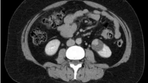
A 44-year-old woman with a history of long-term lower back pain had a contrast-enhanced computed tomography (CT) showing a nodular, solid-cystic lesion in the left kidney, classified as Bosniak IV (Figure1). The mass had a Renal nephrometry score of 8x, measuring 39 mm (1, 54 in).
Due to the clinic-radiologic suspicion of a complex cystic renal neoplasm, a left partial nephrectomy was performed together with an anatomo pathological study of the sample that identified a mesenchymal pattern suggestive of leiomyoma. The sample was then sent to an immuno histochemistry analysis that confirmed the diagnosis of renal leiomyoma.
The patient's condition improved after the surgical intervention.
II. DISCUSSION
Mesenchymal tumors of the kidney, although rare, must be considered in the differential diagnosis of complex kidney lesions. In this context, we reported a case of a patient investigating long-term lower back pain, in which a computed tomography (CT) showed a Bosniak IV lesion. Posteriorly, the patient was submitted to a left partial nephrectomy and the anatomo pathological study revealed a mesenchymal pattern tumor, later confirmed to be a renal leiomyoma by immunohistochemistry.
The renal leiomyoma is a benign mesenchymal tumor that originates from smooth muscle cells found in the renal capsule, renal pelvis, renal calyx and blood vessels. It is a rare neoplasm, with prevalence in autopsies ranging from 4,2% to 5,2%, representing only 1,5% of benign kidney tumors, and 0,3% of all treated kidney tumors .
Leiomyomas mostly affect adults, with peak incidence during the fourth decade of life, with a female-to-male ratio of 2:1. They represent approximately 0,29% of primary renal masses .
In most cases, leiomyomas are asymptomatic. Because of that, the diagnosis is commonly made incidentally during imaging for unrelated conditions. When symptoms are present, they include flank pain, palpable abdominal mass and hematuria.
Macroscopically, they present as firm, nodular masses that are non-capsulated and well-circumscribed, with rare calcifications or cystic features, no necrosis, frequently single and of various sizes . Microscopically, the tumor resembles leiomyomas found in other soft tissue, composed of long, fusiform cells organized in intersecting fascicles with abundant eosinophilic cytoplasm and an elongated, expressionless nucleus, with blunt ends (spindle-shaped cells) . The neoplastic cells present few nuclear polymorphisms and no mitotic activity, hyperchromatism or perilesional invasiveness.
The diagnosis is made based on the histopathological analysis and confirmed by immunohisto chemistry , since the radiologic findings are similar to those of other renal neoplasms.
The immunohistochemistry study is positive for vimentin, smooth muscle actin, myosin, desmin, laminin and type IV collagen . In contrast, tumor cells are negative for low molecular weight keratin, c-Kit and HMB45. The cell proliferation index (Ki-67) is low, which reinforces the benign nature of the neoplasm .
The treatment consists of surveillance or surgical resection, depending on the size, location and symptoms. It has an excellent prognosis after surgical excision.
This case is relevant because most diagnosis of leiomyoma are made in solid tumors, and in this case, the initial presentation was of a complex renal cyst, which makes the diagnosis even more challenging. It underscores the importance of clinical, radiological and histopathological correlation when evaluating renal masses.
List of Abbreviations
CT: Computed Tomography.
RENAL: Categorizes renal masses by complexity. Ki-67: Cell proliferation index.
ACKNOWLEDGMENTS
We thank the medical team at Hospital São Vicente de Paula for the clinical support.
Conflicts of Interest: The authors declare no conflicts of interest.
Conflict of Interest
The authors declare no conflict of interest.
Ethical Approval
Not applicable
Data Availability
The datasets used in this study are openly available at [repository link] and the source code is available on GitHub at [GitHub link].
Funding
This work did not receive any external funding.
References
Cite this article
Related Research


Special Issue
Launch a focused special issue to highlight research, emerging trends, and expert insights in your academic field.