



IntelliPaper
Abstract
Background: Pulmonary hydatid cyst (PHC) is a parasitic infectious disease, that is endemic in various regions worldwide. Thoracoscopic treatmentremains the treatment of choice for PHC.
The objective of this study is to assess the feasibility of video-assisted thoracic surgery (VATS) and compare the outcomes of patients undergoing uniportal VATS (U-VATS)with those undergoing multiportal VATS(M-VATS) for the treatment of PHC.
Methods: A retrospective analysis of medical records from 134 patients whounderwent VATSfor PHC between January 2018 and January 2022 was conducted. Among them, 90 (67%) patients who underwent M-VATS, while 44 (33%) patients underwent U-VATS.
Parameters including patient characteristics, cyst diameter, surgical duration, time to drain removal, length of hospital stay and complications were compared between the M-VATS and U-VATS groups.
Results: There were no significant differences in patient characteristics, cyst diameter and surgical duration between the two groups.
The time to drain removal and length of hospital stay in the U-VATS group were significantly shorter than those of the M-VATS group. Postoperative complications were not significantly different between the two groups. There was no postoperative mortality in either group. Throughout the follow-up period, no recurrence was observed in either group.
Conclusions: VATS with uni or multiportal was determined to be a safe and effective technique for the treatment of PHC and can serve as an alternative to traditional thoracotomy.
Explore Digital Article Text
I. INTRODUCTION
Hydatid disease, also referred to as echinococcosis or hydatidosis, is a parasitic infectious disease that is common in rural regions where cattle and dogs are in abundance. The disease affects all ages, both sexes, and no organs or tissues are spared (1). The causal parasite of the disease is Echinococcus Granulosus, for which humans can serve as intermediate hosts (2).
In Morocco, hydatid disease is endemic with an incidence rate of 4.55/100000 individuals (3).
The liver is the most frequently affected site, accounting for 60-80% of cases, followed by the lungs, which account for 10–30%. Unlike in adults, pulmonary hydatid disease is more common than hepatic hydatid disease in pediatric patients (4).
Pulmonary hydatid cyst (PHC) is often symptomatic and may lead significant morbidity. Surgical intervention remains the gold standard in the treatment of PHC. The aim of surgical management includes complete elimination of the parasite, preservation of the utmost of the healthy tissue, prevention of intraoperative rupture, ligation of bronchial fistula and capitonnage of the residual cavity (5, 6). These can be achieved through either the traditional postero-lateral thoracotomy or a minimally invasive approach.
Indeed, video-assisted thoracic surgery (VATS) appears to be an excellent alternative to thoracotomy, offering well-established advantages (1, 5, 6).
Our goal of this study was to present a series of cases involving the management of PHC using VATS. We aimed to assess the benefits of this approach and compare the outcomes between patients undergoing uniportal VATS (U-VATS) and those undergoing multiportal VATS (M-VATS).
II. MATERIALS AND METHODS
2.1 Patients and Data Collection
We have retrospectively collected and reviewed the medical records of 134 consecutive patients who underwent surgery for PHC at our department between January 2018 and January 2022. We excluded patients with intrathoracic extrapulmonary cysts from this study and no specific selection criteria were applied for the VATS approach.
We divided the patients into two groups: Group A consisted of 44 (33%) patients who underwent U-VATS, while Group B comprised 90 (67%) patients who underwent M-VATS. Conversion to thoracotomy was necessary in 12 patients (13.3%). The reasons for conversion included intolerance to single lung ventilation (n=4), severe adhesions (n=6) and central localized cysts (n=2).
We collected demographic data, cyst's characteristics, intraoperative and postoperative data.
All patients underwent physical examination and routine laboratory tests. In the preoperative evaluation, chest X-rays and thoracic computed tomography (CT) scan (as shown in Figure 1 and 2) were used to determine the location, size, and integrity of the cysts. The presence of hepatic cysts was established through abdominal ultrasonography. Flexible bronchoscopy was selectively performed in patients with a history of hemoptysis to rule out a concomitant endobronchial disease. Serologic testing was used routinely for diagnostic workup.
For complicated hydatid cysts, preoperative preparation based on albendazole treatment, antibiotics and postural drainage is necessary.
2.2 Surgical Technique
Video-assisted thoracoscopic surgery (Figure 2) was performed under general anesthesia with the use of double lumen tube in order to isolate the affected lung during surgery and to avoid the possible spillage of cyst contents into the contralateral bronchus. The patient was placed in a lateral decubitus position.
A M-VATS using a standardized three-port anterior approach. A 3–4 cm utility incision was made in the 4th intercostal space along the mid-axillary line. A 1 cm camera port was made in the 6th or 7th intercostal space along the anterior axillary line to create an access port for observation and a 1.5-cm incision was made in the posterior axillary line at the same level of the camera port.
For U-VATS, a 4–5 cm incision was made in the anterior to mid axillary line usually in the fifth intercostal space, or according to the location of the cystic lesion. The camera was mostly held in the posterior part of the incision by the assistant surgeon.
The utility incision was protected with an incision protector (Ring Protector*). The lung was spared from adhesions of chest wall and the cyst was identified and surrounded by 10% hypertonic saline to prevent dissemination in case of a ruptured laminated membrane.
The choice of surgical technique depended on the location, size, and intact state of the hydatid cyst.
For cysts located deep inside the lung parenchyma, the technique was needle aspiration plus cystotomy plus bronchial fistula suturing and capitonnage of the residual cavity.
For geant superficial cysts, a technique of needle aspiration plus cystotomy plus partial pericystectomy plus bronchial fistula suturing and capitonnage of the residual cavity.
For small superficial cysts, wedge resection was performed.
For complicated cysts with destroyed lungs, the anatomical resections (lobectomy or segmentectomy) were used.
For cysts ruptured into the pleural cavity, the decortication may be needed.
The surgical intervention was completed following chest tube placement into the pleural space and closure of the chest. All cysts were subjected to histopathologic examination to confirm the diagnosis.
2.3 Follow-up
Patients were followed up by chest X-rays at 1, 3, 6 and 12 months after surgery and every year thereafter. All patients received postoperative
Albendazole treatment at a dose of 10 mg/kg per day, administered for three courses of 21 days each. Between each course, patients did not take the drug for 7 days.
Liver function tests were checked at the end of each course. If the tests were abnormal, the next treatment of Albendazole was postponed until the liver enzymes were normal.
2.4 Statistical Analysis
Continuous variables were reported as mean and standard deviation. Categorical variables were reported as frequency and proportion.
The statistical analysis has been performed by Fisher's exact test for categorical variables and Student's t-test for continuous variables, utilizing SPSS version 17.0 software. The difference was considered as significant for values .
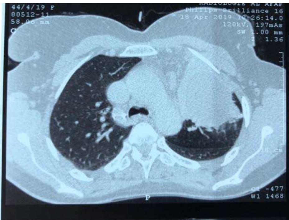 Fig. 1: Preoperative CT Scan Showing Hydatid cyst in left Upper Lobe Destrying Culmen
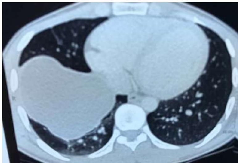 Fig. 2: Preoperative CT Scan Showing Intact Hydatid Cyst in the Right Lower Lobe
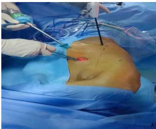
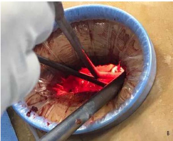
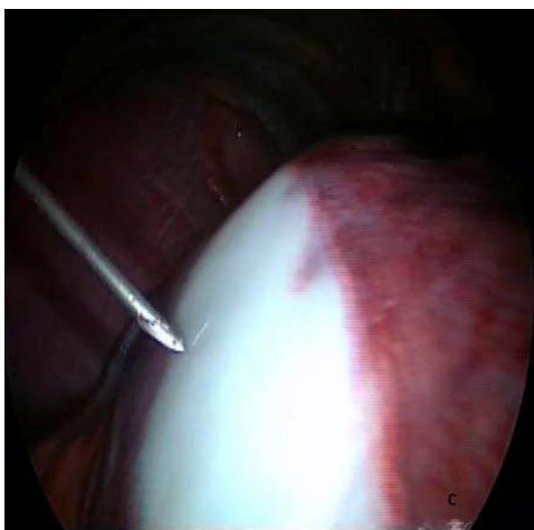
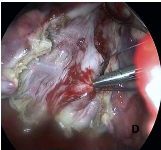 Fig. 3: Incision and Surgical Instrumentation of Multiportal VATS (A) and Uniportal VATS (B). A Needle Aspiration (C). Bronchial Fistula Suturing (D)
III. RESULTS
In this retrospective study, 134 patients with PHC were collected. Group A consisted of 44 (33%) patients of whom 22 were female (50%) and the mean age was 32.25± 17.24 years (range 6-81 years). Among the 90 (67%) patients in Group B, 46 were female (51.1%) and the mean age was 35.14±16.68 years (range 8-78 years). 73 patients (54.5%) were from pastoral areas and had a long history of close and prolonged contact with domestic animals. Liver cystic lesions were found in 95 (70.9%) cases. The characteristics of the patients are shown in Table 1. The demographic characteristics_were not significantly different between the two groups.
115 (85.8%) patients were symptomatic. The presenting signs and symptoms of patients are shown in Table 2. The most common symptom was chest pain, followed by hemoptysis and cough. There was no significant statistical difference in the rate of symptoms between the 2 groups.
In total, 176 cysts operated in both groups. 74 cysts were located in the left lung and 102 cysts in the right lung. Dimensions of the cyst lesions varied from 1.5 to 12 cm. 8 patients (6%) presented with multiple cysts and 36 patients (26.9%) with cyst rupture. Perioperative characteristics of pulmonary hydatid cyst in two groups are shown in Table 3.
There was no significant statistical difference in the average diameter of the cysts, location and number of the cysts, anatomical location and operation type of PHC between the 2 groups (P >0.05). Mean length of surgery was comparable between the two groups (136.74±54.23 minutes versus 98.15±32.75 minutes; p=0.35).
The postoperative complications were not significantly different between the two groups. The time to drain removal and length of hospital stay were significantly shorter in U-VATS group (2.6±1.7 versus 4.7±1 and 3.1±1.4 versus 5.2±1.3; p\<0.05, respectively). Postoperative outcomes are shown in table 4.
No postoperative mortality was seen in either group. The patients were followed up for an average of 14.2 months (range 2-50 months) and no recurrence, no mortality was reported among the operated patients of both groups.
IV. DISCUSSION
Hydatid disease is a major health problem in Morocco. PHC is often symptomatic, as shown in our study. Symptoms are probably due to the mass effect or bronchial opening of the cyst (6).
Medical management may be applied in ruptured hydatid cysts or in preventing recurrences after surgery. In our study, preoperative medical therapy was administered in 36 (26.9%) patients with ruptured cyst, and all patients received Albendazole as postoperative medical treatment for a duration of 6 months.
Surgery remains the gold standard for patients with PHC, associated with low rates of morbidity and mortality (5, 7). VATS treatment of PHC was first described by Becmeur in 1994 (8-10). From that time, several studies were presented (5, 9, 11-13).
The principal objectives of the surgery are to remove the cyst, to prevent the cyst rupture at the operative site, to close the bronchial openings and to manage the residual cavity (2).
Preoperative assessment of a patient with PHC for VATS removal allows determining the number, the diameter and the localization of the cysts (5, 7, 14). In our experience, all types of cyst are most suitable for U-VATS and M-VATS.
In the study by Mehta and al (1), the authors showed that VATS is feasible for the management of single and uncomplicated lung hydatid cyst, and they found shorter operative duration, earlier drain removal and less postoperative analgesia treatment.
Alpay and colleagues (5) found that VATS approach was better in terms of chest pain, cosmetic result, length of surgery, drainage volume and duration of chest tube.
In 2018, Ocakcioglu et al (14) performed the first U-VATS removal for PHC in 18 patients. One year later, Abu Akar et al (8) also reported a comparative evaluation of U-VATS patients (n=23) versus thoracotomy patients (n=16) in the management of lung hydatid disease. They concluded that a single thoracoscopic approach is a safe option that can be used as an alternative to open surgery.
In the literature, the rate of conversion is 12.6% (1, 16). In our study, it was 13.3%.
To summarize, all studies favoring VATS treatment of PHC had significant rates of complication or morbidity, consistent with our study.
Just two recurrences after VATS approach were reported out of 221 patients in the literature (7). No recurrence was seen during the period of follow-up in our study. Boubia et al reported that the long-term prognosis was excellent (15).
V. CONCLUSION
VATS is an effective, feasible and safe surgical approach in the treatment of PHC. It has the advantages of shorter time to drain removal and the hospital stay time. Thoracoscopy can be used as an alternative to thoracotomy, in the hands of experienced surgeons.
ACKNOWLEDGMENTS
None
Declaration of conflicting interests
The author(s) declared no potential conflicts of interest with respect to the research, authorship, and/or publication of this article.
Funding
The author(s) received no financial support for the research, authorship, and/or publication of this article.
Conflict of Interest
The authors declare no conflict of interest.
Ethical Approval
Not applicable
Data Availability
The datasets used in this study are openly available at [repository link] and the source code is available on GitHub at [GitHub link].
Funding
This work did not receive any external funding.
References
Cite this article
Related Research


Special Issue
Launch a focused special issue to highlight research, emerging trends, and expert insights in your academic field.
