



IntelliPaper
Abstract
Congenital hemangiomas are benign vascular tumors fully developed at the time of delivery, in which the proliferative phase occurs exclusively in utero, so they never increase in size (1). The incidence is similar in both sexes and there are four types according to the International Society for the Study of Vascular Anomalies (ISSVA), rapidly involuting congenital hemangioma (RICH), non-involuting congenital hemangioma (NICH) and slowly and partially involuting congenital hemangiomas (SICH and PICH) (2)(3).
Explore Digital Article Text
INTRODUCTION
Congenital hemangiomas are benign vascular tumors fully developed at the time of delivery, in which the proliferative phase occurs exclusively in utero, so they never increase in size (1). The incidence is similar in both sexes and there are four types according to the International Society for the Study of Vascular Anomalies (ISSVA), rapidly involuting congenital hemangioma (RICH), non-involuting congenital hemangioma (NICH) and slowly and partially involuting congenital hemangiomas (SICH and PICH) (2)(3).
We present the case of a newborn male with a vascular tumor in the right knee.
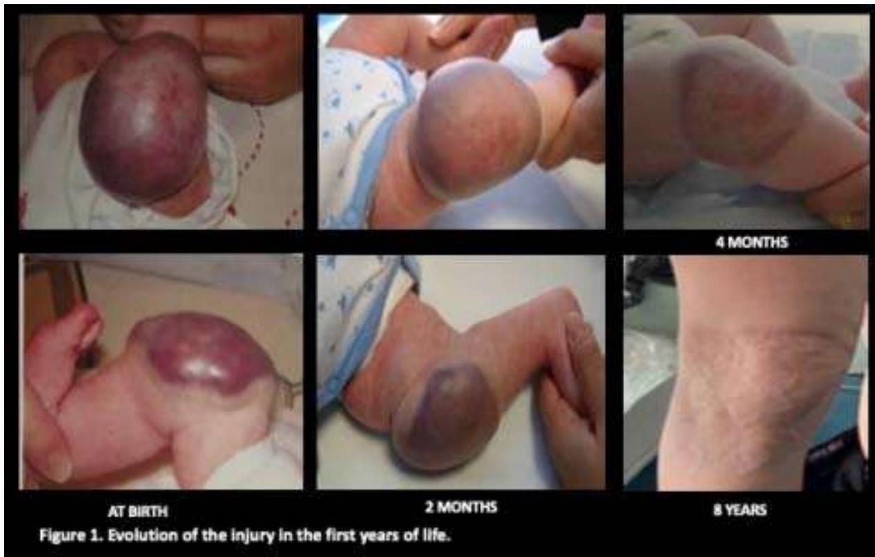
Born at term by eutocic delivery after a controlled pregnancy with normal prenatal ultrasounds, the examination presented a rounded, painless, delimited and purplish mass in the right knee measuring about 5x5 cm (Figure 1). The lesion is neither friable or ulcerated. There are no other skin lesions. Femoral pulses are present and symmetrical. Cardiopulmonary auscultation is normal and there are no signs of heart failure.
Doppler ultrasound was performed at birth, visualizing a hyperechogenic mass with serpingine structures in its thickness with Doppler uptake and high-velocity arterial and venous flow, compatible with a vascular anomaly such as a vascular tumor (4).
In initial blood analysis, moderate thrombopenia and normal coagulation. Assessed by Children's Cardiology, normal electrocardiogram and echocardiogram, minimal shunt from left to right through the foramen ovale in closing pathways with no evidence of ventricular dysfunction.
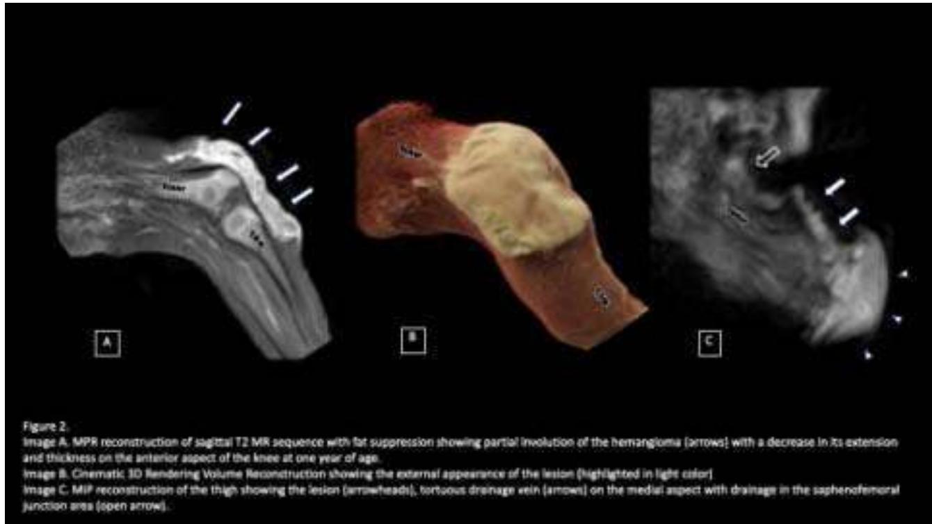
The study was completed with Magnetic Resonance (MRI), confirming a soft tissue tumor circumscribed in the anterior aspect of the right knee measuring 6.8 cm x 6.4 cm x 2.1 cm. The lesion appears isointense on T2 sequences with few serpinginous tubular structures with flow voids inside compatible with blood vessels. It presents varicose veins draining the Greater Saphena (SM) on the anterointernal aspect of the thigh. (Figure 2).
Due to its full development at birth, its location and the absence of T2 hyperintensity and prominent flow voids on MRI, congenital hemangioma was considered the most likely diagnosis.
A wait-and-see attitude was decided, confirming during follow-up the progressive reduction in the size of the hemangioma (Figure 1) with normalization of platelet numbers in control analysis and disappearance of the shunt.
At one year of age, the mother reports that she sometimes has a lump in the right inguinal region. On examination, he presented a smaller vascular lesion in the right knee and a tumor in the root of the right thigh with varicosities on the anteromedial aspect of the thigh.
MID Doppler was performed, verifying that the clinical tumor corresponds to the dilated and incompetent arch of the SM, probably related to hyperflow of the varicosities visualized on MRI.
In subsequent controls, complete involution was confirmed at two years of age with residual skin atrophy on the anterolateral aspect of the knee with collateral vascularization that did not increase with the valsalva. The inguinal lump was reduced until it completely disappeared and the patient is currently leading a normal life without limitation or discomfort in the area.
We can conclude that this is a rapidly involuting congenital hemangioma (RICH). According to the ISSVA classification, it is a benign vascular tumor, which has regressed, with only mild hyperpigmentation and skin atrophy persisting.
They can present as violaceous tumors with telangiectasias and phlebectasias, hard pinkish bulging nodules with pale halos, compact and lobulated tumors with normal superficial skin color, or infiltrated violaceous plaques with a bluish halo.
We must perform a differential diagnosis with the rest of vascular tumors to avoid unnecessary aggressive behavior since their evolution, prognosis and treatment are different (5). Due to the involution of the lesion, we ruled out other benign vascular tumors, such as infantile hemangioma or NICH hemangioma and tufted angioma.
As the thrombopenia resolved and there was no consumption coagulopathy, we rejected the hypothesis of more aggressive vascular tumors such as kaposiform hemangioendothelioma. The MRI characteristics, age, circumscribed nature with absence of congenital lymphedema and rapid progression made the possibility of epithelioid hemangioendothelioma and angiosarcoma unlikely (6).
RICH hemangiomas present more or less complete involution before the first year of life, with conservative treatment being sufficient in most cases. Pharmacological treatment is ineffective and excision exceptional. However, NICH hemangiomas do not present significant changes during their childhood, making their surgical removal necessary. ITS appearance requires an appropriate differential diagnosis with other soft tissue vascular tumors of the newborn of a locally aggressive and even malignant nature to choose the most appropriate management.
Conflict of Interest
The authors declare no conflict of interest.
Ethical Approval
Not applicable
Data Availability
The datasets used in this study are openly available at [repository link] and the source code is available on GitHub at [GitHub link].
Funding
This work did not receive any external funding.
References
Cite this article
Related Research



Special Issue
Launch a focused special issue to highlight research, emerging trends, and expert insights in your academic field.
