


IntelliPaper
Abstract
Introduction: Tibial plateau fractures account for approximately 1% of all fractures; however, they are associated with high morbidity due to a wide spectrum of soft tissue injuries and a significant incidence of concomitant ligamentous and meniscal lesions. Among these, anterior cruciate ligament (ACL) injuries are particularly relevant, as delayed diagnosis or inadequate management of these injuries can compromise joint stability and long-term functional outcomes.
Case presentation: We present the case of a 33-year-old male patient with a lateral tibial plateau fracture (Schatzker type I) associated with a complete anterior cruciate ligament tear and a fibular head fracture in the right knee following a traffic accident. The patient underwent ACL reconstruction using hamstring tendon autograft, along with open reduction and internal fixation of the tibial plateau and fibular head fractures. Postoperative evolution has been satisfactory, with full recovery of range of motion, a stable, pain-free knee, and favorable functional outcomes.
Discussion: Tibial plateau fractures are complex articular injuries with a high incidence of associated ligamentous damage, particularly involving the anterior cruciate ligament. Treatment should be individualized based on the fracture pattern and associated injuries. Arthroscopy-assisted techniques represent a viable option for reduction, fixation, and simultaneous management of intra articular lesions, contributing to joint function preservation and showing satisfactory functional outcomes.
Conclusion: This case highlights the importance of maintaining a high index of suspicion for associated ligament injuries in tibial plateau fractures, regardless of the fracture pattern. Additionally, the arthroscopy-assisted approach is emphasized as a valuable tool for achieving precise anatomic reduction and simultaneous anterior cruciate ligament (ACL) reconstruction in a single surgical procedure. The case presented, along with the available evidence, supports the combined approach as a safe, effective, and minimally invasive strategy that promotes early functional recovery and favorable clinical outcomes.
Explore Digital Article Text
I. INTRODUCCION
Tibial plateau fractures are complex intra-articular injuries that compromise both the articular surface and the ligamentous stability of the knee, in addition to presenting a wide range of clinical manifestations and long-term complications (3, 5). These fractures typically result from high-energy mechanisms in younger patients or low-energy mechanisms in older patients due to underlying bone quality. They are also associated with a significant incidence of concomitant ligament injuries, particularly involving the anterior cruciate ligament (ACL) (2,5).
The Schatzker classification, widely recognized and routinely implemented in clinical practice, is based on the two-dimensional representation of the fracture and provides guidance for classification and treatment according to the fracture pattern; however, it does not assess intra-articular soft tissue involvement (3).
Arthroscopic studies have documented a high frequency of partial or complete ACL injuries in Schatzker type II, IV, and VI tibial plateau fractures, with an incidence ranging from to . It is important to note that these injuries are often underdiagnosed during the initial preoperative evaluation, and their detection requires direct or arthroscopic exploration when clinical suspicion is present (4,6).
Over the years, the development of arthroscopy-assisted surgical techniques for tibial plateau fractures has allowed anatomical reduction under direct visualization, while simultaneously addressing associated ligamentous and/or meniscus injuries, in addition to minimizing soft tissue invasion (1,4).
In this context, and considering reports supporting simultaneous surgical management through fracture reduction and fixation along with ACL reconstruction in a single operative setting, we present the following clinical case with the aim of describing the surgical approach used and the clinical and functional outcomes obtained.
II. OBJECTIVE OF THE REPORT
To describe the clinical findings, diagnosis, treatment, follow-up, and postoperative outcomes in a patient with a Schatzker type I lateral tibial plateau fracture associated with a complete anterior cruciate ligament injury and fibular head fracture in the right knee. Additionally, to provide evidence regarding the surgical management of tibial plateau fractures associated with ligamentous injury.
2.1 Study Population
A 33-year-old male patient with a Schatzker type I lateral tibial plateau fracture associated with a complete anterior cruciate ligament (ACL) injury and a fibular head fracture in the right knee, sustained in a traffic accident, who was treated at Clínica Medicentro in Bogotá, Colombia.
2.2 Case Report
A 33-year-old male patient was admitted following a motorcycle accident, sustaining predominant trauma to the right leg and knee. On initial physical examination, there was significant swelling, functional limitation of the right knee, and an extensive hematoma over the anterolateral aspect of the proximal third of the leg.
Radiographs of the right leg and knee revealed a comminuted and displaced fracture of the fibular head, as well as a lateral tibial plateau fracture, Schatzker type I. Given the clinical context and imaging findings, an associated ligamentous injury of the right knee was also suspected.
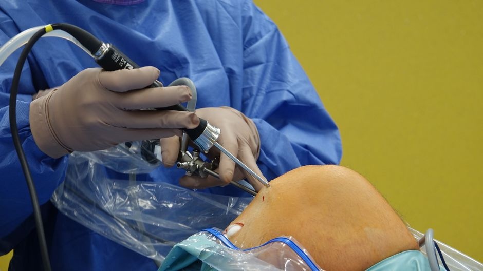
The patient underwent arthroscopically assisted open reduction and internal fixation (ORIF) of the lateral tibial plateau fracture, ORIF of the fibular head fracture and right ACL reconstruction. The procedure was initiated arthroscopically through anteromedial and anterolateral portals, confirming the presence of a complete ACL rupture.
Hamstring tendon harvest was performed through a medial approach for autograft preparation, followed by creation of the femoral tunnel. The graft was inserted using the open socket technique and secured at both proximal and distal ends, achieving adequate anteroposterior stability.
Subsequently, open reduction and internal fixation of the lateral tibial plateau fracture was carried out using three cannulated screws. Finally, reduction and fixation of the fibular head fracture was performed with a cannulated screw.
The patient showed satisfactory postoperative recovery and was discharged from the hospital. Follow up evaluations demonstrated full range of motion, Lachman test 1A, and a stable, pain-free knee.
III. DISCUSSION
Tibial plateau fractures are complex intra-articular injuries that account for approximately of all fractures but represent high morbidity rates, usually associated with soft tissue injuries. Their prevalence is estimated at 10.3 per 100,000 individuals per year (1), with a bimodal distribution: complex fractures secondary to high-energy trauma in young patients, and low-energy fractures in older adults, often favored by osteoporotic changes (5).
A detailed understanding of these fractures is a key element in selecting the most appropriate treatment. Several classification systems have been proposed, among which the Schatzker classification stands out. This system divides fractures into six types (I-VI) and is based on a two-dimensional representation of the fracture. It considers the proximal tibial epiphysis as having two axial columns—medial and lateral—each supporting its corresponding condyle with its articular surface. Types I to III involve the lateral tibial plateau: type I is a cleavage fracture of the lateral column (more common in young individuals), type II is a split fracture associated with articular depression, and type III is a pure articular depression. Conversely, types IV to VI are typically associated with high-energy trauma and joint instability: type IV corresponds to an isolated fracture of the medial column, type V is a bicondylar fracture where continuity between the diaphysis and the overlying joint is preserved, and type VI involves loss of continuity between the diaphysis and the articular surface (3).
Despite its clinical utility, the Schatzker classification is based on a two-dimensional radiological assessment and therefore does not encompass all fracture patterns. For this reason, three-dimensional classifications have been suggested. Kfuri and Schatzker, in their update of the original classification based on computed tomography, established anatomical landmarks to define a "virtual equator," dividing the tibial plateau into anterior and posterior halves, thereby segmenting the tibia into four articular quadrants. This new classification allows identification of discontinuity of the articular rim and subsequent loss of joint stability, thus guiding the choice of surgical approach and fixation method (3).
A frequent but less addressed feature of these fractures is the presence of concomitant soft tissue injuries. Mechanism of injury, trauma force, and degree of osteopenia are factors influencing both fracture pattern and associated meniscal and ligamentous damage. In a retrospective analysis of 98 tibial plateau fractures assessed arthroscopically, Abdel-Hamid et al. identified a high prevalence of associated soft tissue injuries, noting that the ACL was the most frequently affected ligament, thus reinforcing the diagnostic of arthroscopy in these fractures. Similarly, in a series of 31 tibial plateau fractures treated with arthroscopy-assisted osteosynthesis, Shih et al. reported a incidence of ACL injuries, of collateral ligament injuries, and of lateral meniscal injuries (6).
In line with these findings, Deng et al. performed arthroscopic evaluation following closed reduction and internal fixation of tibial plateau fractures, documenting a range of injuries and incidence rates. Among 185 fractures assessed, ACL injuries were diagnosed in of cases and posterior cruciate ligament (PCL) injuries in . ACL injuries were most frequently associated with Schatzker type IV fractures, presumably due to the classic injury mechanism of lateral plateau subluxation, which generates rotational and shear loading forces on the ligament (4). Although high-energy fractures such as Schatzker type IV-VI, account for the majority of ligamentous injuries, a high index of suspicion should be maintained in all tibial plateau fractures.
Treatment of tibial plateau fractures may be conservative or surgical; however, most are displaced and unstable, thus requiring surgical management. The choice of approach and fixation method depends on the fracture pattern. In general, unicondylar fractures (Schatzker I-IV) can be managed with compression screws or buttress plates, whereas bicondylar fractures (Schatzker V-VI) require separate incisions and fixation with plates on both condyles. In addition, minimally displaced fractures (types I-III) are suitable for treatment with percutaneous techniques, assisted by either arthroscopy or fluoroscopy (5).
When comparing open reduction with arthroscopy-assisted surgery, a reduction in morbidity and lower complication rates, including infection, nonunion, and reoperation, have been demonstrated. With complication rates ranging from to (1). However, its use is not recommended in complex and comminuted fractures (types IV-VI), due to the high risk of fluid extravasation that may lead to compartment syndrome (1). Regardless of the chosen surgical method—open, fluoroscopy-assisted, or arthroscopy-assisted—the primary goal is to restore the articular surface to prevent sequelae such as osteoarthritis or its rapid progression.
There is still no consensus on the ideal approach to tibial plateau fractures associated with ligamentous injuries. Nevertheless, the main rationale for arthroscopic evaluation is that it allows direct visualization of articular surface reduction as well as concomitant intra-articular meniscal and ligamentous injuries, which can be diagnosed and treated simultaneously (4). While some authors recommend delaying reconstruction to avoid additional tissue damage, the minimally invasive nature of arthroscopy allows these procedures to be performed effectively in a single stage. Reported outcomes are favorable, with fewer procedures required and shorter recovery times (2). Postoperatively, patients tend to resume weight-bearing at 6 to 12 weeks and achieve functional ranges of motion within the first months. Scheerlinck et al., reported that of patients regained their pre-injury sports level after arthroscopy-assisted treatment, and in a subsequent study by Holzach et al., the return-to-sport rate reached (1).
In this context, we report the case of a young patient with a Schatzker type I tibial plateau fracture and a comminuted, displaced fibular head fracture, with additional suspicion of ligamentous injury. An arthroscopy-assisted procedure confirmed an ACL tear, leading to fracture fixation and ligament reconstruction. The patient achieved satisfactory recovery, with follow-up showing a stable, pain-free knee and full range of motion. This case highlights an uncommon and underreported injury, successfully managed through an arthroscopy-assisted surgical technique, resulting in favorable functional outcomes.
IV. CONCLUSION
This case highlights the importance of suspecting associated ligamentous injuries in tibial plateau fractures, even in patterns considered simple, such as Schatzker type I. The arthroscopic approach enabled precise anatomical reduction and simultaneous ACL reconstruction, achieving a satisfactory functional outcome. Current evidence suggests that, in selected patients, a combined single-stage surgical approach is a safe and effective strategy that promotes earlier functional recovery. However, further clinical evidence is required to establish more standardized therapeutic protocols for the management of these injuries, thereby optimizing long-term functional outcomes.
Ethical Considerations
Written informed consent was obtained from the patient for publication of the clinical case and the associated images, in accordance with the Declaration of Helsinki, international ethical guidelines, and the editorial policy of the journal.
Expected Results/Products and Potential Beneficiaries
- Present the clinical and diagnostic findings obtained in the study with the aim of publishing the case report for academic dissemination.
- Share the experience gained in the management of this specific pathology.
- Serve as a reference for future similar studies in Colombia and internationally.
- Contribute to medical training through locally generated, context-specific clinical evidence.
Expected Impacts Derived from the Use of the Results.
Conflict of Interest
The authors declare no conflict of interest.
Ethical Approval
Not applicable
Data Availability
The datasets used in this study are openly available at [repository link] and the source code is available on GitHub at [GitHub link].
Funding
This work did not receive any external funding.
Cite this article
Related Research


Special Issue
Launch a focused special issue to highlight research, emerging trends, and expert insights in your academic field.
