





IntelliPaper
Abstract
As an important product of glycolysis, lactate plays a crucial role in mitochondrial oxidative metabolism and gluconeogenesis. Lactate not only provides energy as a substrate to support cell growth and development but also acts as an important signaling molecule to affect the biochemical functions of proteins in cells, thereby regulating the corresponding biological functions. An important feature of energy metabolism in tumor cells is known as the Warburg effect, which is characterized by a heavy reliance on glycolysis and the production of large amounts of lactate. Scientists have successively reported that lactic acid (lactate with extra protons) is associated with cancer growth and immune suppression. But a new study shows for the first time that lactic acid promotes anti-tumor immunity by increasing CD8+ T cells in multiple tumor models1. This interesting discovery has led to a whole new understanding of the role of lactate in tumors. In this review, we not only summarize the role of lactate in the pathophysiological process of tumors but also further discuss new therapeutic modalities.
Explore Digital Article Text
I. INTRODUCTION
Lactate has long been thought to be a metabolic waste product produced by skeletal muscle during carbohydrate fermentation or anaerobic glycolysis during exercise. When cells have an excessive demand for oxygen and ATP, such as during strenuous exercise and infection, lactic acid is produced in large quantities . Lactate is a product of glycolysis during glucose metabolism, and the glycolysis pathway is activated to produce ATP when hypoxia inhibits the tricarboxylic acid (TCA) cycle, which is presented in Figure 1.
Specifically, cells produce pyruvate through glucose metabolism, and in the absence of mitochondrial oxidation, pyruvate is directly reduced to lactic acid by a process called lactate dehydrogenase (LDH) . In addition to glycolysis, glutamine catabolism is another source of lactate in cancer cells . Excessive accumulation of lactate in the human body can lead to lactic acidosis . In the early 1920s, Warburg found that cancer cells rapidly produce lactic acid even in the presence of oxygen, a process known as aerobic glycolysis . In proliferative cells, glycolysis is so highly vigorous to ensure that intracellular and extracellular concentrations of lactate is higher than those found in cells at the resting state .
Notably, lactate accumulation often occurs in the tissue microenvironment in inflammatory diseases and cancers. . In addition, many researchers report that the Warburg effect occurred in nontumor cells and noncancerous diseases, such as pulmonary hypertension, heart failure, atherosclerosis, pulmonary fibrosis, and polycystic kidney disease . The main factors affecting the formation of lactate are the low oxygen environment, the concentration of glycolytic product - pyruvate, and the PH of the surrounding environment. However, most cancer cells have a relatively low oxygen microenvironment, which is conducive to the stable production and expression of hypoxia-inducing factor-1 (HIF-1 ). HIF-1 has the effect of activating glucose uptake, improving glycolysis, increasing pyruvate production, and promoting the conversion of pyruvate to lactate. A RASSF1A-HIF1 loop drives the Warburg effect in cancer and pulmonary hypertension . As a glycolytic metabolite, lactate is not a waste product of cancer cell proliferation, but has a broad effect that plays an important role in tumor progression and metastasis . To avoid intracellular acidification, cancer cells rapidly export lactate via monocarboxylate transporters (MCTs) . This mechanism avoids excessive lactate concentration in the cell and ensures that aerobic glycolysis and lactate production continue while increasing lactate concentration in the extracellular tumor microenvironment (TME).
High lactate concentrations in the TME are associated with a poor prognosis . In this review, we will detail the biological role of lactate in cancer cells and TME, including their influence on cancer progression, metastasis, and anti-tumor therapy.
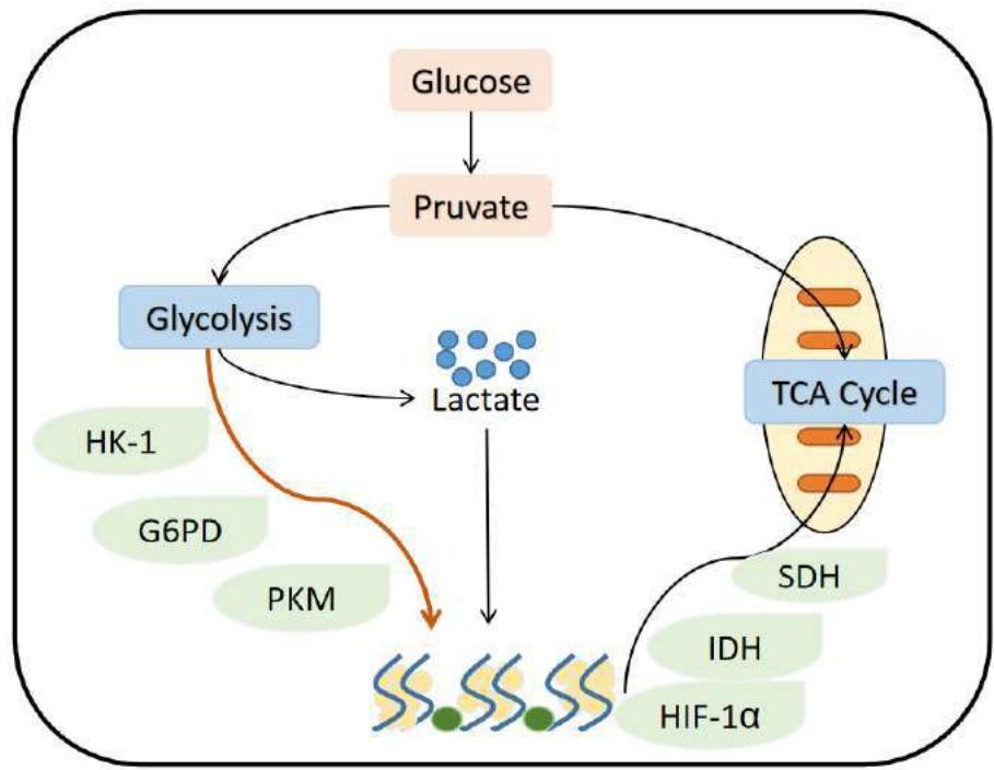 Figure 1: The Process of Lactate Metabolism
II. EFFECT ON TUMOR MICROENVIRONMENT
The tumor microenvironment (TME) is a microenvironment formed by the accelerated metabolism of tumor cells and cancer-related fibroblasts, which promotes tumor growth . Lactate transport is crucial for the survival of cancer cells in the TME. Lactate, produced by the Warburg metabolism of cancer cells, is secreted into the extracellular environment, forming an active niche for tumor pathogenesis and evolution, which plays a key role in promoting cancer progression . Because tumors grow faster than blood vessels form, cancer cells close to blood vessels are normoxic, while cancer cells far from blood vessels are hypoxic . Specifically, hypoxic tumor cells produce lactate via lactate dehydrogenase A (LDHA), which is exported out of the cells, and normoxic tumor cells will use this lactate for ATP production with the conversion of lactate into pyruvic acid via lactate dehydrogenase B (LDHB) . Monocarboxylate transporters (MCTs) belong to the SLC16 gene family, facilitating bidirectional symport of monocarboxylates and H+ across cell membranes . Based on the differential regulation by hypoxia-related genes, MCT4 can modulate the release of lactate from hypoxic tumor cells and MCT1 can modulate the uptake of lactate in normoxic tumor cells . Hypoxia is one of the main features of most tumors and tends to produce large amounts of lactate mediated by HIF1α . HIF1α can promote invasion and resistance to chemotherapy by activating Snail and Twist, two transcription factors involved in E-cadherin modulation . Numerous studies have shown that proton-coupled lactate efflux from cancer cells or stromal cells contributes to the preservation of acidic phenotypes and promote tumor progression by modulating TME, including cell invasion, survival signaling, angiogenesis, metastasis development, and evasion of immune surveillance . Cell surface lactate receptor GPR81 is crucial for cancer cell survival, presenting in the colon, breast, hepatocellular, lung, gland, cervical, salivary, and pancreatic carcinoma cell lines . Studies have shown that lactic acid secreted by tumor cells activates GPR81 on tumor cells, resulting in a carcinogenic phenotype .
Extracellular lactate levels can affect cells and modulate their function in TME, including cancer cells, T cells, NK cells, macrophages, and dendritic cells, which can trigger intracellular signaling that fine-tunes cell behavior . Because extracellular acidosis can suppress T-cell-mediated immunity, neutralizing tumor acidity can improve antitumor response to immunotherapy . Acidosis will affect the potential function of T cells. In TME, low pH reduces the expression of iNOS, CCL2, and IL-6 in M1 macrophages, but increases the expression of M2 macrophage markers, and also inhibits the anti-tumor activity of NK cells . Lactate high levels in TME also reduce lactate efflux from T cells, resulting in their decreased cytokine production and cytotoxic activity .
III. EFFECT ON TUMOR ANGIOGENESIS
Lactate can promote angiogenesis in TME, like a trauma-related microenvironment, stimulating endothelial cell activation and angiogenesis via HIF-independent and HIF-dependent pathways . Some evidence shows that the proangiogenic factor, vascular endothelial growth factor (VEGF), can be induced via both HIF-dependent and HIF-independent pathways . In the HIF-independent pathway, lactate is transferred into cells through MCT1, oxidized to pyruvate to produce NADH that can activate ROS production, which can stimulate angiogenesis . Moreover, lactate can facilitate angiogenesis by directly binding the NDRG3, which activates Raf-ERK signaling to mediate lactate-triggered hypoxia responses . With the low oxygen tension and high lactate concentrations, NDRG3 will bind c-Raf to activate RAF-ERK signaling and promote angiogenesis . The mechanism of the HIF-dependent pathway is that the relatively constant level of lactic acid can maintain the constant level of HIF-1α under normal oxygen conditions. Aberrant activation of the HIF pathway causes overexpression of angiogenic genes, like VEGF . HIF-1α is a vital regulator of metabolic reprogramming in hypoxic cancer cells . As the main oxygen sensors in cells, proline hydroxylases (PHDs) can hydroxylate HIF-1α at specific proline residues to promote the subsequent degradation of HIF-1α by the UPS under normoxic conditions . However, PHD is devitalized under hypoxic conditions. This inactivation of PHD suppresses the proteasomal degradation of HIF-1α, making HIF-1α migrate to the nucleus and facilitates the transcription of some tumor-promoting genes . Pyruvate, which is converted by tumor cells through lactic acid, competes directly with α-KG, inhibiting PHD activity and thus stabilizing HIF-1α levels . Therefore, stable HIF-1α levels can ensure high lactate concentrations in TME, thus guaranteeing transcriptional activation of tumor-promoting genes in all tumor cells .
VI. EFFECT ON TUMOR IMMUNITY
In some previous studies, lactate in the TME inhibits NK cell function and prevents the activation of NFAT in both T cells and NK cells, resulting in diminished IFNγ production . The decrease in NK cell function and IFNγ production will affect the anti-tumor effect. In the TME, the effects of lactic acid on cancer and immune cells are complex and difficult to decipher, which is further confused by acidic protons (a byproduct of glycolysis). Interestingly, there is the latest finding that has upended our conventional understanding of lactate, which can increase stemness of CD8+ T cells and augments anti-tumor immunity . This finding reveals metabolic reprogramming of immune function and distinguishes the role of lactic acid and tumor acidity in anti-tumor immunity. The experimental group injected lactate subcutaneously into colon cancer mice, and the control group injected glucose solution into tumor-bearing mice, and it was found that lactate treatment significantly inhibited tumor growth . Further single-cell RNA sequencing analysis showed that lactic acid treatment increased the number of infiltrated CD8+ T cells in the tumor . When the same experiment was performed in mice genetically engineered to lack CD8+ T cells, the tumor inhibitory effect of lactic acid was blocked . The researchers also found that lactic acid alone did not completely clear tumors; But when histone deacetylase (HDAC, a commonly used immune checkpoint inhibitor) was added, the tumors disappeared completely in about half of the mice .
In addition, lactic acid significantly improved the efficacy of anti-cancer vaccines and increased the anti-cancer response of CD8+ T cells cultured in tumor-bearing mice .
V. CLINICAL SIGNIFICANCE AND THERAPEUTIC TARGETS
Previously, lactic acid is considered an indicator of high malignancy and poor prognosis in several cancers. On the one hand, lactic acid inhibits immune cell action in TME. On the other hand, lactic acid can be directly absorbed and metabolized by tumor cells to promote the TCA cycle. The immunoprotective effect of lactic acid has not been fully appreciated before because the immunosuppressive effect of tumor acidity predominates. New research reveals the special immunoprotective role of lactic acid in anti-tumor immunity, thus suggesting that lactic acid could be used to supplement existing cancer immunotherapies . Lactate is widely used clinically as sodium lactate Ringer's or Hartmann's solution, and there is sufficient evidence that it can be safely used in fluid resuscitation and to reduce metabolic acidosis.
Current Ringer's fluid or modified solutions with increased lactate concentrations can protect CD8+ T cell function during immunotherapy in cancer patients. The addition of lactic acid to the expansion of isolated T cells may be beneficial for chimeric antigen receptor T cell (CAR-T) therapy.
Footnotes
Availability of data and materials
Not applicable.
Authors' contributions
KCX contributed to the manuscript writing and revision.
Funding statement
This work was supported by Jinhua Science and Technology Bureau (NO.2020-4-177). The funders have no influence on study methods, interpretation, or publication of results.
Competing interests statement The authors declare no conflict of interest.
Patient consent for publication Not required.
Conflict of Interest
The authors declare no conflict of interest.
Ethical Approval
Not applicable
Data Availability
The datasets used in this study are openly available at [repository link] and the source code is available on GitHub at [GitHub link].
Funding
This work did not receive any external funding.
References
Cite this article
Related Research


Special Issue
Launch a focused special issue to highlight research, emerging trends, and expert insights in your academic field.
