




IntelliPaper
Abstract
An aneurysm at the fenestration position of the basilar artery is an extremely rare condition that presents challenges in treatment. Aneurysm recurrence is a complication that can occur after interventions for aneurysms in this location. We report the case of a 43-year-old male patient with a ruptured basilar artery aneurysm treated with coils. After 30 months of follow-up, the aneurysm recurred and was treated with a flow diverter stent combined with coils.
Explore Digital Article Text
I. INTRODUCTION
Basilar artery fenestration has a reported incidence ranging from 0.6% to 2.33%, depending on the modality used to study this variant. When basilar artery fenestration is present, associated aneurysms are rare, with a reported incidence of 0.33% of all intracranial aneurysms . Currently, using coils to treat ruptured brain aneurysms has been proven to be a safe and effective treatment method. The main problem with this treatment method is the significant rate of incomplete occlusion of the aneurysm or the possibility of the aneurysm recurring after intervention over time, with the risk of aneurysm rupture causing recurrent bleeding . According to the literature, aneurysm recurrence or aneurysm neck reopening occurs in approximately 20% of patients, requiring retreatment in approximately half of them . However, there is no consensus or specific recommendation regarding retreatment after aneurysm coiling treatment. The use of flow-diverting stents in the treatment of posterior circulation aneurysms in general, and in the treatment of recurrent posterior circulation aneurysms after coils, is still controversial.
We discuss the case of a 43-year-old male patient who underwent coil intervention for a ruptured basilar artery aneurysm in August 2021. Thirty months after the initial treatment, an MRI scan showed aneurysm recurrence (approximately 7 mm in size), confirmed by DSA. The patient was then successfully treated with flow-diverting stents combined with coils.
II. CASE REPORT
A 43-year-old male patient was admitted to the hospital in August 2021 because of a subarachnoid hemorrhage around the brainstem and in the fourth ventricle (Fisher grade 3) due to a ruptured aneurysm at the basilar artery fenestration site (Figure 1).
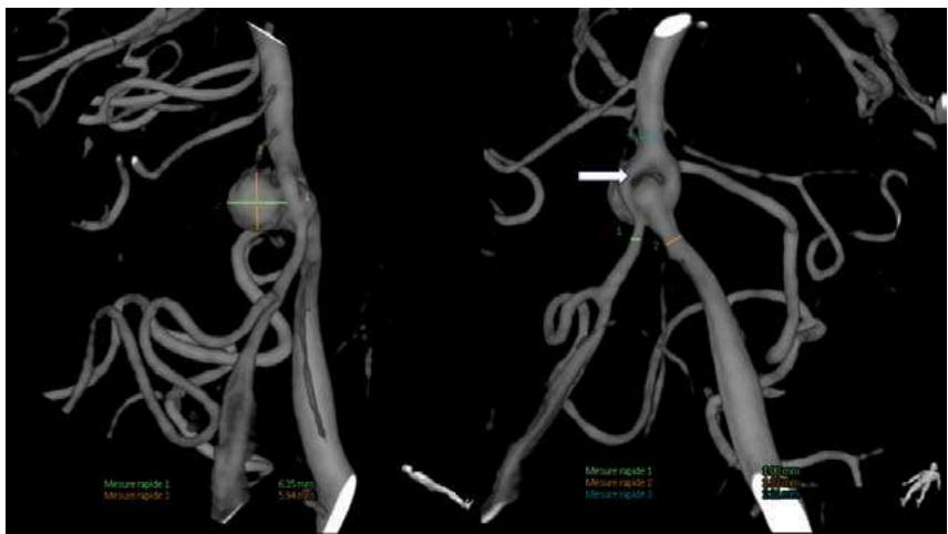 Figure 1: Aneurysm at the Basilar Artery Fenestration Site (arrow)
The patient was subsequently treated with coils under digital subtraction angiography (DSA). The procedure immediately showed complete occlusion of the aneurysm (Figure 2).
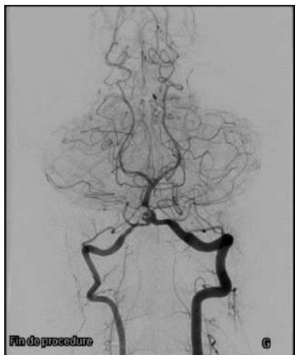
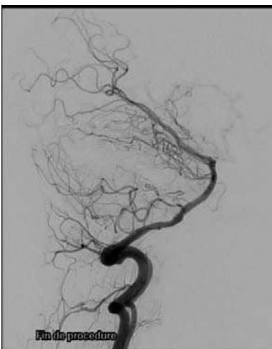
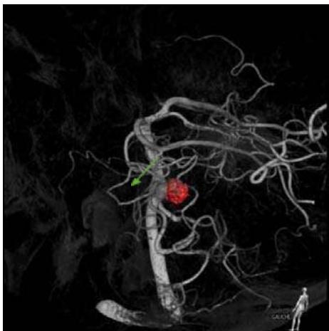 Figure 2: Dsa Scan Checked after the First Coil Button in August 2021, No Residual Bulge was Seen Thirty months after initial treatment, MRI revealed a recurrence of the aneurysm (approximately 7 mm in size) confirmed by DSA (Figure 3). Figure 3: Dsa Scan Shows A Recurrence of the Aneurysm at the Neck of the Old Aneurysm (Blue Arrow) and the Old Aneurysm has Been Occluded with Coils (Highlighted In Red)
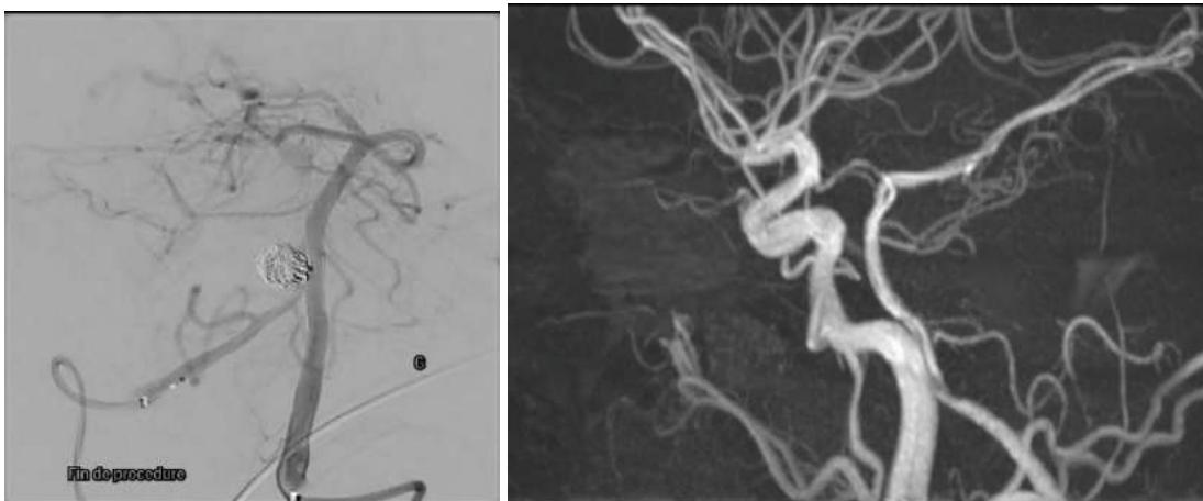
The patient was then successfully treated with a flow-reversing stent combined with coils. Endovascular intervention with access through the bilateral femoral artery, placing 01 8F sheath in the right femoral artery and 01 6F sheath in the left femoral artery. Place Phenom 21 into the basilar artery and Echelon 14 into the aneurysm. We chose the p64-MW 3.5 x 15 mm flow diverter following simulations with Sim and Cure. This is placed at the lower end of the basilar artery up to the left V4 segment to cover the aneurysm neck. Place 2 Optima coils into the aneurysm. DSA check immediately after intervention and cranial MRI 3 months after intervention showed complete occlusion of the lesion (Figure 4).
 Figure 4: Dsa Scan after Stent and Coil Placement Shows No Residual Bulge (A). Magnetic Resonance Image After 3 Months on Tof 3d Pulse Sequence Shows No Residual Bulge (B)
III. DISCUSSION
After the anterior communicating artery, the basilar artery is the second most common site of fenestration, with the rate of basilar artery fenestration ranging from 1% to 2.07% on magnetic resonance imaging, 0.28% to 6% at autopsy, and 0.02% to 0.6% on digital subtraction angiography . When basilar artery fenestration is present, associated aneurysms are rare, with a reported incidence of 0.33% of all intracranial aneurysms . Posterior circulation aneurysms in general, and basilar artery aneurysms in particular, pose significant challenges for interventionalists and neurosurgeons due to the complexity and special anatomical location of these lesions.
In recent years, endovascular treatment has been considered the "gold standard" for intracranial posterior circulation aneurysms due to its lower procedural complication rate compared with surgery . However, long-term post-intervention follow-up studies of posterior circulation aneurysms embolized with coils show a high rate of aneurysm recurrence or aneurysm neck reopening, estimated at about 20% of patients, with retreatment required in about half of them .
In our case, the patient initially had a ruptured aneurysm at the proximal end of the basilar artery, right at the proximal fenestration site of the basilar artery. The patient was then successfully treated with coils, and it seemed that the aneurysm was completely occluded. No residual aneurysm was seen on the DSA scan after the first intervention. However, 30 months later, there was a recurrence of the aneurysm at the neck of the old aneurysm. Choosing a treatment plan in this case was very difficult. In the literature, there is no consensus or specific recommendation regarding the retreatment of recurrent aneurysms after aneurysm coil treatment.
Endovascular treatment seems to be a more optimal option than surgery in this case. The use of flow-diverting stents in the treatment of recurrent posterior circulation aneurysms after coils is controversial. Using stents alone or combining stents and coils helps increase treatment effectiveness and reduce the risk of recurrence, but the risk of stent occlusion and early bleeding after intervention increases in these patients .
In our case, the patient was successfully treated with endovascular intervention, combining coils and flow-diverting stents, with no complications of embolism or early aneurysm rupture after intervention. Post-intervention check-up and follow-up after 3 months showed that the recurrent aneurysm had been completely occluded.
The progression of a brain aneurysm after intervention is unpredictable, making post-treatment follow-up crucial yet challenging. There is ongoing debate about whether long-term follow-up imaging should be performed after intervention and which imaging modality should be used. Digital subtraction angiography (DSA) is considered the gold standard for monitoring, but it has several disadvantages, including the risk of complications during the procedure due to its invasive nature and the risk of radiation exposure to the patient.
Magnetic resonance imaging (TOF 3D) is a suitable technique for monitoring post-intervention cerebral aneurysms. The Clinical Practice Guideline Committee of the KoNES (2024) recommends that CE-MRI or TOF MRA imaging be used to monitor patients 3-6 months after intervention, then at 1, 2, 4, and 6 years after treatment. Computed tomography angiography (CTA) is commonly used to evaluate intracranial arteries due to its lower cost and shorter scanning time compared to MRA. However, CTA involves the use of a contrast agent and radiation exposure, and post-intervention materials such as coils and stents can interfere with imaging .
Regardless of the technique, it is important to provide morphological information, including whether the aneurysm is completely occluded and whether there is any remaining neck or part of the aneurysm. There is no optimal imaging tool to evaluate residual or recurrent aneurysms fully. Regarding timing, follow-up imaging should be performed 3–6 months after treatment, and then over a range of 12 to 18 months. Subsequent follow-ups vary from center to center and must be tailored to the condition of the lesion after intervention.
IV. CONCLUSION
In summary, recurrence after treatment of brain aneurysms, particularly in the posterior cerebral artery system, is a risk that requires long-term monitoring. Treating recurrent aneurysms with flow-diverting stents combined with coils is a feasible, safe, and effective method, although it carries certain risks, such as premature aneurysm rupture or stent blockage after intervention.
Conflict of Interest
The authors declare no conflict of interest.
Ethical Approval
Not applicable
Data Availability
The datasets used in this study are openly available at [repository link] and the source code is available on GitHub at [GitHub link].
Funding
This work did not receive any external funding.
References
Cite this article
Related Research



Special Issue
Launch a focused special issue to highlight research, emerging trends, and expert insights in your academic field.
