




IntelliPaper
Abstract
Introduction: Pulmonary thromboendarterectomy (PEA) is the main method of treatment for patients with chronic thromboembolic pulmonary hypertension (CTEPH). The residual pulmonary hypertension (PH) after CTEPH surgical treatment is a risk factor with increasing of hospital mortality.
Objective: To analyze and evaluate the results of PEA in patients with different persistent time of medical history as a possible prognostic factor of residual PH and outcome.
Methods: Retrospective and prospective analysis of the PEA results in 87 patients operated on from April 2012 to February 2022 was conducted. The patients were divided into 3 groups. The 1st group - 45 patients with a medical history from 3 months to 1 year, the 2nd - 20 patients from 1 year to 3 years and the 3rd - 22 patients with long-term history more than 3 years.
Results: The average age of the patients was 48.7 ± 13.5 years, including 56.7 % males and 43.3 % females. Due to New York Heart Association (NYHA), 7 (8.0%) patients preoperatively belonged to class II, 60 (69.0%) to class III and 20 (23.0%) to class IV. Postoperatively the mean pulmonary artery pressure (mPAP mmHg) and pulmonary vascular resistance (PVR dyn·s·cm⁻⁵) significantly decreased in all groups (mPAP :1st group from 45±13 to 23±6, 2 nd – from 49±14 to 25±6 and 3 rd – from 58±12 to 31±7; PVR: 1st group from 797±262 to 290±135, 2 nd – from 925±383 to 376±159 and 3 rd – from 1248±332 to 505±189). Hospital mortality after PEA was 0 in patients with medical history less than 1 year, 5% from 1 to 3 years and 31% in patients with long- term anamnesis.
Conclusions: PEA is an effective surgery with mPAP and PVR decreasing in the early postoperative period. However, long-term medical history and PVR >1000 dyn·s·cm⁻⁵ are very important risk factors with increasing of hospital mortality.
Explore Digital Article Text
I. INTRODUCTION
Chronic thromboembolic pulmonary hypertension is a severe cardiovascular disease with a highly unfavorable prognosis if not treated in time. CTEPH is a complication of pulmonary embolism (PE) and a treatable cause of PH. The pathology is a combination of mechanical obstruction due to failure of clot resolution, and a variable degree of microvascular disease. The first decision remains assessment of operability, and the best improvement in symptoms and survival is achieved by the mechanical therapies, pulmonary endarterectomy and balloon pulmonary angioplasty. With the advances in multimodal therapies, excellent outcomes can be achieved with 3-year survival of >90%. This is a rare form of PH develops due to obstruction of the elastic pulmonary arteries by thrombi or emboli and usually occurs in 1–4% of cases as a late complication of acute PE [1, 2]. Its incidence, which had been estimated at 5–6 cases per million inhabitants per year, is reaching 13 cases per million inhabitants per year when a systematic PE follow-up is organised [3].
The haemodynamic thresholds defining PH in CTEPH are adopted from the revised and now accepted thresholds based on definition of normal versus abnormal pulmonary haemodynamic parameters . That is why a complete haemodynamic evaluation by right heart catheterisation (RHC) including cardiac output is recommended, because the calculated PVR is important to assess the prognosis and the risks associated with PEA .
Factors contributing to the development of chronic thromboembolism of the pulmonary artery (PA) include ineffective use of anticoagulants, recurrent PE episodes, thrombi formation in the PA in patients with thrombophilia, or due to changes in the vessel wall after a prior PE. The disease progresses with development of microvasculopathy and gradual remodeling of the right heart chambers with elevation of mPAP and PVR and decreasing cardiac output (CO). PH is considered severe when mPAP is more than 45–50 mmHg and PVR is greater than 1000–1200 dyn·s·cm . [7, 8]
Nowadays PEA is recognized as the best treatment for CTEPH. Long-term surgical outcomes have demonstrated significantly higher survival rates among patients who underwent surgery compared to those who did not. The outcome and early postoperative results depend on several factors: the degree and nature of PA lesions, the level of cardiovascular insufficiency, and the experience of the surgical team . One of the major risk factors is the baseline value of the PVR. Studies have shown that mortality among patients with PVR >1000 dyn·s·cm was 3–4 times higher than in patients with lower PVR. Extremely high PVR that does not correlate with imaging and angiographic findings may indicate irreversible microvascular changes, which can ultimately lead to residual PH after surgery, postoperative complications, or even death .
In order to assess potential perioperative risks, a multidisciplinary team (MDT) must identify correlation of the type and severity of PA lesions and the level of PH. Unfortunately, there is no currently unified algorithm capable of predicting the scope of surgical intervention and its possible complications. We hypothesize that evaluation of the CTEPH medical history may be a useful method for predicting and assessing the risks of surgical intervention.
II. METHODS
2.1 Study Design
A retrospective and prospective study was conducted in 87 patients after a CTEPH surgical treatment undercardiopulmonary bypass (CPB), moderate hypothermia, cardioplegia and without deep hypothermic circulatory arrest from April 2012 to February 2022 at the Heart Disease with Progressive Pulmonary Hypertension Surgical Treatment Department in the Bakulev National Medical Research Center for Cardiovascular Surgery under the Russian Federation Health Ministry (Moscow, Russian Federation) were collected and thoroughly analyzed. Written and informed consents from the patients were received. The study protocol was approved by the local ethics committee at the institution where the research was conducted. All the patients were examined before the surgery, immediately after operations and at the hospital discharge. After operation the patients were evaluated with clinical assessment, transthoracic echocardiogram, computed tomography angiogram (CTA), invasive PA pressure (PAP) measurement; the data received were collected. The inclusion criteria suggested by the American College of Chest Physicians were the following: 1) NYHA class symptomatology; 2) preoperative PVR > 300 dyn·s·cm ; 3) surgical accessibility of thrombi in main, lobar, segmental PA as seen in CTA; 4) no debilitating comorbidities. All the patients with acute PE did not fit CPB undergoing and only after adequate anticoagulant therapy for at least 3 months after confirmed PE. Demographic variables, PAP (systolic (PAPs), mPAP, diastolic (PAPd)), right ventricle (RV) dysfunction measured by tricuspid annular peak systolic excursion (TAPSE) on 2D echo, central venous pressure, pulmonary arterial wedge pressure (PAWP), oxygen saturation (SatO2), CPB time, location of thrombi as described by Jamieson's classification as well as the University of California, San Diego (UCSD), surgical classification and any postoperative complications were studied. PAPs, mPAP and PAPd, as well as CO, were invasively measured before and after the operation with the subsequent PVR calculation.
Patients were divided into three groups depending on the time of medical history. The first group consisted of 45 patients with a medical history from 3 months to 1 year; the second group - 20 patients with a history from 1 to 3 years; and the third group - 22 patients with a long-time history more than 3 years. We also analyzed the results of PEA in patients with PVR less or more than 1000 dyn·s·cm in all three groups.
2.2 Surgical Technic
Methods and peculiarities of CPB, anesthetic management and surgical technic were described previously [9, 10]. The surgical technique remained unchanged throughout the study period. CPB was established in a standard fashion with moderate hypothermia, cardioplegia and without deep hypothermic circulatory arrest. An arteriotomy of the right pulmonary artery (RPA) was performed between the aorta and superior vena cava, followed by thromboendarterectomy from the lobar and segmental branches of the RPA. After thrombi removal was completed, the arteriotomy was closed. Access to the left pulmonary artery was achieved by opening the main pulmonary artery trunk and extending the incision into the left branch, followed by thrombus removal. Restoration of cardiac activity occurred spontaneously or after defibrillation. CPB was discontinued under monitoring of PAP and cardiac filling. During surgery, repeated measurements of pulmonary and systemic hemodynamics were performed using a Swan-
Ganz catheter. Cardiac output was measured using the thermodilution method. The type of pulmonary artery lesion was clarified according to Jamieson's classification. After surgery, patients were transferred to the intensive care unit (ICU) for stabilization.
2.3 Intensive Care Unit Management
The outcomes of the surgical intervention were analyzed, including CPB duration and aortic cross-clamp time. In the early postoperative period, hemodynamic parameters obtained via Swan-Ganz catheter, duration of mechanical ventilation (MV), length of stay in the ICU and in hospital, echocardiographic results, functional tests, laboratory parameters at discharge, complication rate, and mortality were evaluated. All patients received correction of infusion and sedative therapy and other medical procedures during the first 24 hours after surgery. Patients with elevated PAP received prophylactic therapy with iloprost at a dose of , administered via nebulizer during MV, 4-8 times per day. After the restoration of normal external respiration and absence of signs of heart failure, tracheal extubation was performed. If severe PH persisted (mean , ), extubation was delayed for several days. Postoperatively, all patients were prescribed anticoagulant therapy in therapeutic doses.
2.4 Statistical Analysis
For statistical analysis of the results, IBM SPSS Statistics version 26 was used. Quantitative variables following a normal distribution were presented as the mean ± standard deviation; quantitative variables with a non-normal distribution were presented as the median and interquartile range (25th–75th percentiles). To assess the normality of the distribution, the Kolmogorov–Smirnov and Shapiro–Wilk tests were used. Qualitative variables were presented as absolute values and percentages. To compare groups of quantitative variables with a normal distribution, Student's t-test was used. Groups of quantitative variables with a non-normal distribution or with different types of distribution were compared using the Wilcoxon and Mann–
Whitney tests. Qualitative variables were analyzed using Pearson's Chi-square test and Fisher's exact test. Differences were considered statistically significant at p < 0.05. To compare more than two groups of quantitative variables with a normal distribution and equal variances, one-way ANOVA for independent groups was used. When more than two groups of quantitative variables with a non-normal distribution and/or unequal variances had to be compared, the Kruskal–Wallis test was applied. Levene's test was used to check for homogeneity of variances. If differences between groups were found (p < 0.05), pairwise comparisons between these groups were conducted. For variables with a normal distribution, Tukey's test was used for pairwise comparison, while for variables with a non-normal distribution, the Mann–Whitney test was applied. In the case of pairwise comparisons, the Bonferroni correction was used to determine the new significance threshold.
2012 to February 2022. The average age of the patients was years, including 56.7% males and 43.3% females. Due to NYHA, 7 (8.0%) patients preoperatively belonged to class II, 60 (69.0%) to class III and 20 (23.0%) to class IV. The patients had some classical symptoms, i.e. dyspnea on exertion (DOE) (in all 87 patients), palpitations (in 62 patients, 71%), pedal oedema (in 56 patients, 64%), and a cough (in 16 patients, 18%). The main CTEPH cause was deep venous thrombosis in 69 patients (79.3%), thrombophilia in 9 patients (10.3%) and was unidentified in 10 patients (11.5%). Location and extent of thrombi were evaluated by CTA. According to Jamieson's classification 49 (56.3%) patients had type I (Figure 1) and 38 (43.7%) patients had type II thrombi. None of the patients had type III or IV thrombi. The baseline demographic and preoperative clinical characteristics of the study population are presented in Table 1 and 2.
III. RESULTS
The present data analysis included 87 patients who underwent surgery in the period from April
Table 1: The Baseline Demographic and Preoperative Clinical Characteristics
| Comparative Characteristics of Patients | ||||
| Parameters | Group 1 (from 3 months to 1 year; n = 45) | Group 2 (from 1 to 3 years; n = 20) | Group 3 (>3 years; n = 22) | p |
| Male, n (%) | 26 (58%) | 13 (65%) | 9 (41%) | |
| Female, n (%) | 19 (42%) | 7 (35%) | 13 (59%) | |
| Age, years | 46 ± 14 | 47 ± 12 | 50 ± 13 | >0.05 |
| BSA, m2 | 1.98±0.63 | 1.96±0.5 | 1.95±0.4 | >0.05 |
| NYHA Functional Class, n (%) | ||||
| II | 7 (15.5%) | 0 (0%) | 0 (0%) | |
| III | 35 (77.78%) | 16 (80%) | 9 (40.9%) | |
| IV | 3 (6.67%) | 4 (20%) | 13 (59.1%) | |
Table 2: Preoperative Clinical and Hemodynamic Parameters
| Comparative Characteristics of Patients | ||||
| Parameters | Group 1(n = 45) | Group 2(n = 20) | Group 3(n = 22) | |
| Functional Tests | ||||
| 6MWT, m | 259 ± 64 | 230 ± 65 | 216 ± 82 | |
| DOEBorg Dyspnea Scale, score | 4.1 ± 1,0 | 4.4 ± 1,1 | 6.7 ± 0,9 | |
| Echocardiography | ||||
| RV EDD, cm | 5.0 ± 0,7 | 5.2 ± 0,6 | 5.5 ± 0,7 | |
| RV EDV, mL | 127 ± 24 | 135 ± 35 | 171 ± 42 | |
| RA width, mm | 56 ± 11 | 62 ± 11 | 62 ± 13 | |
| RA length, mm | 58 ± 11 | 61 ± 11 | 65 ± 11 | |
| RV SV, mL | 26 ± 6 | 29 ± 5 | 33 ± 3 | |
| RV DV, mL | 38 ± 7 | 42 ± 6 | 46 ± 4 | |
| RA SV, mL | 25 ± 6 | 28 ± 5 | 32 ± 3 | |
| RA DV, mL | 34 ± 7 | 39 ± 5 | 42 ± 3 | |
| TAPSE, mm | 15.2 ± 3,3 | 14.1 ± 1,7 | 12.0 ± 1,7 | |
| RV EF,% | 41 ± 8 | 41 ± 7 | 37 ± 6 | |
| sPAP, mmHg | 75 ± 23 | 92 ± 23 | 97 ± 17 | |
| Right Heart Catheterization | ||||
| PVR, dyn·s·cm-5 | 797 ± 262 | 925 ± 383 | 1248 ± 332 | |
| mPAP, mmHg | 45 ± 13 | 49 ± 14 | 58 ± 12 | |
| PAWP, mmHg | 15.7 ± 2,5 | 17.0 ± 3,8 | 18.0 ± 2,8 | |
| CO, L/min, median (IQR) | 4.0(3.4-5.2) | 5.1 (3.6-8.1) | 3.5 (3.1-4.4) | |
| CI, L/min/m2 | 2.3 ± 0.5 | 2.6 ± 0.9 | 2.0 ± 0.3 | |
| Laboratory Diagnostics | ||||
| NT-pro BNP, pg/mL, median (IQR) | 138 (81-223) | 243 (151-345) | 503 (324-705) | |
| Pulmonary Vascular Resistance | ||||
| PVR < 1000 dyn·s·cm-5, n (%) | 37 (82) | 12 (60) | 7 (32) | |
| PVR ≥ 1000 dyn·s·cm-5, n (%) | 8 (18) | 8 (40) | 15 (68) | |
There was no statistical difference in CPB time, aortic cross-clamp duration, or body temperature between the groups. Figure 2 shows the thrombotic masses removed at the time of surgery. The most patients with PVR > 1000 dyn·s·cm were in group 3 (68%) while in group 1 only 18%.
Table 3: Postoperative Clinical and Hemodynamic Parameters
| Comparative Results | ||||
| Parameters | Group 1(n=45) | Group 2(n=20) | Group 3(n=22) | p |
| Functional Tests | ||||
| 6MWT, m | 434 ± 55 | 415 ± 36 | 373 ± 47 | $p_{1-2}$ =0.291 $p_{1-3}$ =0.005 $p_{2-3}$ =0.013 |
| DOEBorg Dyspnea Scale, score | 1.8 ± 0.5 | 2.2 ± 0.4 | 2.8 ± 0.4 | $p_{1-2}$ =0.004 $p_{1-3}$ <0.0001 $p_{2-3}$ =0.001 |
| Echocardiography | ||||
| RVEDD, cm, median (IQR) | 4.5 (4.0-5.1) | 4.3 (4.1-5.1) | 5.0 (4.4-5.3) | >0.05 |
| RVEDV, ml | 93 ± 11 | 97 ± 15 | 102 ± 22 | >0.05 |
| RA width, mm | 48 ± 8 | 51 ± 11 | 55 ± 10 | >0.05 |
| RA length, mm | 46 ± 8 | 49 ± 7 | 51 ± 7 | >0.05 |
| RV SV, ml | 16 ± 4 | 17 ± 4 | 20 ± 2 | $p_{1-2}$ =0.048 $p_{1-3}$ <0.0001 $p_{2-3}$ =0.007 |
| RV DV, ml | 24 ± 5 | 26 ± 3 | 28 ± 3 | $p_{1-2}$ =0.045 $p_{1-3}$ =0.003 $p_{2-3}$ =0.099 |
| RA SV, ml | 14 ± 4 | 16 ± 2 | 18 ± 2 | $p_{1-2}$ =0.009 $p_{1-3}$ <0.0001 $p_{2-3}$ =0.059 |
| RA DV, ml | 21 ± 4 | 23 ± 3 | 25 ± 2 | $p_{1-2}$ =0.008 $p_{1-3}$ <0.0001 $p_{2-3}$ =0.117 |
| TAPSE, mm | 18.5 ± 2,2 | 18.4 ± 2,1 | 17.2 ± 1,7 | >0.05 |
| RV EF,% | 51 ± 6 | 49 ± 5 | 47 ± 4 | >0.05 |
| sPAP, mmHg | 37 ± 16 | 45 ± 18 | 53 ± 9 | $p_{1-2}$ =0.047 $p_{1-3}$ =0.001 $p_{2-3}$ =0.11 |
| Right Heart Catheterization | ||||
| PVR, dyn·s·cm−5 | 290 ± 135 | 376 ± 159 | 505 ± 189 | $p_{1-2}$ =0.037 $p_{1-3}$ <0.0001 $p_{2-3}$ =0.071 |
| mPAP, mmHg | 23 ± 6 | 25 ± 6 | 31 ± 7 | $p_{1-2}$ =0.179 $p_{1-3}$ <0.0001 $p_{2-3}$ =0.01 |
| dPAP, mmHg | 10.7 ± 4,0 | 10.5 ± 3,3 | 9.6 ± 4,2 | >0.05 |
| CO, L/min, median (IQR) | 5.4(4.6-7.6) | 6.2 (4.7-25.0) | 4.0 (3.8-7.0) | >0.05 |
| CI, L/min/m2, median (IQR) | 2.7 (2.5-3.4) | 3.5 (2.7-4.1) | 2.5 (2.3-3.5) | >0.05 |
| Laboratory Diagnostics | ||||
| NT-pro BNP, pg/ml, median (IQR) | 39 (13-63) | 67 (46-75) | 94 (87-105) | $p_{1-2}=0.01$ $p_{1-3}<0.0001$ $p_{2-3}=0.001$ |
| Stay in ICU | ||||
| MV, hours, median (IQR) | 13 (8; 19) | 18 (8; 32) | 24 (11; 264) | $p_{1-2}=0.086$ $p_{1-3}=0.002$ $p_{2-3}=0.271$ |
| ICU, hours, median (IQR) | 21 (18; 23) | 22 (21; 67) | 43 (20; 288) | $p_{1-2}=0.012$ $p_{1-3}=0.002$ $p_{2-3}=0.205$ |
| Stay in Hospital | ||||
| Length, days, median (IQR) | 10 (8-12) | 14 (9-18) | 20 (17-36) | $p_{1-2}=0.004$ $p_{1-3}<0.0001$ $p_{2-3}=0.013$ |
Decreasing of PVR after surgical intervention was statistically significant in all groups. In group 1, PVR decreased to (p < 0.001), in group 2 to (p < 0.001), and in group 3 to dyn·s·cm (p < 0.001). Decreasing of NT-pro BNT was also observed in all three groups. In group 1 – to 39 pg/ml, in group 2 – to 69 pg/ml and in group 3 – to 94 pg/ml. Simultaneously, the 6MWT and TAPSE showed a significant increase. In group 1 to 434 m and 18.5 mm, in group 2 – to 415 m and 18.4 mm and in group 3 – to 373 m and 17.2 mm respectively. There were no significant differences between groups 1 and 2 in the time of mechanical ventilation or length of stay in the
ICU. However, these indicators showed statistically significant differences between groups 1 and 3 (Table 3).
Postoperative incidence of complications is presented in Table 4. We also analyzed an affecting of baseline PVR >1000 dyn·s·cm on the developing a composite endpoint reflecting all identified postoperative complications within the groups (Tables 4, 5). Non-lethal complications, such as moderate heart failure, pneumonia, and arrhythmias (paroxysmal form of atrial fibrillation) were revealed in group 1.
Table 4: Complications in Early Postoperative Period
| Comparative Results | |||
| Complications, n (%) | Group 1(n = 45) | Group 2(n = 20) | Group 3(n = 22) |
| Reperfusion pulmonary oedema | 0 (0) | 3 (15) | 9 (41) |
| Pneumonia | 1 (2.2) | 1 (5) | 2 (9.1) |
| Heart failure | 7 (16) | 10 (50) | 11 (50) |
| Cardiac arrhythmia | 1 (2.2) | 2 (10) | 1 (4.5) |
| Residual pulmonary hypertension | 2 (4.4) | 4 (20) | 8 (36) |
| Mortality | 0 (0) | 1 (5) | 7 (32) |
| Composite endpoint | 10 (22) | 12 (60) | 20 (91) |
In group 3 the complication rate was significantly higher, and the early postoperative period was notably more severe. No cases of reperfusion pulmonary oedema were observed in group 1, whereas its incidence was 15% and 41% in groups 2 and 3, respectively (p < 0.0001). Additionally, residual PH was observed in 2 patients in group 1,
Table 5: Development of Postoperative Complications based on the Composite end Point in Patients with PVR More and Less than 1000 dyn·s·cm and with Different Time of Medical History
| Groups (n) | PVR $(dyn·s·cm^{-5})$ | Complication rate(composite endpoint), n (%) |
| 1 (37) | PVR<1000 | 7 (19.0) |
| 1 (8) | PVR≥1000 | 3 (37.5) |
| 2 (12) | PVR<1000 | 5 (41.6) |
| 2 (8) | PVR≥1000 | 7 (87.5) |
| 3 (7) | PVR<1000 | 6 (85.7) |
| 3 (15) | PVR≥1000 | 14 (93.3) |
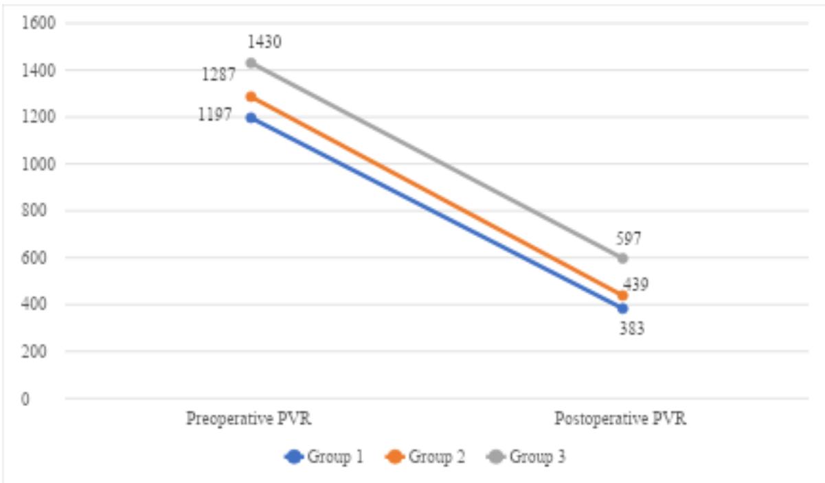
Surgical outcomes of all patients were thoroughly analyzed and compared with data of PVR changes before and after surgery in all three groups (Figures 3, 4). Residual PH and outcome depend on the preoperative level of PVR.
IV. DISCUSSION
PEA performed in specialized centers contributes to the normalization of the main parameters of the pulmonary circulation, improves long-term prognosis in patients with CTEPH, and is the preferred treatment method for patients with proximal pulmonary artery lesions. However, there is no universal algorithm for patient selection for surgery that could predict outcomes, especially with different times of medical history. Due to the nonspecific clinical presentation of CTEPH, misdiagnosis is common in clinical practice. As a result, there is often a significant delay in referring patients to expert centers for diagnosis confirmation and assessment for surgical treatment . In our center, as well as in other clinics worldwide, candidates for pulmonary PEA are selected by a multidisciplinary team, including a cardiovascular surgeon, anesthesiologist-intensivist, and cardiologist. As mentioned earlier, candidate selection is based on the overall patient condition, the characteristics and extent of pulmonary vascular lesions, and the presence of comorbidities
Anatomically, two different vascular lesions participate in the increasing of PVR in patients with CTEPH: an obstruction of pulmonary arteries by unresolved thrombi and a microvasculopathy observed in both obstructed and nonobstructed lung areas [12]. A microvasculopathy is also observed in completely obstructed lung areas and is attributed to the development of systemic bronchial arteries anastomosing with pulmonary arterioles and venules [13]. Simonneau G. and colleagues presented similar data regarding the possibility of collateral blood flow development bypassing pulmonary vascular obstructions in patients with long-term medical history, which increases the likelihood of residual PH postoperatively and an unfavorable surgical outcome [14]. The presence of microvasculopathy is suspected when mechanical obstruction does not correlate with the haemodynamic severity. The microvasculopathy may explain the persistence of PH after PEA [15]. Microvasculopathy is suspected when mechanical obstruction does not correlate with the haemodynamic severity and may explain the persistence of residual PH after PEA [15].
Historically, high PVR was considered as a potential contraindication to surgery , however, even in patients with the most severe forms of disease, characterised by a preoperative PVR of more than , the operative mortality has decreased to . Madani M. and colleagues in their study reported that a baseline PVR value over leads to a fourfold increase in preoperative mortality . According to data from an international registry, the mortality rate was 10.6% in patients with a baseline PVR above . Elevated PVR values exceeding , regardless of the lesion type, indicate a severe form of CTEPH and may be a contraindication for pulmonary PEA. However, a clearly expressed lesion in the proximal pulmonary artery, even with a high PVR, indicates potential effectiveness and safety of surgical intervention .
Bergin C. and colleagues confirmed that in patients with similar characteristics, unfavorable postoperative outcomes were associated not only with PVR >1000 dyn·s·cm but also with the presence of secondary microvasculopathy [19]. Residual PH rates ranged from 8.2% [20] to 41.9% [21]. Some previous studies provided solid evidence that patients with persistent or residual PH immediately after PEA have an increased risk of in-hospital death [18, 22]. These studies showed that mPAP >30 mmHg after PEA was relevant for the prognosis. On the other hand, the United Kingdom National Cohort showed that mPAP >38 mmHg measured 3 - 6 months postoperatively correlated with a poor long-term survival and a higher risk of CTEPH related death [23]. In our previous study we described, that it is rational to use 35 mmHg as a cut-off value, which is a mean data between 30 mmHg and 38 mmHg [10].
A long time before correct diagnosis may play a crucial role in the development of microvasculopathy and right heart failure. This leads to a worse prognosis with increasing complications rate and operative mortality . Hsieh W. and colleagues conducted a meta-analysis dedicated to residual PH after surgical treatment of CTEPH. The study showed that after PEA PAP and PVR usually decrease even in patients with distal-type lesions . Our results confirm this conclusion, demonstrating significant improvement in the hemodynamic parameters of the pulmonary circulation in all groups. All patients in the third group were included in study retrospectively, and at the time of surgery they were identified as potential candidates considering the proximal type of the pulmonary vascular lesions, despite the high PVR. This means that there was a relative correspondence between the severity of the arterial lumen obstruction and the degree of PH. In our study, we also referred to the work from the Meshalkin E. N. Institute of Circulation Pathology . Authors concluded that the time of medical history does not affect the possibility of PH reduction in the postoperative period. We confirmed that PEA significantly reduces PVR and improves hemodynamic parameters in all patients including those with long-term medical history. Nevertheless, complications such as residual PH, reperfusion pulmonary oedema and severe heart failure in the early postoperative period were significantly higher in patients with long-term medical history (anamnesis more than 3 years), with high hospital mortality.
The extent of microvasculopathy significantly affects the development of disease severity and postoperative outcomes. Therefore, analysis of small vessel damage in the lungs may be key to achieve the best surgical results . Further research in this field will help identify patients who achieve the best treatment outcomes using all available strategies and develop new therapies to prevent disease progression to irreversible right ventricular dysfunction.
V. CONCLUSION
PEA is an effective and safe treatment of CTEPH in patients with PVR less than 1000 dyn·s·cm . In patients with PVR more than 1000 dyn·s·cm and a clearly established time of medical history more than 3 years, PEAmay be effective in reducing of right heart failure and PH, however, the early postoperative period is associated with a higher frequency of complications and a high surgical mortality.
Limitations
Our research cannot definitively determine whether the above-mentioned patients with long-term medical history of CTEPH (more than 3 years) and with PVR more than 1000 dyn·s·cm were unsuitable for surgical intervention, leaving the question of operability open for further study.
What is Already Known?
After PEA PAP and PVR usually decreases even in patients with distal-type lesions.
What Does This Study Adds?
The long-term medical history of CTEPH (more than 3 years) is a risk factor for severe residual PH after PEA with high hospital mortality.
ACKNOWLEDGEMENTS
We express our gratitude to Bakulev National Medical Research Center for Cardiovascular Surgery for the support of our investigation and all the patients who took part in this research.
Conflicts of Interest
The authors declare no conflicts of interest regarding the publication of this paper.
Funds
Neither fund nor financial support have been received from anybody.
Conflict of Interest
The authors declare no conflict of interest.
Ethical Approval
Not applicable
Data Availability
The datasets used in this study are openly available at [repository link] and the source code is available on GitHub at [GitHub link].
Funding
This work did not receive any external funding.
References
Cite this article
Related Research



Special Issue
Launch a focused special issue to highlight research, emerging trends, and expert insights in your academic field.
