




IntelliPaper
Abstract
Oral manifestations accompanying the syndromes may help dentists in diagnosing this syndrome. This article reports the case of a Syrian 4-year-old girl with Kabuki syndrome and the oral/dental aspects of this syndrome; including hypodontia with interdental spacing, abnormal tooth morphology.
Explore Digital Article Text
I. INTRODUCTION
Kabuki syndrome (KS) was described in 1981 at two Japanese centers in the Kanto area and Hokkaido [1] [2]. It is a rare genetic disorder (congenital distortion syndrome) [3] which characterized by multiple congenital anomalies and mental disability [4] [5]. Proportion of Kabuki Syndrome is around 1/32,000 of births [3]. The main cause of Kabuki Syndrome is unknown. However, X-linked and autosomal dominant gene have been suggested [6] [7] [8].
Whereas KMT2D (MLL2) gene mutations were observed in most patients with Kabuki Syndrome, and a few have mutation or deletion of KDM6A [3]; thus the first pathogenic gene recognized in Kabuki Syndrome patients was KMT2D according to Ng, S.B., et al., study [9], But nearly in 30% of patients with Kabuki Syndrome, the Potential genetic defects are still unknown [3].
Diagnosis of the syndrome is based on 5 main clinical features: (1) a special face (100%) which characterized by protruding ears, depressed nasal tip and long palpebral fissures with eversion of the lateral third of the lower eyelids, higharched eyebrows with sparse lateral one-third, (2) Growth deficiency (83%) with short stature, (3) moderate to severe mental disability (92%), (4) skeletal anomalies (92%) and (5) abnormalities of dermatoglyphic (93%) [7] [10].
The face of patients with Kabuki syndrome is similar to the makeup worn by actors of Kabuki; a Japanese traditional play. That's why it's called Kabuki syndrome [8].
Other important clinical features have been reported include: early puberty, premature breast development in girls, anal atresia, congenital heart disorder, craniofacial anomalies, gastrointestinal anomalies, fingers abnormalities (Short fifth fingers), dental anomalies, [6] [7] [4] [5] [10] and renal and vertebral anomalies [3].
The most frequent oral manifestations reported were: cleft lip/palate; bifid tongue and uvula; malocclusion (micrognathia, severe maxillary recession, mid-facial hypoplasia, high-arched palate, widely spaced teeth); delayed tooth eruption pattern; dental abnormalities (hypodontia, conical teeth, neonatal teeth, large pulp chamber); diastema and lower lip pits [11] [12] [8].
Other oral manifestations reported in Do Prado Sobral et al., Study are: Developmental Enamel Defects (DED), Dental crowding, Atypical crown shape, Taurodontia, Microdontia, and Retained primary teeth [4].
This article documents the case of a Syrian girl diagnosed with Kabuki syndrome; addressing the clinical features observed, and the dental treatment submitted. Comparing oral manifestations previously described in the literature and those observed in this present case.
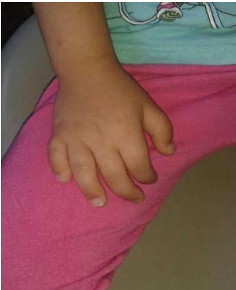 Figure 1: Short Fingers (absence of the third phalanx from the fifth finger)
II. CASE REPORT
A 4-years-old female child diagnosed with Kabuki syndrome by Pediatric Hospital (Damascus University), was referred to the department of pediatric dentistry (Faculty of Dentistry of, Damascus University) for dental care. Chief complaint was the presence of teeth caries. The girl was born to healthy parents after full-term pregnancy and had been diagnosed with Kabuki syndrome. Examination and syndrome diagnosis were done by pediatric specialist and genetic clinic in pediatric hospital and the hospital's report shows stature incompatible with her chronological age; craniosynostosis with Microcephaly. Physical examination shows short fingers (absence of the third phalanx from the fifth finger) (Figure. 1), special facies consisting of narrow front, high-arched eyebrows with scattered lateral one-third, elongated palpebral fissures, eyes with eversion of the lateral one-third of the lower eyelids, small eyeball, prominent ears, broad depressed nasal root with flat nasal tip. (Figure. 2) Anal atresia which was treated surgically within a few days of giving birth delivery.
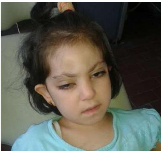
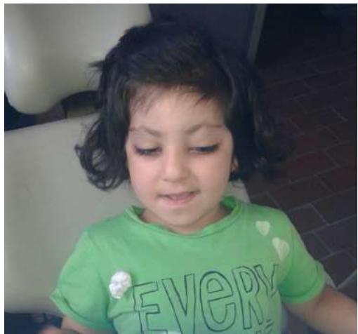 Figure 2: Special Face Consisting of Narrow Front, High-Arched Eyebrows With Sparse Lateral One-Third, Eyes with Eversion of the Lateral One-Third of the Lower Eyelids, Small Eyeball, Prominent Ears, Broad Depressed Nasal Root With Flat Nasal Tip
The girl showed a noticeable improvement acquired psychomotor skills, she is able to run and go up the stairs, formulate sentences of three words, count until number 5, and Play with dolls. She has a moody and shy behavior.
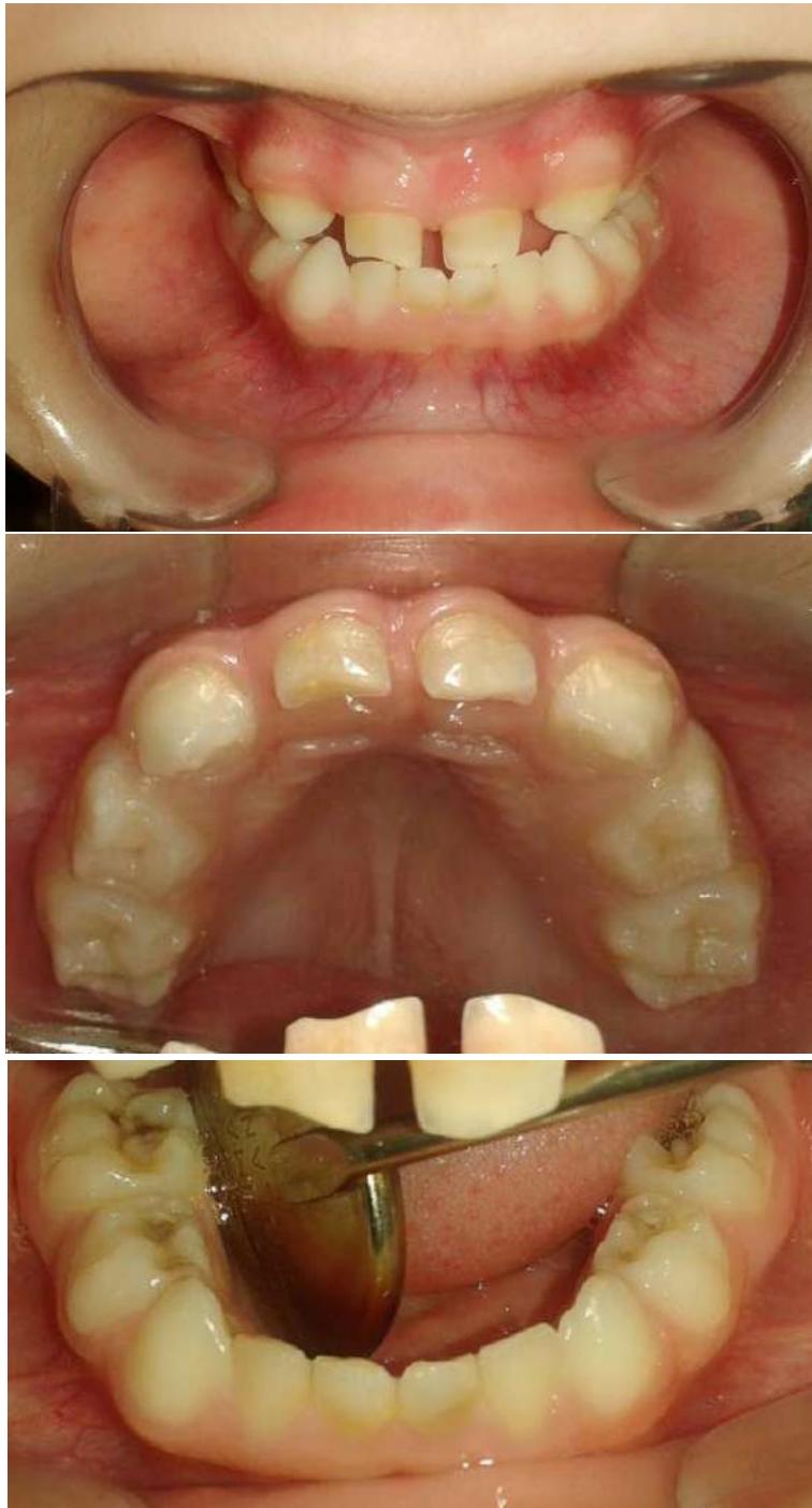 Figure 3: (a) Intra-Oral View of Anterior Teeth, (b) Maxillary Occlusal View, (c) Mandibular Occlusal View
2.1 Intra-Oral Examination
In intraoral examination; no abnormalities were observed in lips, tongue and oral mucosa.
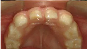
The patient was in primary dentition stage; with edge-to-edge bite, high-arched palate, in the upper arch the present teeth were primary central incisors, primary canines, first and second primary molars, the upper incisors as 'flat head' screwdriver-shaped, in the lower arch the present teeth were primary central and lateral incisors, primary canines, first and second primary molars.
The primary maxillary lateral incisors were absent (hypodontia) with interdental spacing.
Carious cavities were seen in the mandibular first and second primary molars.
2.2 Radiographic Findings
A panoramic photo (Figure. 4) showed carious mandibular second primary molars. The primary maxillary lateral incisors (previously noted as absent), the maxillary permanent lateral incisors buds and mandibular permanent central incisors buds, and the right mandibular permanent lateral incisors buds were absent, while the maxillary permanent second and third molars and the mandibular third molars buds were not considered as absent because they need time to develop.
According to the American Association of Pediatric Dentistry (AAPD), this case is classified as Early Childhood Caries (ECC) [13].
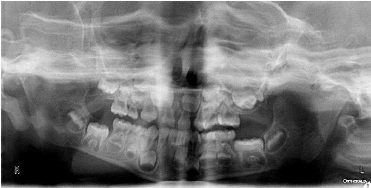 Figure 4: Panoramic Photo
2.3 Treatment
The girl's behavior was Negative (Reluctance to accept treatment, uncooperativeness, some evidence of negative attitude but not pronounced (sullen, withdrawn) according to the Frankl behavior rating scale [14].
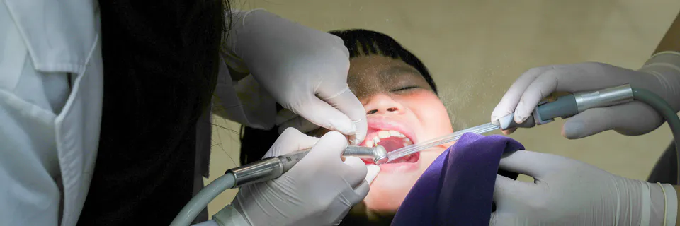
The dental treatment was accomplished under intravenous sedation (one session) by an anesthesiologist in the oral and maxillofacial surgery hospital, Damascus University.
Whereas a medical specialist consultation for sedation had been requested, and pre-sedation dietary instructions which determined by the American Academy of Pediatric Dentistry had been given: (1). Clear liquids: up to 2 hours before the procedure. (2). Breast milk up to 4 hours before the procedure. (3). Infant formula, nonhuman milk or a light meal up to 6 hours before the procedure [15]. And the medication used in IV sedation was (Midazolam 1.5 mg, Fentanyl, 7 Microgram and Propofol 30 Mg) (girl weighed 15 kilograms), while the treatment session lasted 45 minutes. Dental treatment included:
-
Pulpotomy of the left primary second mandibular molar and applying of Stainless-steel crown (SSC).
-
Restoration of the right and left primary first mandibular molars with amalgam.
-
Hall Technique on right primary second mandibular molar.
-
Sealing of the left and right primary maxillary molars.
-
Parent was given oral hygiene instruction.
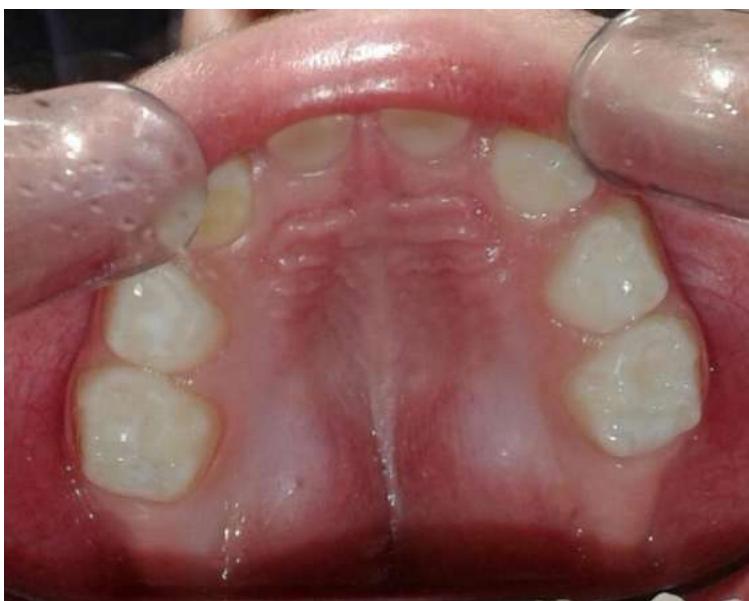
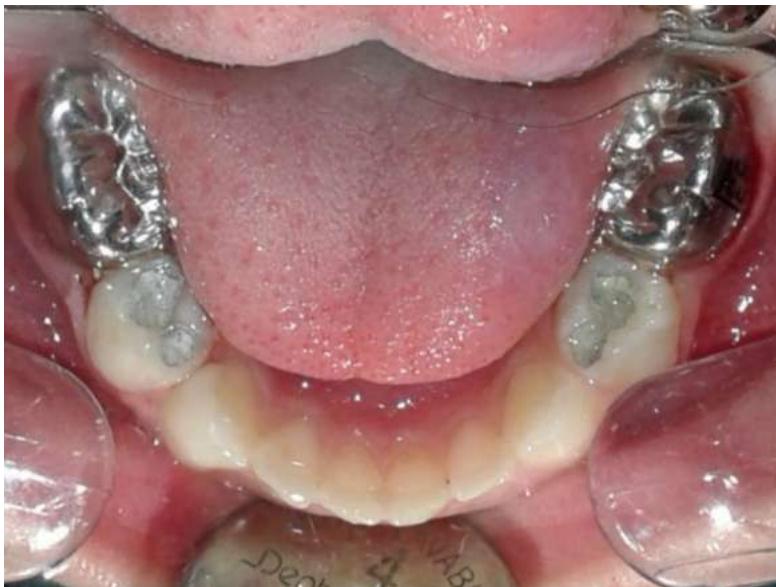 Figure 5: Maxillary Occlusal View after Treatment Figure 6: Mandibular Occlusal View after Treatment
IV. DISCUSSION
Kabuki syndrome is considered as a rare condition; although dentists may find difficult to understand the case, but they may contribute to the diagnosis, so it is important to know the facial and oral clinical manifestations accompanying this syndrome to request further examinations when noticing any changes in the normal state.
The etiology of the Syndrome is unclear, and diagnosis is clinically and mainly based on facial features in addition to other clinical features: Growth deficiency, mental disability, skeletal anomalies, abnormalities of dermatoglyphic [4].
Typical facial features can be identified from an early age to help in clinical diagnosis. However; clinical identification of the syndrome in the neonate is difficult, maybe the phenotype is developed by the time .
The patient in this case has a short stature, craniosynostosis with Microcephaly, short finger, special facies consisting of narrow Front, high-arched eyebrows with sparse lateral one-third, elongated palpebral fissures, eyes with eversion of the lateral one-third of the lower eyelids, Small eyeball, prominent ears, broad depressed nasal root with flat nasal tip. These manifestations also reported by Petzold et al.,
2003, Lung and Rennie, 2006, Dos Santos et al., 2006, Cudzilo and Czochrowska, 2018, Shangguan et al., 2019, and Santos et al., 2019 [16], [12], [17], [18], [10], [19].
Dental abnormalities have been reported in over 60% of patients with Kabuki Syndrome [11] [17] [20]. The most common dental finding was the hypodontia [8], in this case Maxillary primary lateral incisors, the maxillary permanent lateral incisors buds, mandibular permanent central incisors buds, and the right mandibular permanent lateral incisors buds were absent, these findings about missing teeth are in agreement with the literature reported by Mhanni et al., Matsune et al., Petzold et al., Lung and Rennie, dos Santos et al., Teixeira et al., and Tuna et al., [21], [11], [16], [12], [17], [8], [20]. The finding of absent premolars or molars as described by Mhanni et al., Tuna et al., and do Prado Sobral et al., were not observed [21], [20], [4].
Space between maxillary teeth that was found in this case is associated with hypodontia, and this characteristic was reported previously by Petzold et al., [16], in addition to the high-arched palate which also observed by Matsune et al., and do Prado Sobral et al., [11], [4].
The upper incisors were as 'flat head' screwdriver-shaped, this finding was reported by Mhanni et al., Petzold et al., Lung and Rennie, Do Prado Sobral et al., and Cudzilo and Czochrowska [21], [16], [12], [4], [18], whereas Dental shape abnormalities were not observed in Teixeira et al., who study dental examination and panoramic radiography of nine patients [8]. There are no lips, oral mucosa or tongue abnormalities observed in this present case.
Pediatric dentists should choose the best behavioral management technique that fit the patient status as well as the procedure nature which needs to be accomplished, and they have often found that anxiety and behavioral assessment to be helpful in determining the behavioral management technique to be chosen for each child [15].
Sedation requires an accurate medical history accomplishment to determine whether the patient a good candidate to it or not. The American Society of Anesthesiologists (ASA) guidelines are considered as the most accurate method when taking patients medical history , and case in this report is classified as ASA class II that is frequently considered appropriate candidate for minimal, moderate, or deep sedation. However, counsel with an anesthesiologist is often desired .
While IV Sedation can be a suitable alternative to general anesthesia for children with ECC and the equipment to provide general anesthesia is far more expensive than what is required for IV sedation , in addition to the girl's behavior in this case was negative according to the Frankl behavior rating scale as well as the treatments were required enough time to be accomplished, therefore dental treatment in this case was done under intravenous sedation.
V. CONCLUSIONS
The dental manifestations observed in this case were hypodontia, abnormal teeth morphology and high-arched palate.
These dental abnormalities in addition to another clinical features may help in the clinical diagnosis of the syndrome, so It is important that dentist be aware of this syndrome and its facial manifestations and oral/dental findings to recognize children who may be affected by this disorder.
Conflict of Interest
The authors declare no conflict of interest.
Ethical Approval
Not applicable
Data Availability
The datasets used in this study are openly available at [repository link] and the source code is available on GitHub at [GitHub link].
Funding
This work did not receive any external funding.
References
Cite this article
Related Research



Special Issue
Launch a focused special issue to highlight research, emerging trends, and expert insights in your academic field.
